Abstract
Objectives
We wanted to analyze the radiological features of degenerative lumbar spondylolisthesis and retrolisthesis, and we wanted to verify what radiological factors are related to the development of the retrolisthesis. We also wanted to determine these radiological factors' clinical significance.
Summary of the literature review
There is little information about the pathological mechanism and the clinical and radiological aspects of degenerative lumbar retrolisthsis.
Materials & methods
Sixty patients were reviewed and divided into three groups. The degenerative lumbar retrolisthesis patients were in group A. The degenerative lumbar spondylolisthesis patients were in group B. Group C patients had no vertebral shift in any direction. The factors we measured were the facet joint angle, the disc height of L3- 4, L4- 5 and L5- S1, and the lordosis of the lumbar spine. The evaluation of the clinical results was then quantified.
Results
The facet joint angle showed no statistical significance between the two groups. The disc height of group A at L4- 5 and L5- S1 was more decreased in group A than in group B (p<0.05). Lumbar lordosis was decreased significantly in group A (p<0.05). The preoperative pain was improved at the final follow up, but preoperative pain was significantly higher in group A than in group B (p<0.05). The clinical results were improved in each group, but there was no statistically significant difference between the two groups.
Conclusions
The disc height and lumbar lordosis were considerably reduced in the patients with retrolisthesis, especially compared to those patients with spondylolisthesis. Preoperative pain was higher for the retrolisthesis patients than for the spondy-lolithesis patients, but there was no significant difference.
REFERENCES
1). Farfan HF, Huberdeau RM, Dubow HI. Lumbar intervertebral disc degeneration. The influence of geometrical features on the pattern of disc degeneration. J Bone Joint Surg Am. 1972; 54:492–510.
2). Farfan HF. Mechanical disorders of the low back. Lea and Febiger;Philadelphia: 1973. 33-40.
3). Inoue S, Watanabe T, Goto S, Takahashi K, Takada K, Sho E. Degenerative spondylolisthesis. Pathophysiology and results of anterior interbody fusion. Clin Orthop. 1988; 227:90–98.

4). Knutsson F. The instability associated with disc degeneration in the lumbar spine. Acta Radiol. 1944; 25:593–609.
5). Kummer B. Funktionelle und Pathologische Anatomie der Lendenwirbelsaule. Orthop. Praxis. 1982; 18:84–90.
6). Lorenz R, Patwardhan A, Vanerby R Jr. Load-bearing characteristics of lumbar facets in normal and surgically altered spinal segments. Spine. 1983; 8:122–130.

7). Yang KH, King AI. Mechanism of facet load transmission as a hypothesis for low-back pain. Spine. 1984; 9:557–565.

8). Farfan HF, Sullivan JD. The relation of facet orientation to intervertebral disc failure. Can J Surg. 1967; 10:179–185.
9). Grobler LJ, Robertson PA, Novotny JE, Pope MH. Etiology of spondylolisthesis. Assessment of the role played by lumbar facet joint morphology. Spine. 1993; 18:80–91.
10). Boden SD, Riew KD, Yamaguchi K, Branch TP, Schellinger D, Wiesel SW. Orientation of the lumbar facet joints: Association with degenerative disc disease. J Bone Joint Surg Am. 1996; 78A:403–411.
11). Berlemann U, Jeszenszky DJ, Buhler DW, Harms J. Mechanisms of retrolisthesis in the lower lumbar spine. A radiologic study. Acta Orthopaedica Belgica. 1999; 65:472–477.
12). Vogt MT, Rubin DA, Palermo L, Christianson L, Kang JD, Nevitt MC, Cauley JA. Lumbar spine listhesis in older African American women. Spine. 2003; 3:255–261.

13). van Akkerveeken PF, O'Brien JP, Park WM. Experimental induced hypermobility in the lumbar spine: A pathologic and radiologic study of the posterior ligament and annulus fibrosus. Spine. 1979; 4:236–241.
14). Rosenberg NJ. Degenerative spondylolisthesis. Predis -posing factors. J Bone Joint Surg Am. 1975; 57:467–474.
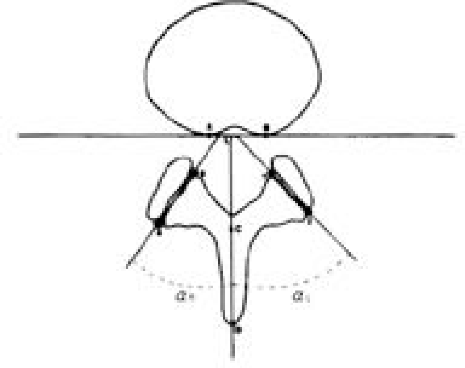
Fig. 1.
Measurement of facet joint angulation in the transverse plane. A line parallel to the posterior vertebral body wall serves as reference.

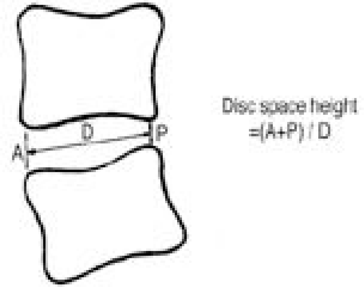
Fig. 2.
Measurement of disc height as Farfan index. The sum of anterior disc height A and posterior height B is divided by sagittal disc width D.

Table 1.
Average values of facet joint angle in each group
| Group A | Group B | |
|---|---|---|
| L3-4 (°) | 37.25± 6.22° | 43.57± 10.84° |
| L4-5 (°) | 41.61± 6.19° | 44.89± 8.94°0 |
| L5-S1 (°) | 45.82± 7.59° | 48.16± 9.6°00 |
Table 2.
Average values of disc height in each group
| Group A | Group B | Group C | |
|---|---|---|---|
| L3-4 | 47.55± 11.57 | 54.17± 6.500 | 55.23± 5.340 |
| L4-5 | 46.28± 14.82 | 60.55± 12.39 | 62.89± 10.92 |
| L5-S1 | 47.63± 13.77 | 65.63± 12.30 | 66.19± 9.750 |
Table 3.
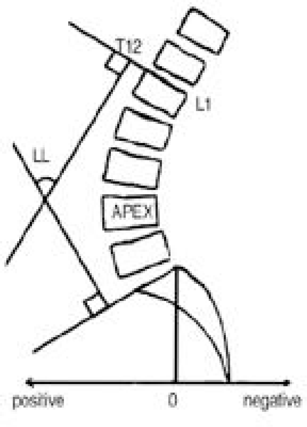
Average values of lumbar lordosis in each group
| Group A | Group B | |
|---|---|---|
| lumbar lordosis | 24.68± 9.85° | 48.35± 9.28° |


 PDF
PDF ePub
ePub Citation
 Print
Print


 XML Download