Abstract
Study Design
We retrospectively evaluated the value of magnetic resonance imaging (MRI) in the diagnosis of lumbar lateral disc herniations.
Summary of Literature Review
MRI is known to be a reliable study for the diagnosis of a lumbar disc herniation. However, recent studies of its diagnostic value for lateral disc herniation have been rare.
Objectives
We aimed to assess the diagnostic value of simple MRI, to determine the need for additional imaging studies and to investigate mimicking lesions.
Materials and Methods
In a lateral herniation group composed of 21 cases, including 10 foraminal and 11 extraforaminal herniations, the diagnostic value of simple MRI was evaluated, and the potential requirement for additional studies investigated. In a mimicking lesion group(5 cases), the entity of each lesion was identified.
Results
All 10 foraminal disc herniation cases were able to be confirmed with simple MRI, six of which were confirmed using sagittal images alone. In contrast, for the eleven extraforaminal disc herniations, sagittal MR images were not at all helpful in the diagnosis; however, six(55%) were confirmed from axial images, but the other five could not be confirmed until additional studies, such as enhanced MRI(4 cases), 1mm- sliced CT (1) and CT-discography (3), were carried out. All 5 mimicking lesions were upper endplates of the lower vertebrae.
Conclusions
Simple MRI is useful in the diagnosis of foraminal herniations, but not so helpful for extraforaminal herniations; particularly, sagittal images are of little use. Therefore, whenever a patient complaining of severe radiating pain presents with no causative finding on simple MRI, the extraforaminal regions on the axial images should be diligently scrutinized again, and additional studies considered when necessary. Conversely, mimicking lesions, such as an upper endplate, should be differentiated when a lateral disc herniation is suspected.
Go to : 
REFERENCES
1). Abdullah AF, Ditton EW III, Byrd EB. Extreme-lateral lumbar disc herniations: clinical syndrome and special problems of diagnosis. J Neurosurg. 1974; 41:229–234.
2). Antuaco EJC, Holder JC, Boop WC, Binet EF. Computed tomographic diagnosis in the evaluation of extreme lateral disc herniation. Neurosurgery. 1984; 14:350–352.
3). Benini A. Der Zugang zu den lateralen lumbalen diskush -ernien am beispiel einer hernie L4/L5. Operat Orthop Traumatol. 1988; 10:103–116. (cited from Greiner-Perth R, Bohm H, Allam Y: A new technique for the treatment of lumbar far lateral disc herniation: technical note and preliminary results. Eur Spine J. 2003; 12:320–324. .).
4). Jackson RP, Clah JJ. Foraminal and extraforaminal lumbar disc herniation: Diagnosis and treatment. Spine. 1987; 12:577–585.

5). Kornberg M. Extreme lateral lumbar disc herniations: clinical syndrome and computed tomography recognition. Spine. 1987; 12:586–589.
6). Lindblom K. Protrusions of disks and nerve compression in the lumbar region. Acta Radiol. 1944; 25:195–212.

7). Macnab I. Negative disc exploration: an analysis of the causes of nerve-root involvement in sixty-eight patients. J Bone Joint Surg. 1971; 53-A:891–903.
8). Maroon JC, Kopitnik TA, Schulhof LA, Abla A, Wilberger JE. Diagnosis and microsurgical approach to far-lateral disc herniation in the lumbar spine. J Neurosurg. 1990; 72:378–382.

9). O'Hara LJ, Marshall RW. Far lateral lumbar disc herniation. The key to the intertransverse approach. J Bone Joint Surg. 1997; 79-B:943–947.
10). Ohmori K, Kanamori M, Kawaguchi Y, Ishihara H, Kimura T. Clinical features of extraforaminal lumbar disc herniation based on the radiographic location of the dorsal root ganglion. Spine. 2001; 26:662–666.

11). Osborn AG, Hood RS, Sherry RG. CT/MR spectrum of far lateral and anterior lumbosacral disk herniations. Am J Neuroradiol. 1998; 9:775–778.
12). Patrick BS. Extreme lateral ruptures of lumbar intervertebral discs. Surg Neurol. 1975; 3:301–304.
13). Schlesinger SM, Fankhauser H, de Tribolet N. Microsurgical anatomy and operative technique for extreme lateral lumbar disc herniations. Acta Neurochir (Wien). 1992; 118:117–129.

14). Segnarbieux F, Van de Kelft E, Candon E, Bitoun J, Frerebeau P. Disco-computed tomography in extraforaminal and foraminal lumbar disc herniation: influence on surgical approaches. Neurosurgery. 1994; 34:643–647.
Go to : 
Figures and Tables%
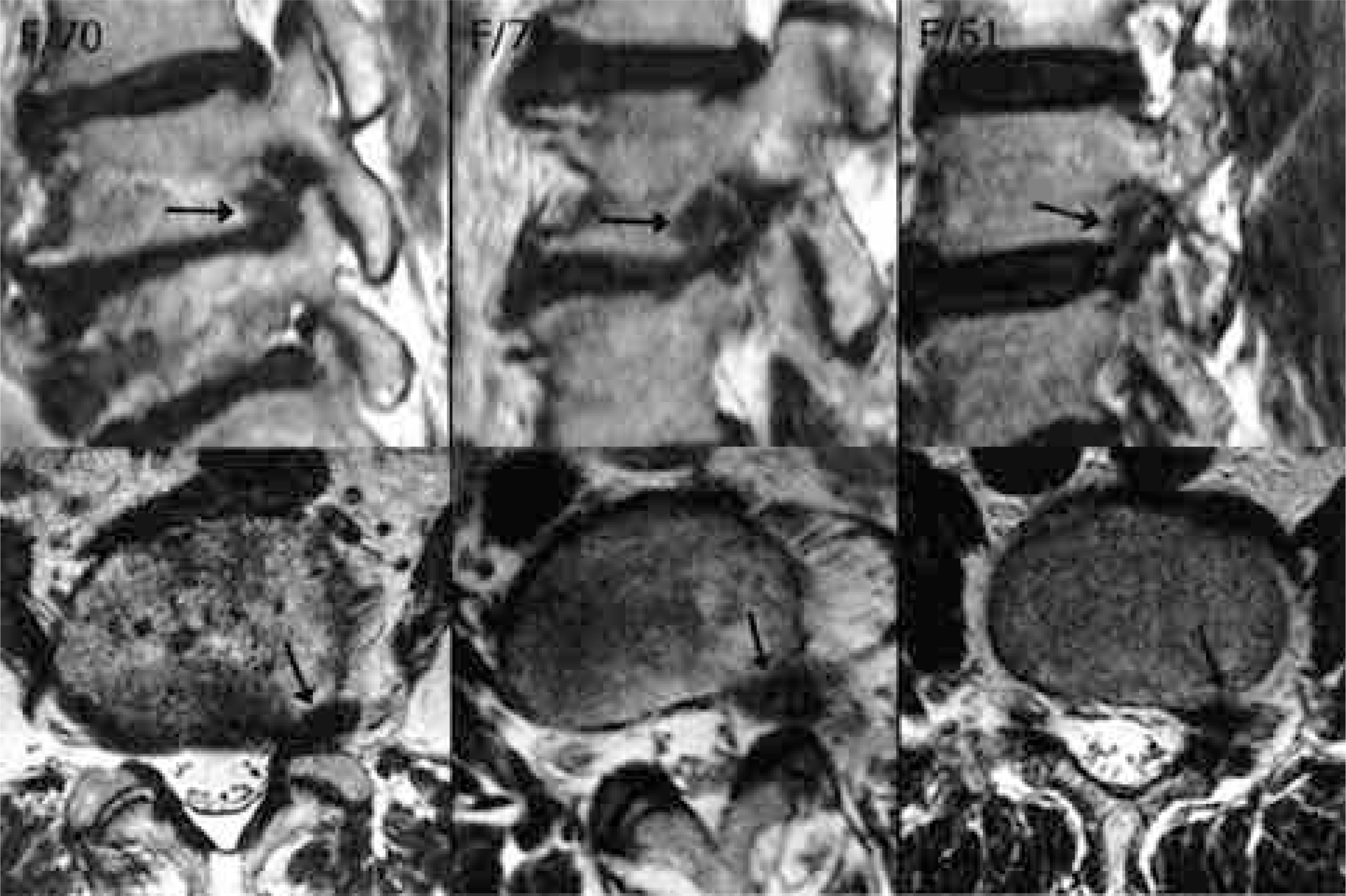 | Fig. 1.MR images of foraminal disc herniation. These three cases could be confirmed using sagittal images alone. |
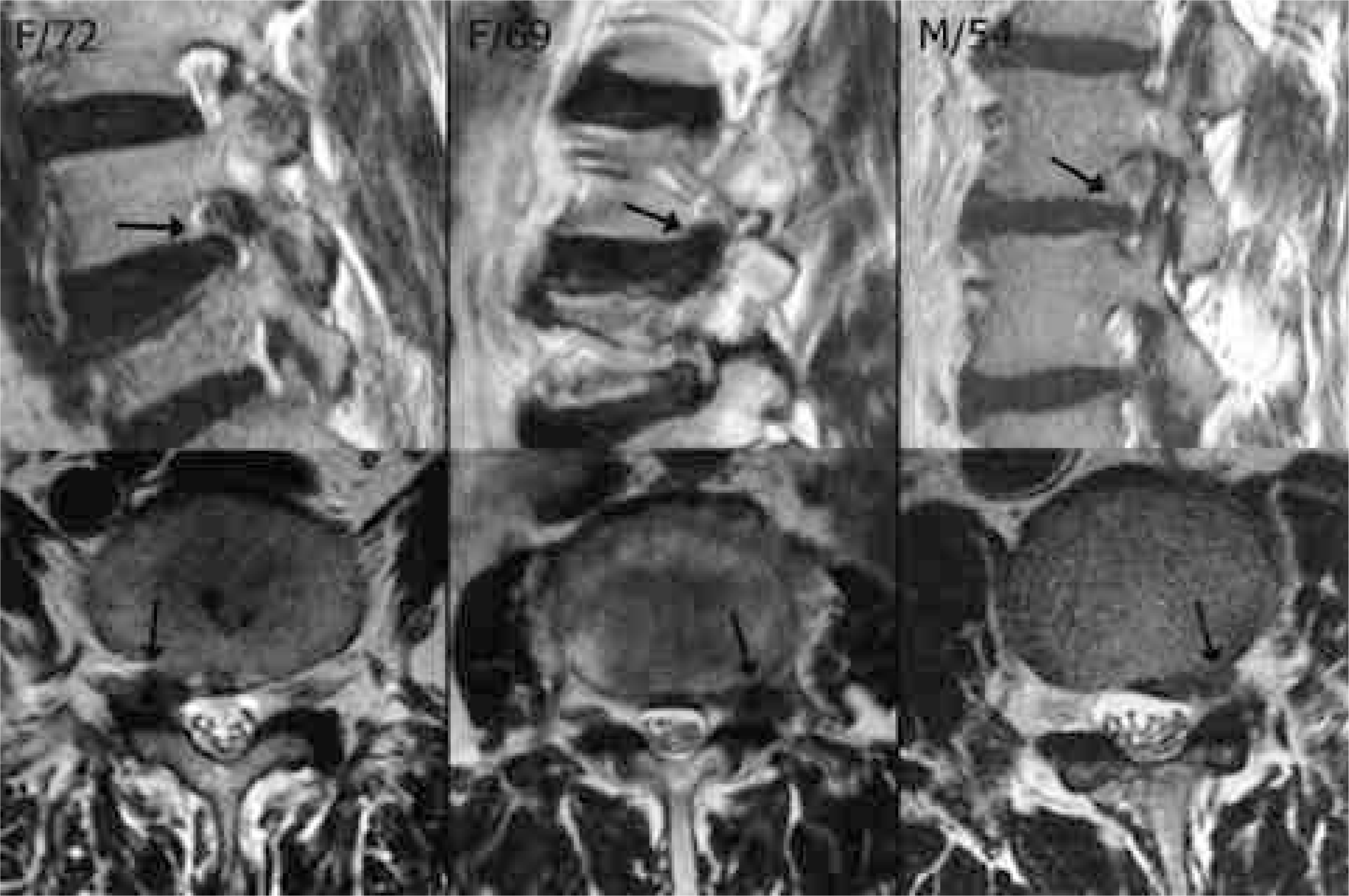 | Fig. 2.MR images of foraminal disc herniation. These three cases could not be confirmed with sagittal images alone, but together with axial images. |
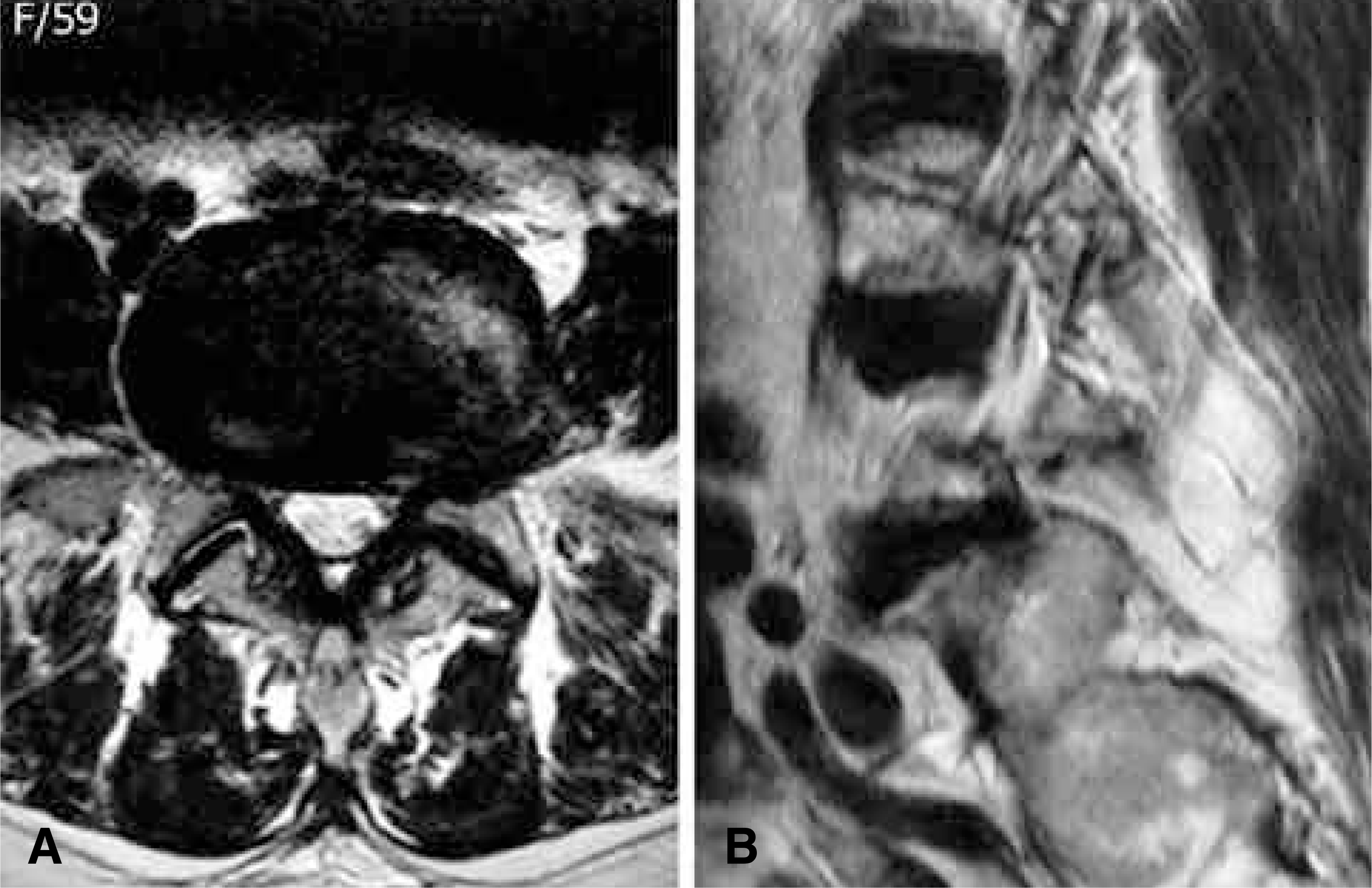 | Fig. 3.Axial and sagittal images of extraforaminal disc herniation. Extraforaminal herniation on the left side of L5-S1 is clearly observed on an axial image (A). However, it is not confirmative on the sagittal image the position of which is lateral to the pedicles. (B). This is not because the position of the sagittal image is not lateral enough, but because it does not have reliable anatomical landmarks. |
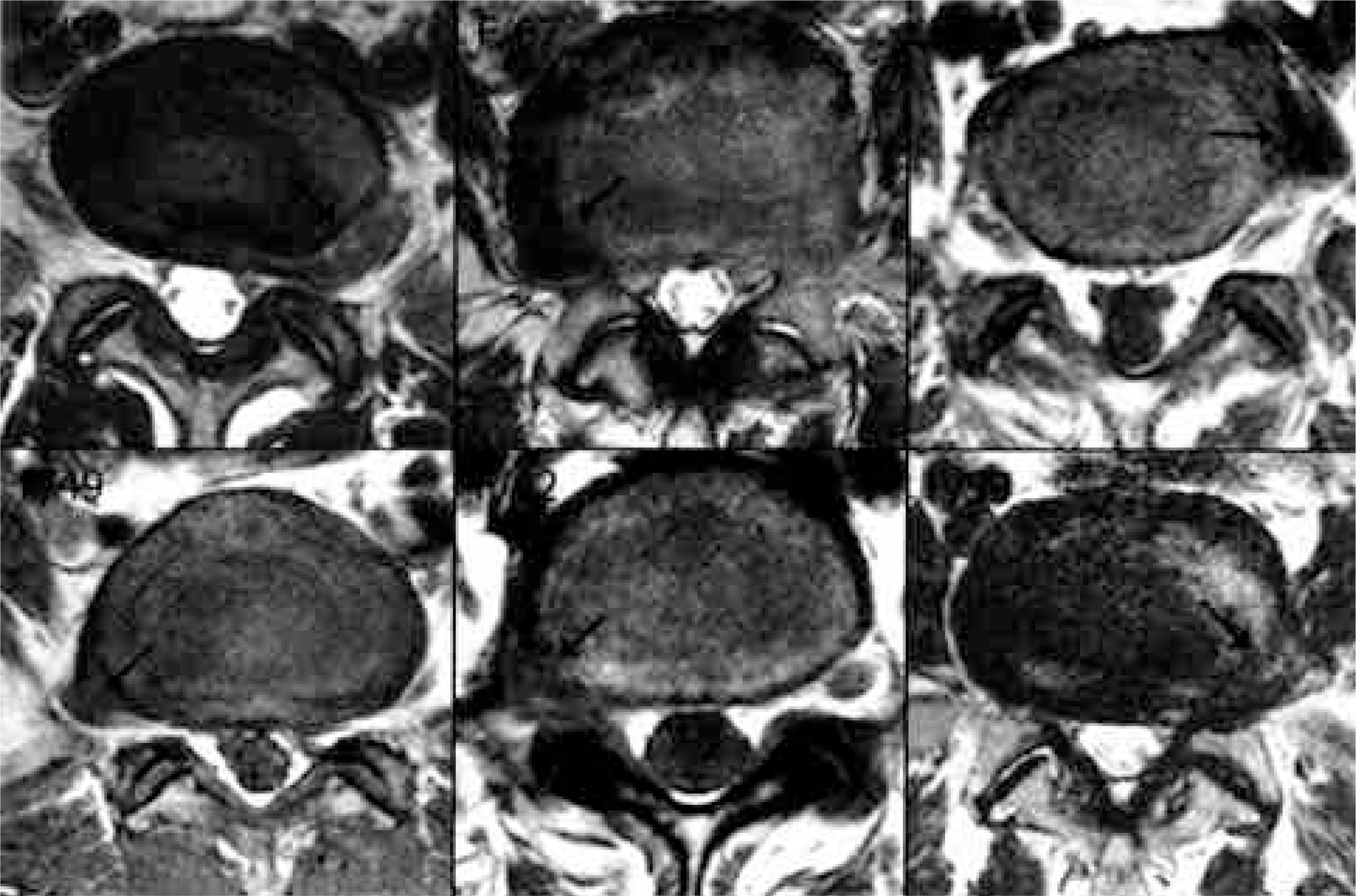 | Fig. 4.MR images of extraforaminal disc herniation. These six cases could be confirmed using simple MR images alone. |
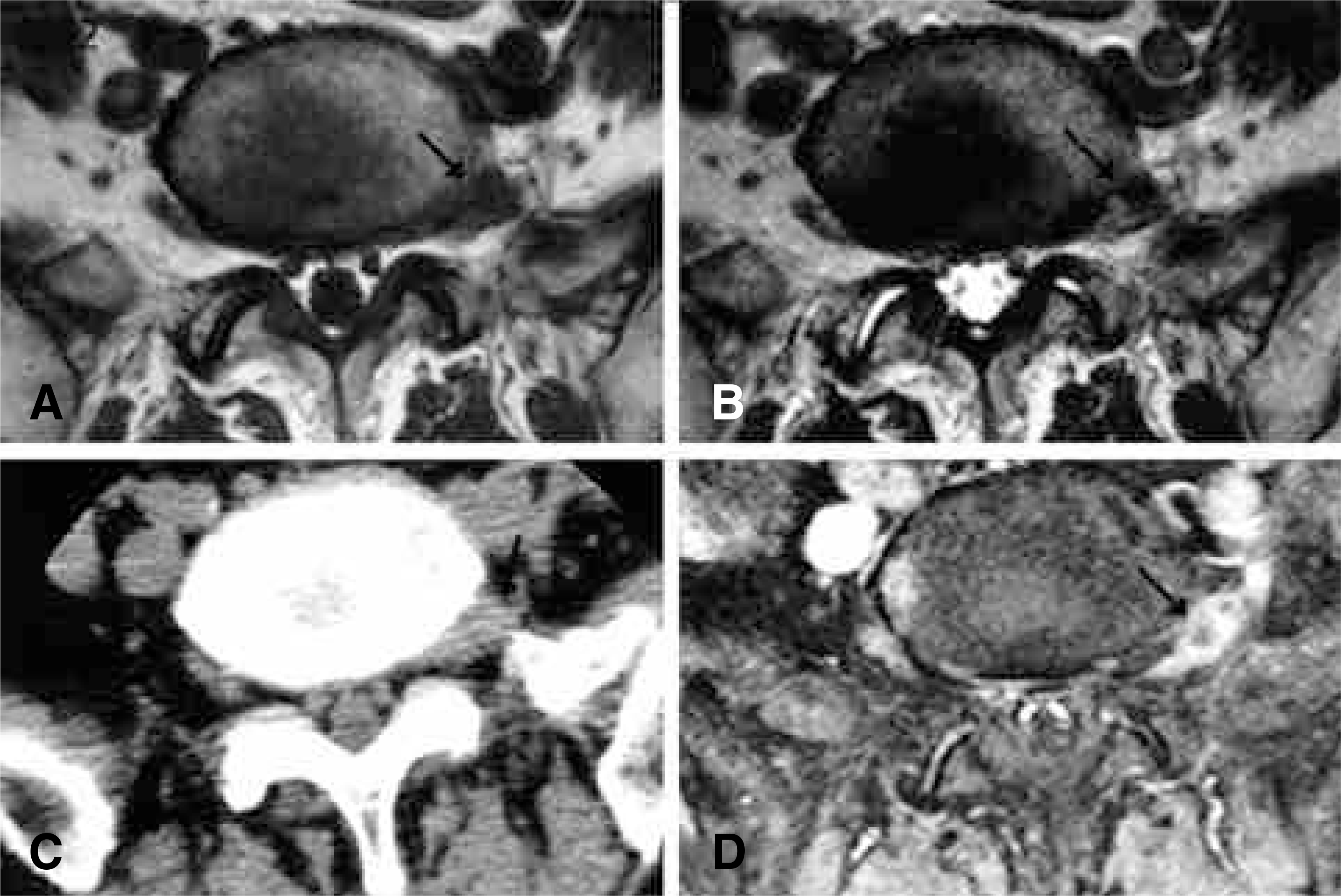 | Fig. 5.MR and CT scan images of a patient with extraforaminal disc herniation. It was suspected on T1WI (A), T2WI (B) and CT scan images (C), and confirmed with enhanced MR images showing peripheral enhancement (D). |
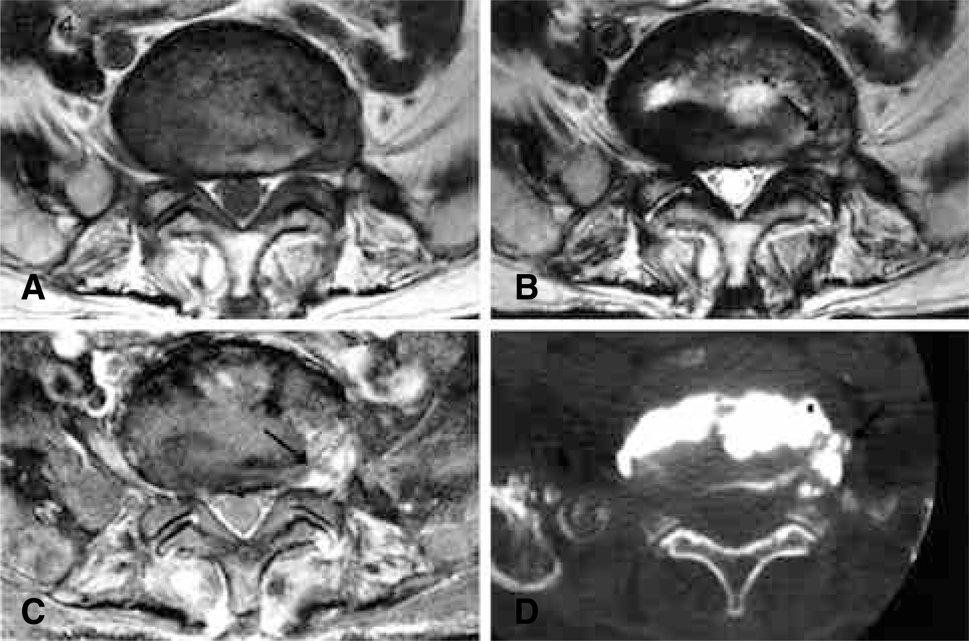 | Fig. 6.MR and CT-discography images of a patient with extraforaminal disc herniation. A suspected lesion on T1WI (A) and T2WI (B) showed heterogeneous enhancement (C), which is not a typical finding of disc herniation. Therefore CT-discography was performed (D), which confirmed the diagnosis. |
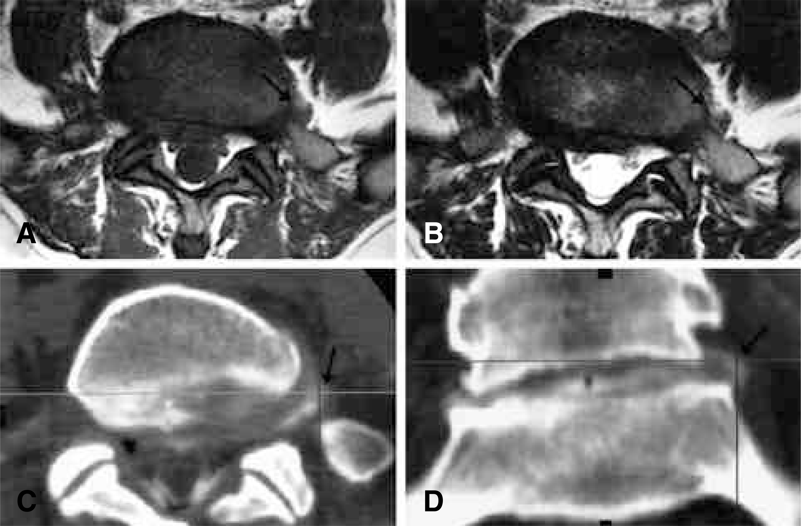 | Fig. 7.MR and CT-discography images of a patient with extraforaminal disc herniation. She had extremely severe radiating pain, but MR findings (A, B) were almost normal. Extraforaminal herniation was found on axial and coronal CT-discography images (C, D). |
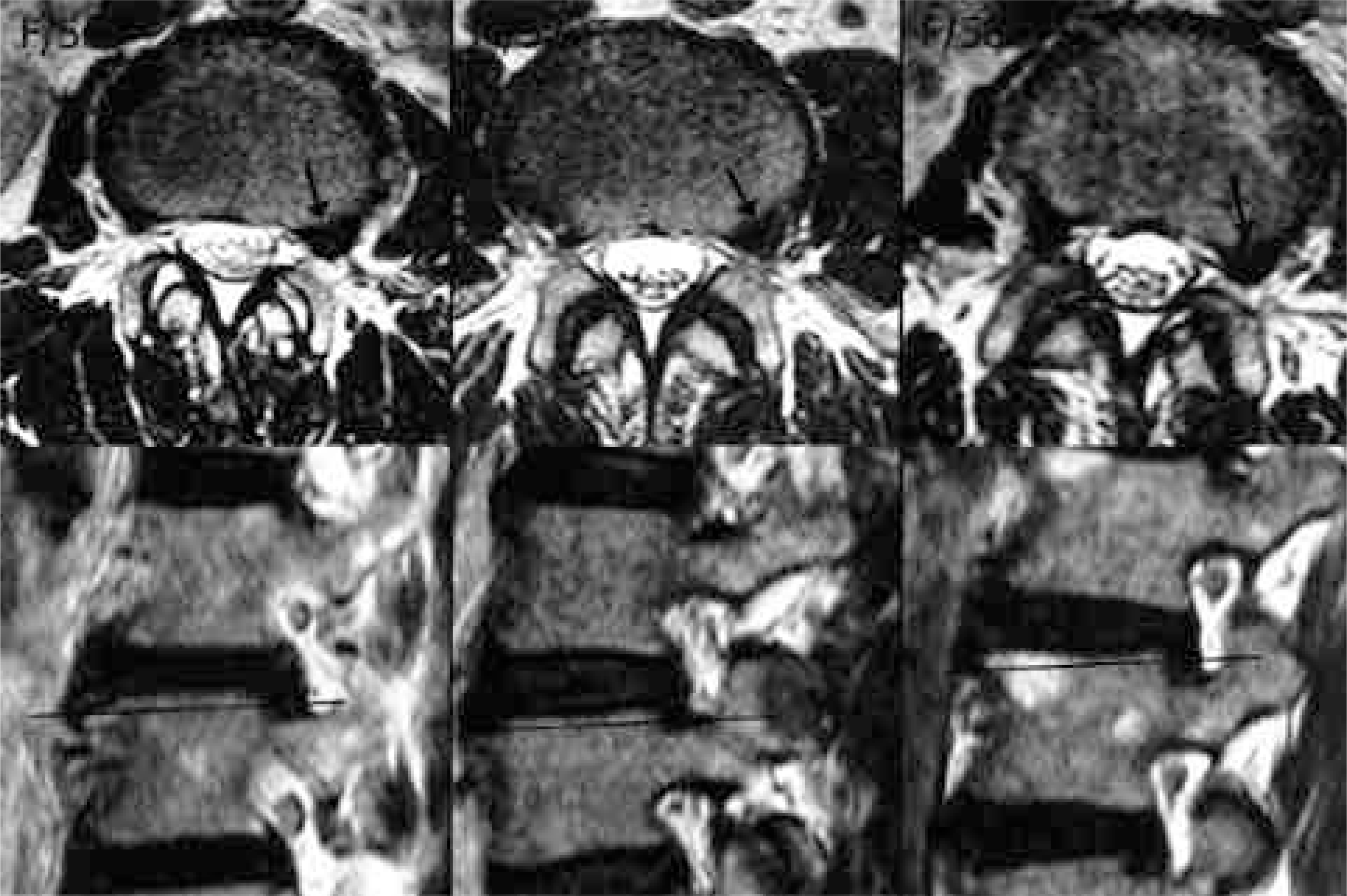 | Fig. 8.MR images of mimicking lesions. Every lesion was an upper endplates of lower vertebra. |
Table 1.
Cases Confirmed as Foraminal Herniations
Table 2.
Cases Confirmed as Extraforaminal Herniations


 PDF
PDF ePub
ePub Citation
 Print
Print


 XML Download