

 PDF
PDF ePub
ePub Citation
 Print
Print


Abstract
Study Design
This was a retrospective study to evaluate anterior cervical interbody fusion with plates.
Objective
To examine the degree of angulation and translation after an anterior interbody fusion, using anterior plate fixation, upon the fusion rate and clinical outcome.
Summary of Literature Review
A nterior cervical interbody fusion with plate allows immediate rigid internal fixation after decompression and bone grafting
Materials and Methods
65 cases had an anterior interbody fusion on the cervical spine, using an anterior approach and Smith-Robinson’s method, between January 1998 and A ugust 2003. Of these, 41cases, which could be followed up for at least one year, were selected. There were 26 and 15 males and females, respectively, with an average age of 43.5 and mean follow up period of 2.1years. 15 cases underwent an operation due to dislocation or fracture of the cervical spine due to trauma, and 26 due to cervical diseases. The angulation and translation of the plate was measured by postoperative X - rays. The fusion rate was also determined by the follow up X - rays. The Chisquared test was used to analyze the data.
Results
Bony fusion was obtained in all cases. Two patients developed hoarseness and one showed torticollis, but all had recov-ered by the follow up. The average angulation of the plate and translation were 6.2 degrees and 3.21mm, respectively, but there was no significant difference of the interbody fusion period due to angulation and translation of the plate or in the improvement of the clinical outcomes.
REFERENCES
1). Robinson RA, Smith SW. Anterolateral cervical disc removing and interbody fusion for cervical disc syndrome. Bull John Hopkins Hospital. 1955; 96:223–224.
2). Bohlmann HH, Emery SE, Goodfellow DB. Robinson anterior cervical discectomy and arthrodesis for cervical radiculoapthy. Instr Course Lect Am Acad Orthop Surg. 1993; 75:1298–1307.
3). Bohlmann HH. Cervical spondylosis with moderate to severe myelopathy: A report of seventeen cases treated by Robinson anterior cervical discectomy and fusion Spine. 1977; 2:151–6.
4). Bohler J, Gaudernak T. Anterior plate stabilization for fracture-dislocation of the lower cervical spine. J Trauma. 1980; 20:203–205.
5). Brener AM, Nguyen TQ. Internal metal plate fixation combined with anterior interbody fusion in cases of cervical spine injury. Neurosurgery. 1983; 12:649–653.

6). Casper W, Barbier DD, Klara PM. Anterior cervical fusion and Casper plate stabilization for cervical trauma. Neurosurgery. 1989; 25:491–502.
7). Vaccaro AR, Falatyn SP, Scuderi GJ. Early failure of long segment anterior cervical plate fixation. J Spinal Disord. 1998; 11:410–415.
8). Brantigan JW. Pseudarthrosis rate after allograft posterior lumbar interbody fusion with pedicle screw and plate fixation. Spine. 1994; 19:1271–1280.
9). Bell GD, Bailey SI. Anterior cervical fusion for trauma. Clin Orthop. 1977; 128:155–158.
10). Wang JC, McDonough PW, Endow K, Kanim LE, Delamarter RB. The effect of cervical plating on single-level anterior cervical discectomy and fusion. J Spinal Disord. 1999; 12:467–471.
11). Griffith SL, Zobgi SW, Guyer RD, Shelokov AP, Con-tiliano JH, Geiger JM. Biomechanical comparison of anterior instrumentation for the cervical spine. J Spinal Disord. 1995; 8:429–438.
12). Karasick D. Anterior cervical spine fusion. Struts, plugs and plates. Skeletal Radiol. 1993; 22:85–94.
13). Brunton FJ, Wilkinson JA, Wise KSH, Simmonis RB. Cineradiography in cervical spondylolysis as a means of determining the level for anterior fusion. J Bone Joint Surg. 1982; 64-B:399–404.
14). Clements DH, O’ leary PF. Anterior cervical discectomy and fusion. Spine. 1990; 15:1023–1025.
15). Gore DR, Sepic SB. Anterior cervical fusion for degenerated or protruded disc. A review of one hundred forty-six patients. Spine. 1984; 9:667–671.
16). Stauffer S, Kelly EG. Fracture-dislocation of the cervical spine instability and recurrent deformity following treatment by anterior interbody fusion. J Bone Joint Surg. 1977; 59-A:45–47.
17). Brodsky AE, Khalili MA, Sassard WR, Newman BP. Repair of symptomatic pseudarthrosis of anterior cervical fusion. Posterior versus anterior repair. Spine. 1992; 17:1137–1143.
18). Farey ID, McAfee P, Davis RF, Long DM. Pseudarthrosis of the cervical spine after anterior arthrodesis. Treatment by posterior nerve root decompression, stabilization, and arthrodesis. J Bone Joint Surg. 1990; 72:1171–1177.
19). Wang JC, McDonough PW, Kanim LE, Endow KK, Delamarter RB. Increased fusion rates with cervical plating for three-level anterior cervical plating for three-level anterior cervical discectomy and fusion. Spine. 2001; 26:643–646.
20). Emery SE, Fisher JR, Bohlman HH. Three level anterior cervical discectomy and fusion. Radiographic and clinical results. Spine. 1999; 22:2622–2624.
21). Robinson RA, Walker AE, Ferlic DC, Wiecking DK. The results of anterior interbody fusion of the cervical spine. J Bone Joint Surg. 1962; 44:1569–1587.
22). Zdeblick TA, Ducker TB. The use of freeze-dried allograft bone for anterior cervical fusions. Spine. 1991; 16:726–729.
Figures and Tables%
Fig 1.
The measurement of angulation and translation of the anterior plates. (a: angulation, b: translation)
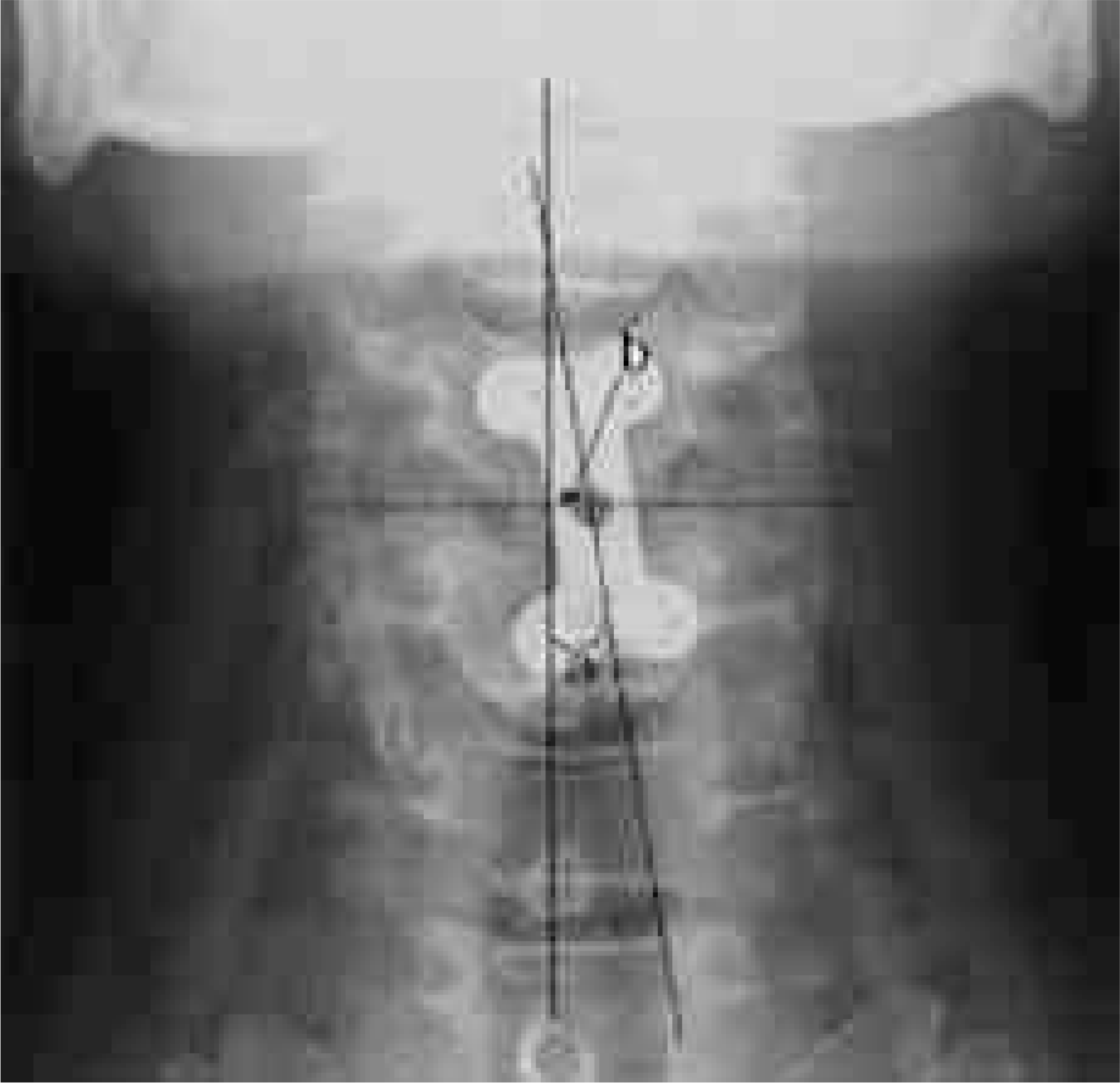
Table 1-1.
The relationship of the angulation of plates and fusion period.
| mean fusion period(weeks) | |
|---|---|
| 0-5 deg | 13.2 |
| 5-10 deg | 12.8 |
| 10-15 deg | 12.6 |
| 15-20 deg | 13.0 |
Table 1-2.
The relationship of the translation of plates and fusion period.
| mean fusion period(weeks) | |
|---|---|
| 0-3 mm | 12.8 |
| 3-6 mm | 9.3 |
| 6-9 mm | 14.1 |
| 9-12 mm | 11.5 |
 XML Download