

 PDF
PDF ePub
ePub Citation
 Print
Print


REFERENCES
1). 왕진만, 김동준. 척추불안전성이 동반된 척추 분리증에 대한 후방기기 및 전방추체 유합술. 대한척추외과학회지. 1:233–239. 1994.
2). Bagby GW. Arthrodesis by the distraction - compression using a stainless implant. Orthopedics. 11:931–933. 1988.
3). Cloward RB. The teatement of ruptured intervertebral disc by vertebral body fusion. Ann Surg. 136:987. 1952.
4). Cotrel Y and DuBousset J. New segmental posterior instrumentation of the spine. Proceedings at the 19th Annual Meeting of the Scoliosis Research Society, Orlan -do. 1984.
5). Dunn HK. Anterior stabilization of thoracolumbar injuries. Clin Orthop. 189:116–124. 1984.

6). Dwyer AF, Newton NC, Sherwood AA. An anterior approach to scoliosis. Clin Orthop. 62:192–202. 1969.
7). Harrington PR. Surgical instrumentation for management of scoliosis. J Bone Joint Surg. 42-A:1448. 1960.
8). Hodgson AR, Stock FE, Fang HSY and Ong GB. Anterior spinal fusion: The operative approach and pathologic findings in 412 patients with Pott's disease of the spine. Br J Surg. 48:172–178. 1960.
9). John W. Frymoyer. The adult spine principle and prac -tice, Lippincott. Raven. 3-40:1997.
10). Luque ER. The anatomic basis and development of segmental spinal instrumentation Spine. 7:256–259. 1982.
11). Wang JM, Kim DJ and Yun YH. Posterior pedicular screw instrumentation and anterior interbody fustion in adult lumbar spondylolysis or Grade I spondylolisthesis with segmental instability, J Spinal Disorder. 9:83–8. 1996.
12). White AA and Panjabi MM. Biomechanical considerations in the surgical management of the spine. White AA and panjabi MM, editor. Clincial biomechanics of the spine. 2nd ed.Philadelphia: JB Lippincott;p. 511–634. 1990.
13). Zielke K, Stunkat R and Beaujean F. Ventral derotation spondylodesis. Arch Orthop Trauma Surg. 85:257–260. 1976.
Go to : 
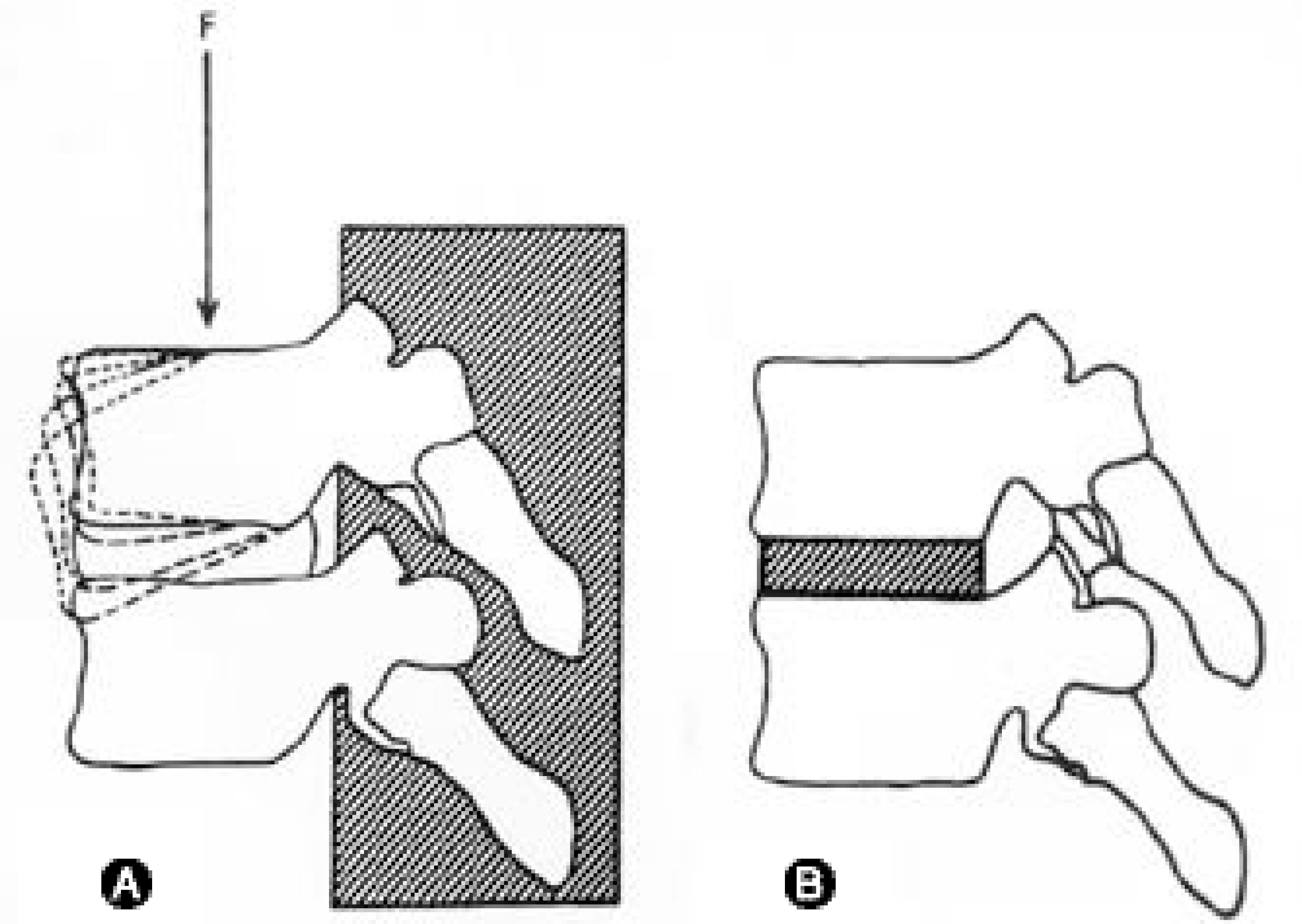 | Fig. 1.(A) Constant elastic motions in the anterior part of motion segment even in solid posterior fusion (B) Anterior interbody fusion eliminate elastic moton by take out disc. |
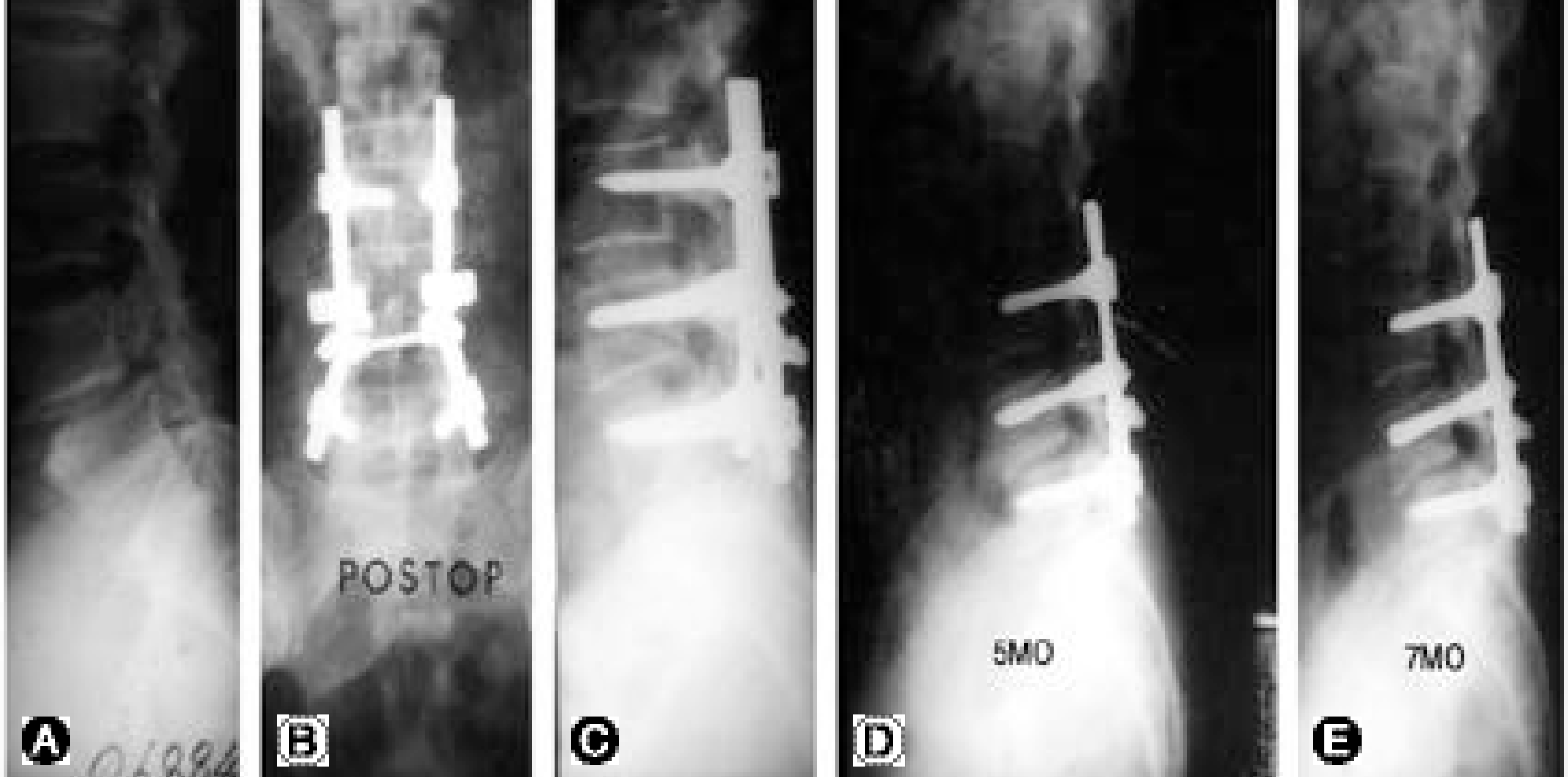 | Fig. 2.(A) Preoperative radiogaphy of 54 year old female under the diagnosis of isthmic spondylolisthesis. (B, C) Postoperative radiography of the posterolateral fusion and posterior instrumentation (D, E) Postoperative radiography in 5 and 7 month of inter-val with gradual flexon deformity |
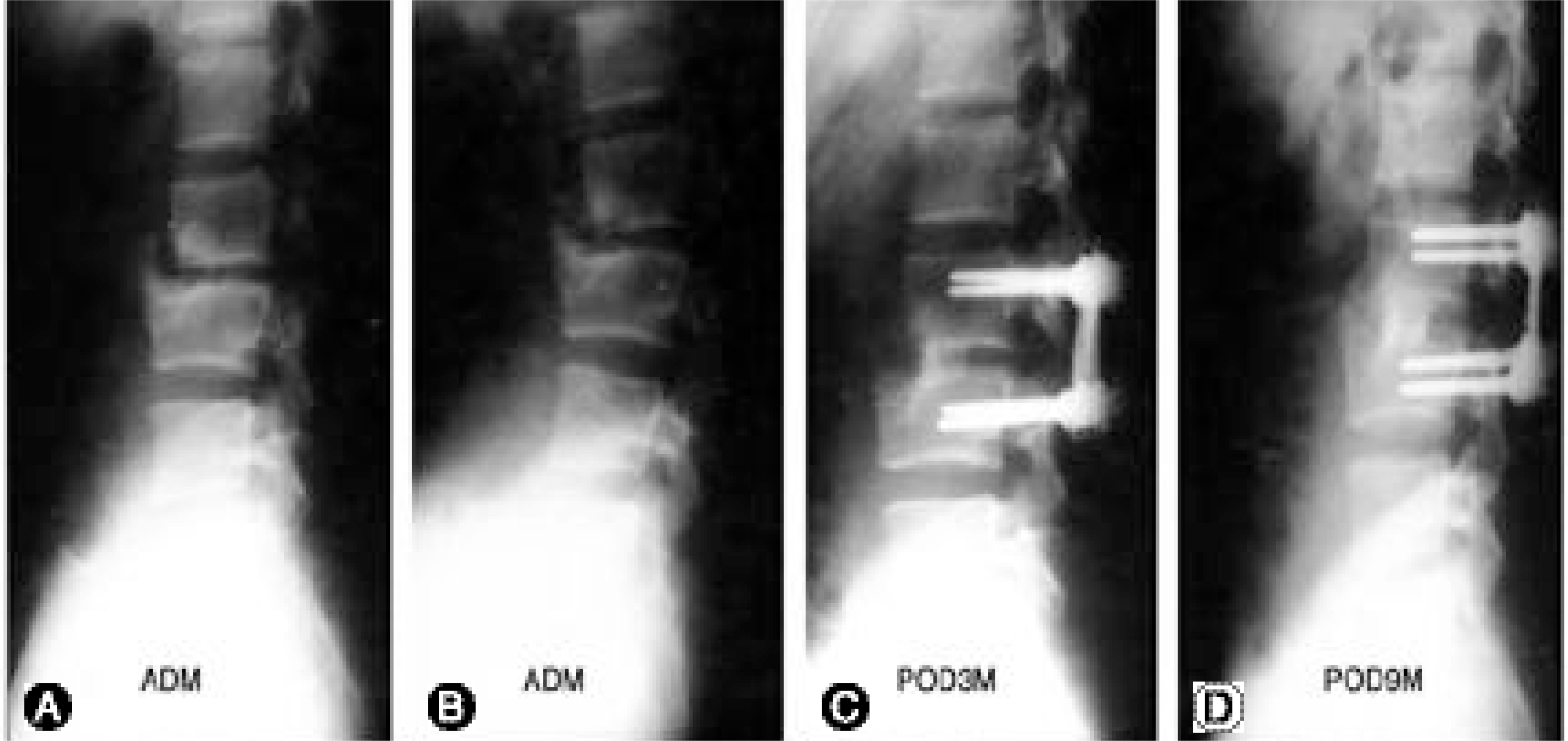 | Fig. 3.(A, B) This 18 year old male had back pain and limit of spinal motion due to segmental instability. (C, D) Postoperative 3month & 6month of lateral radiography shows complete fusion of anterior interbody fusion with good sagittal balance. |
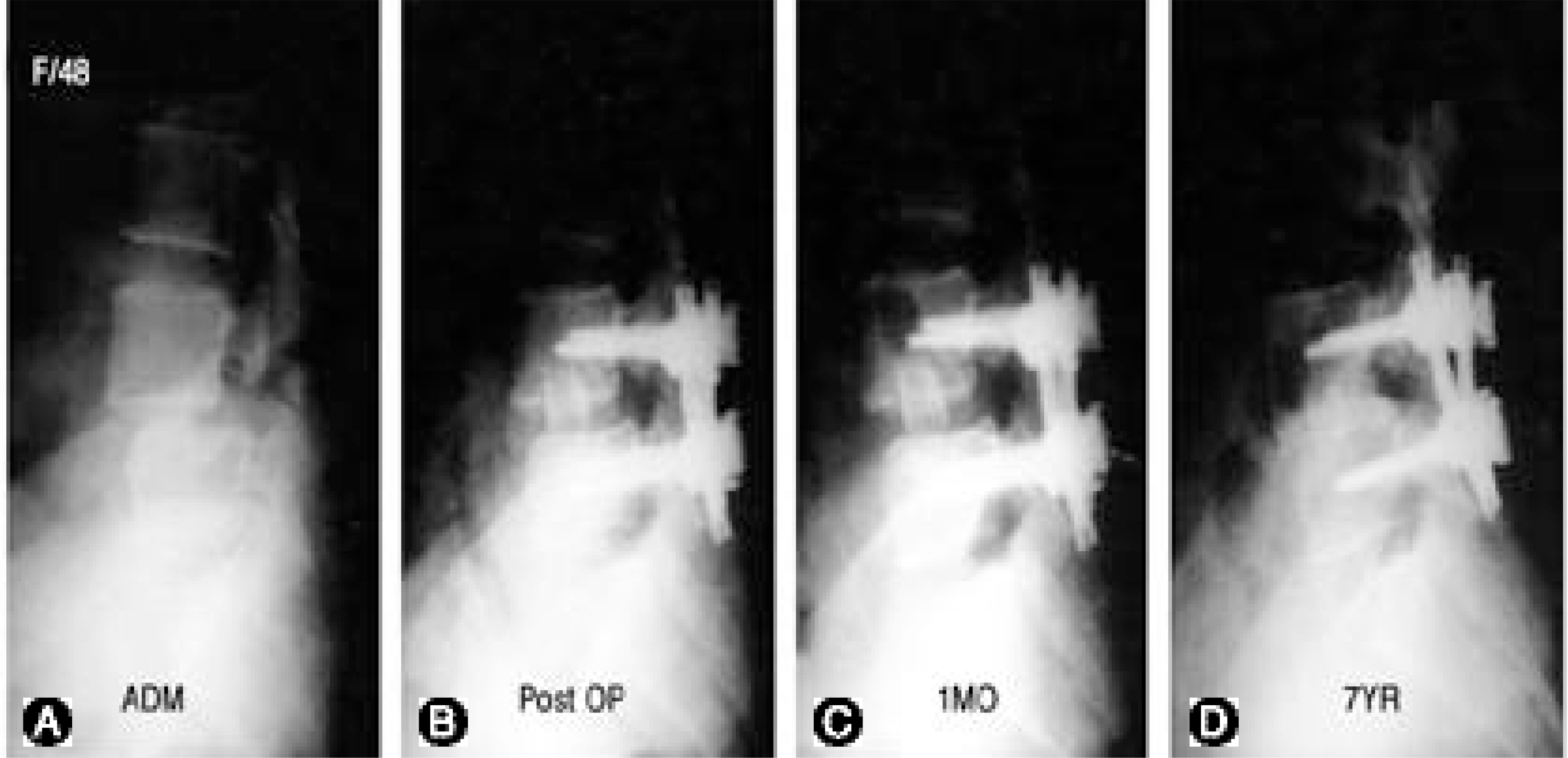 | Fig. 4.(A) Preoperative radiography of 48 year old female under the diagnosis of isthmic spondylolisthesis. (B) Postoperative radiography of the L 4-5 by doing posterior instrmentation and anterior interbody fusion. (C, D) Postoperative radiography 1 month and 7years follow up with complete full width fusion of L4-5 disc. |
Table 1.
Summary of spinal surgery (1984. 3.~2000. 4.)
 XML Download