Abstract
Ob j e ctives
To evaluate the results of posterior stabilization of a thoracolumbar fracture, without fusion, followed by the removal of metal implants within an appropriate period. Changes in the sagittal alignment and the restoration of segmental motion were also investigated.
Summary of Literature R eview
In managing unstable thoracolumbar and lumbar fractures, posterior fusion, using a transpedicular screw system, has been the treatment of choice, but results in the permanent loss of segmental motion.
Materials and Methods
Twelve patients with thoracolumbar and lumbar spine fractures, under 40 years of age (mean 28.4 years), were managed using this nonfusion method. Implants were removed at mean 9.2 months after the initial fixation of the fracture. For metal- fixed segments, the sagittal alignment, such as the angle of kyphosis, height of body, recovered motion range in flexionextension and rightleft bending view, were measured radiologically and compared with a control group. Clinical aspects, such as gross deformity and functional ability, were also investigated.
Results
The average sagittal angle at the time of injury was average 17.2°, but became 1.7° post-fixation, which increased after removal of the implants, reaching 9.8° at the final follow up. The height of the fractured body was maintained until the final follow- up. The mean segmental motion measured in the sagittal and coronal planes were 11.7 and 9.5°, respectively. Most patients were satisfied with the final gross appearance and functional outcomes. Only one patient showed considerable development of kyphotic angulation, but the functional outcome was good.
Conclusion
The author's nonfusion method seems to be effective in achieving stability and sagittal alignment, as well as in regaining segmental motion of the fixed segments. The nonfusion method seems to be an effective method for managing thoracolumbar fractures, especially for young active persons.
Go to : 
REFERENCES
1). Koh YD, Kim JO. Risk factors in progression of deformity in compression fracture of thoracolumbar junction. J Kor Soc Fracture. 1999; 12:372–378.

2). You JW, Lee SH, Park JK. Operative treatment of burst fracture on the thoracolumbar junction. J Kor Orthop Assoc. 1995; 30:364–374.
3). Aebi M, Etter C, Kehl T, Thalgott J. Stabilization of the lower thoracic and lumbar spine with the internal spinal skeletal fixation system: indication, techniques and first results of treatment. Spine. 1987; 12:5454–551.
4). Ahn JS, Lee JK, Hwang DS, Kim YM, Kim WJ, Byun KH. The change of kyphotic angle and anterior vertebral height after posterior or posterolateral fusion with transpedicular screw for thoracolumbar bursting fracture. J Kor Soc Fracture. 2003; 12:379–387.
5). Knop C, Fabian HF, Bastian L, Blauth M. Late Results of Thoracolumbar Fractures After Posterior Instrumentation and Transpedicular Bone Grafting. Spine. 2001; 26:88–99.

6). Shin BJ, Kim DS, Choi CU. Analysis of loss of correction in spinal fractures treated by pedicle screw system. J Kor Spine Surg. 1994; 1:223–232.
7). Oner FC, Van der Rijt RR, Ramos LM, Dhert WJ, Verbout AJ. Changes in the disc space after fractures of the thoracolumbar spine. J Bone Joint Surg. 1998; 80-B:833–839.

8). Lindsey RW, Dick W, Nunchuck S, Zach Z. R esidual intersegmental spinal mobility following limited pedicle fixation of thoracolumbar spine fractures with the fixateur interne. Spine. 1993; 15:474–478.
9). Gardner VO, Amstrong GD. Long term lumbar facet joint change in spinal fracture patients treated with Har -rington rods. Spine. 1990; 15:479–484.
Go to : 
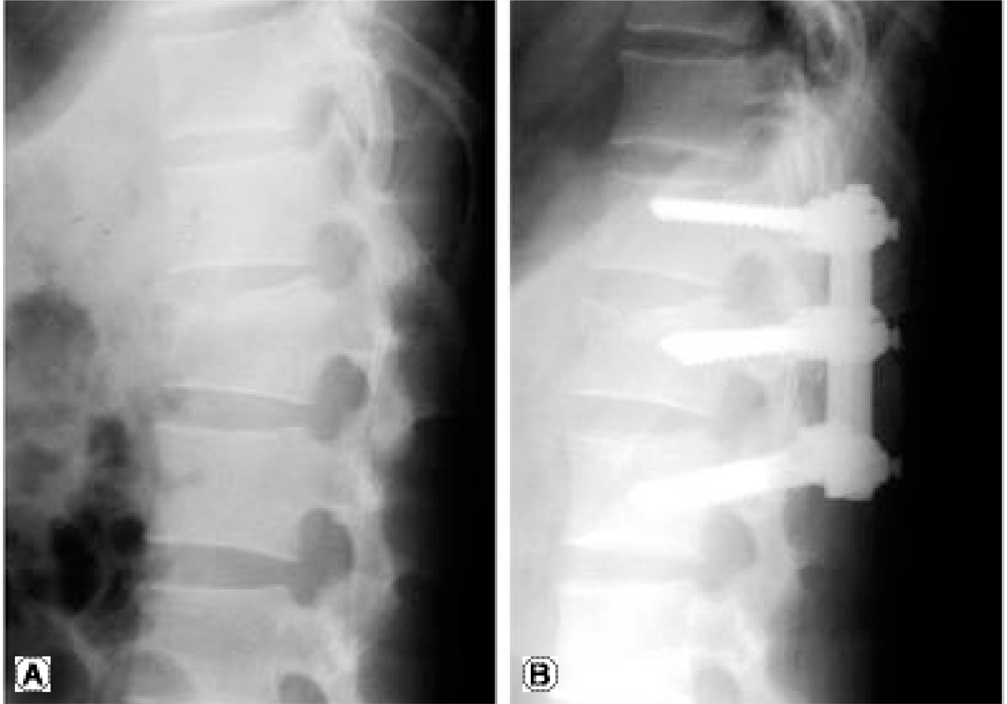
 | Fig. 1.(A) 32 year old male was injured at L1 by fall from height. Anteroposterior and lateral view at injury shows decrease of L1 body height and widening of interspinous space. (B) Kyphotic angle at injury(17°) between T12 and L2 was improved to 2° after post fixation. |
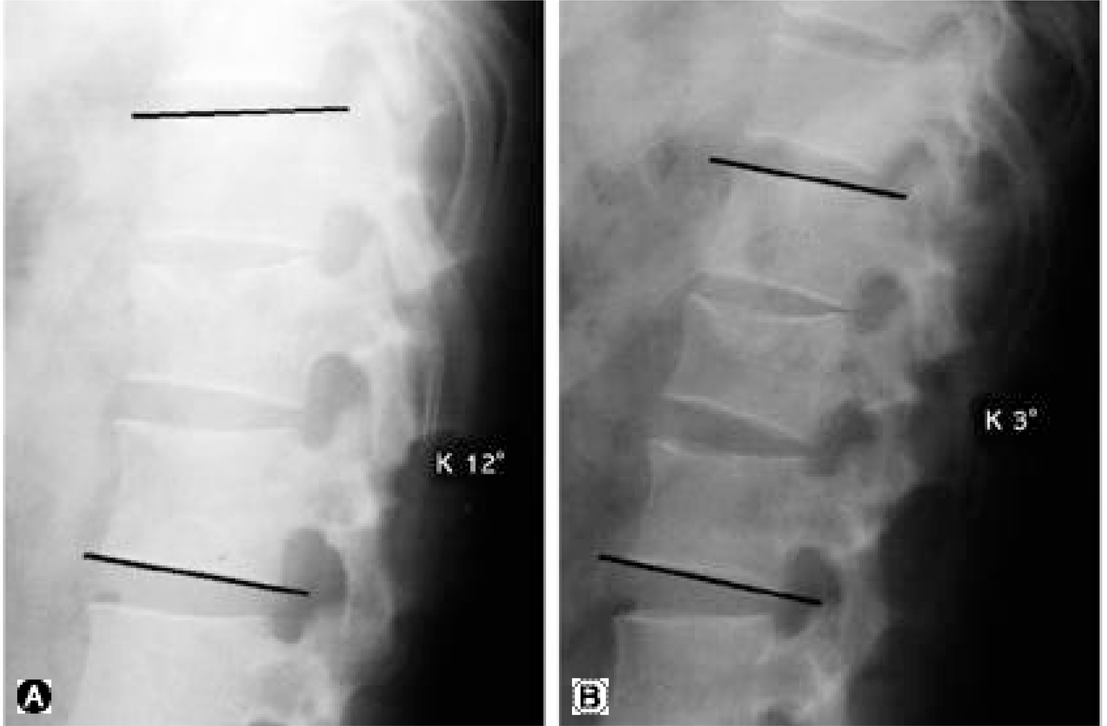
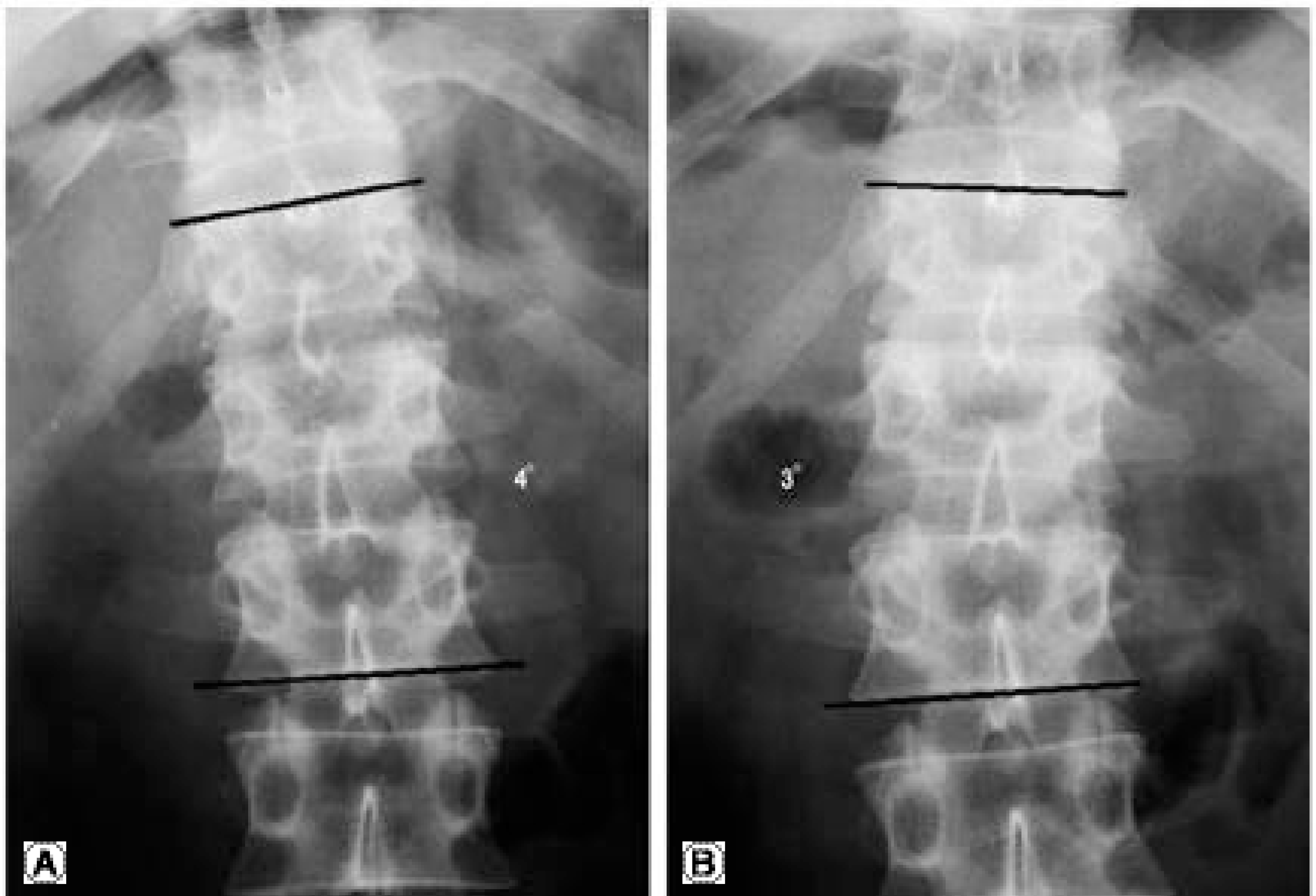
 | Fig. 2.(A) Flexion view. (B) Extension view at last follow up shows 9° of motion angle in the segments fixed previously. |
Table 1.
Mean motion angle of the patient group according to fixed segments.
| Range of posterior fixation | Average | |||||
|---|---|---|---|---|---|---|
| T11-L1 | T12-L2 | L1-L3 | L2-L4 | L1-L4 | ||
| Flexion | K∗26° | K15.5° | K17.5° | L† 1° | K25° | K20° |
| Extension | K19.6° | K4.8° | K5.5° | L15° | K9° | K8.3° |
| MA‡ | 6.3° | 10.7° | 12° | 14° | 16° | 11.7° |
| Rt. bending | 2.3° | 5.0° | 3.5° | 26° | 27° | 24.5° |
| Lt. bending | 2.3° | 4.5° | 6.5° | 26° | 27° | 25.0° |
| MA | 5.3° | 9.5° | 10° | 12° | 14° | 29.5° |
| LL§ | 35.5° | |||||
Table 2.
Percentage of recovered motion compared to normal control group.
| Motion angle | ||||||
|---|---|---|---|---|---|---|
| Patients | T11-L1 Control | % | Patients | T12-L2 Control | % | |
| Flexion-Extension | 6.3° | 10.8° | 58.3 | 10.7° | 15.1° | 70.9 |
| R-L∗ bending | 5.3° | 08.2° | 64.6 | 09.5° | 12.9° | 88.8 |


 PDF
PDF ePub
ePub Citation
 Print
Print


 XML Download