Abstract
Objectives
To clarify the clinical significance of the radiolucent zones surrounding transpedicular screws that occasionally appears following lumbar spinal instrumented fusion.
Summary of Literature Review
that the formation of radiolucent zones surrounding pedicular screws are significantly frequent after transpedicular fixation.
Materials and Method
88 cases, age 50 or older, which underwent lumbar spinal fusion with transpedicular screws, between January 1999 and December 2002, were included in this investigation. The postoperative radiographs of all patients were analyzed for radiolucent zones around the transpedicular screws. These radiolucent zones were evaluated in relation the number of fusion levels, the existence of osteoporosis, and the fusion status and satisfaction rates. Results: Radiolucent zones were observed in 30 cases (34%, 30/88), 13 (43%, 13/30) of which disappeared during the follow- up period. The average number of fixation levels in the cases with and without radiolucent zones were 2.33 (range 1- 4, SD 0.94) and 1.74 (range 1- 4, SD 0.82), respectively. Osteoporosis was found to accompany 43.3 and 20.7% of the cases with and without radiolucent zones, with the latter cases showing a statistically significant higher fusion rate and greater patient satisfaction.
Conclusion
Radiolucent zones, a frequent finding following pedicle screw fixation, resulted in less favorable outcomes. Surgeons should be alert to radiolucent zones and their transformation during follow- up. Methods for improving the stability of the interface between the pedicle screw and vertebral bone will require further research.
Go to : 
REFERENCES
1). Cook SD, Barbera J, Rubi M, Salkeld SL, Whitecloud TS 3rd. Lumbosacral fixation using expandable pedicle screws. an alternative in reoperation and osteoporosis. Spine J. 2001; 1:109–114.
2). Gibson JN, Grant IC, Waddell G. The Cochrane review of surgery for lumbar disc prolapse and degenerative lumbar spondylosis. Spine. 1999; 24:1820–1832.

3). Gibson JN, Waddell G, Grant IC. Surgery for degenerative lumbar spondylosis. Cochrane Database Syst Rev. 2000; 3:CD001352.

4). Jang EC, Seo JH, Song KS, Ryu HS. A biomechanical study of two kinds of tapered pedicle screws in osteoporot -ic lumbar spine. J Kor Orthop Assoc. 1999; 34:955–962.
5). Jansson KA, Blomqvist P, Granath F, Nemeth G. Spinal stenosis surgery in Sweden 1987-1999. Eur Spine J. 2003; 12:535–541.

6). Kim EH, Cho DY, Kim JH. A clinical analysis of long segment fusion with pedicle screw in degenerative lumbar spine. J Kor Spine Surg. 1999; 6:388–396.
7). Kim NH, Lee HM, Lee WS. The effect of bone mineral density on instrumented spine fusion. J Kor Spine Surg. 1994; 1:133–139.
8). Kirkaldy-Willis WH, Paine KWE, Cauchoix J, McIvor G. Lumbar spinal stenosis. Clin Orthop. 1974; 99:30–52.
9). Lenke LG, Bridwell KH, Bullis D, Betz RR, Baldus C, Schoenecker PL. Results of in situ fusion for isthmic spondylolisthesis. J Spinal Disord. 1992; 5:433–442.

10). Lu WW, Zhu Q, Holmes AD, Luk KD, Zhong S, Leong JC. Loosening of sacral screw fixation under in vitro fatigue loading. J Orthop Res. 2000; 18:808–814.
11). Ohlin A, Karlsson M, Duppe H, Hasserius R, Redlund-Johnell I. Complications after transpedicular stabilization of the spine. A survivorship analysis of 163 cases. Spine. 1994; 19:2774–2779.
12). Okuyama K, Abe E, Suzuki T, Chiba M, Sato K. Can insertional torque predict screw loosening and related failures? An in vivo study of pedicle screw fixation augmenting posterior lumbar interbody fusion. Spine. 2000; 25:858–864.
13). Okuyama K, Abe E, Suzuki T, Tamura Y, Chiba M, Sato K. Influence of bone mineral density on pedicle screw fixation: a study of pedicle screw fixation augmenting posterior lumbar interbody fusion in elderly patients. Spine J. 2001; 1:402–407.
14). Pihlajamaki H, Myllynen P, Bostman O. Complications of transpedicular lumbosacral fixation for non-traumatic disorders. J Bone Joint Surg. 1997; 79-B:183–189.
15). Reindl R, Steffen T, Cohen L, Aebi M. Elective lumbar spinal decompression in the elderly: Is it a high-risk oper -ation? Can J Sur. 2003; 46:44–46.
16). Renner SM, Lim TH, Kim WJ, Katolik L, An HS, Anderson GB. Augmentation of pedicle screw fixation strength using an injectable calcium phosphate cement as a function of injection timing and method. Spine. 2004; 29:212–219.

17). Sanden B, Orelud C, Johansson C, Larsson S. Improved bone-screw interface with hydroxyapatite coating, Spine. 2004; 26:2673–2678. 2001.
18). Sanden B, Orelud C, Petren-Malmin M, Johansson C, Larsson S. The significance of radiolucent zones surrounding pedicle screws. J Bone Joint Surg. 2004; 86-B:457–461.
19). Shin BJ, Know YB, Suh YS, et al. Complications and problems related to pedicle screw fixation of spinal column. J Kor Spine Surg. 1994; 1:206–215.
20). Tokuhashi Y, Matsuzaki H, Ishikawa H. Clear zone around pedicle screw on radiograph after posterior stabi - lization. 1st Meeting of Advanced Technologies in Spinal Treatment. 2001.
21). Wimmer C, Gluch H. Aseptic loosening after CD instrumentation in the treatment of scoliosis: A report of eight cases. J Spinal Disord. 1998; 11:440–443.
Go to : 
 | Fig. 1.(A) Clearly visible radiolucent zones surrounding both S1 screws at postoperative 1 year. (B) Radiolucent zones become less clearly visible at two and half years after surgery. (C)Radiolucent zones disappear completely at postoperative 4 years. |
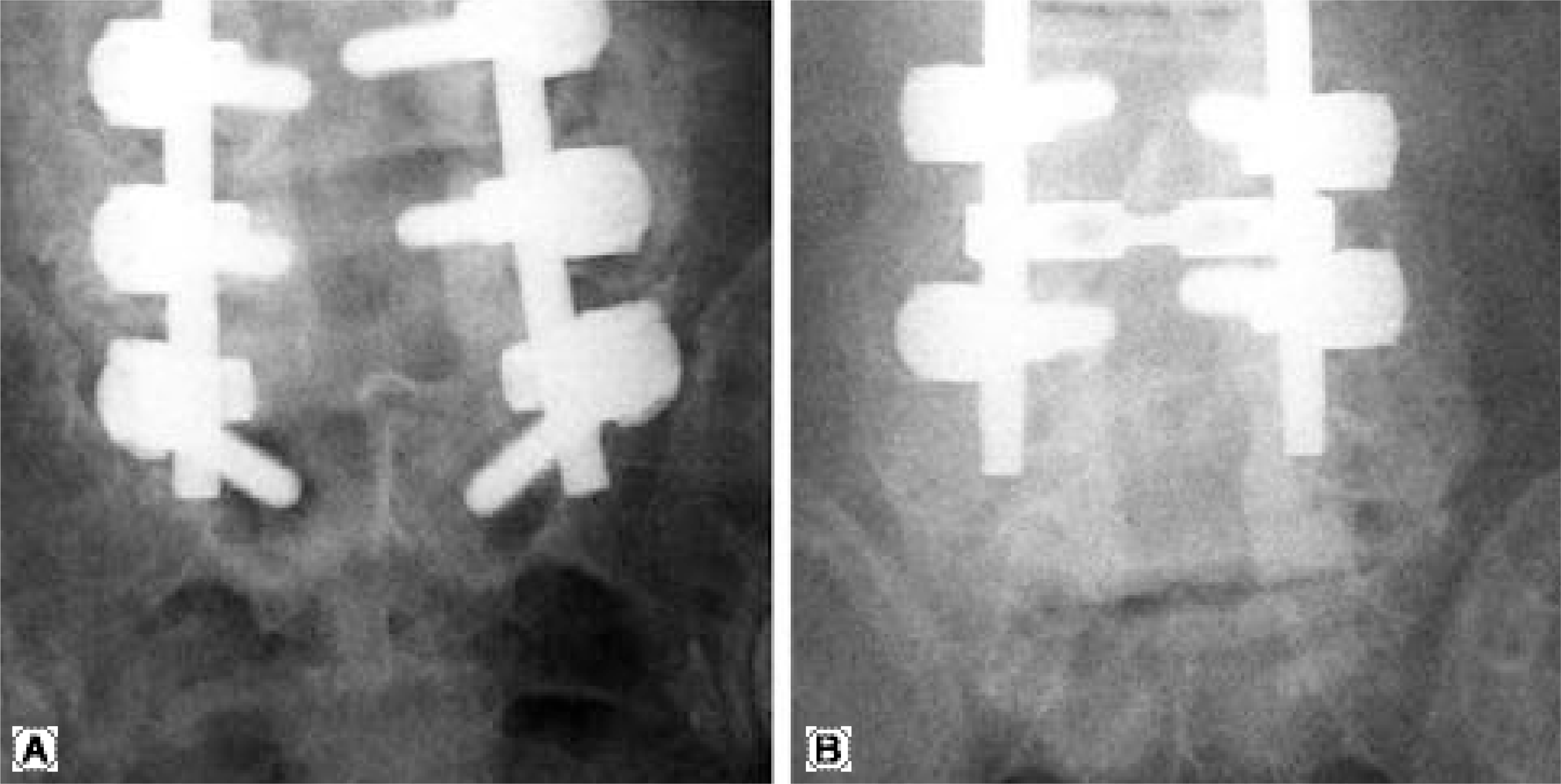
 | Fig. 2.(A) Wide radiolucent zones around the both sacral screws. (B) Radiograph after removal of sacral pedicle screws, which had shown severe loosening in the operating field. |
Table 1.
Disappearance of radiolucent zones according to the widths
| Appearance | Disappearance | |
|---|---|---|
| 1~2 mm | 12 | 08 |
| 2~3 mm | 13 | 04 |
| > 3 mm | 05 | 01 |
| Total | 30 | 13 |
| % | 100% | 43.3% |
Table 2.
Radiolocent zones according to number of fusion segments
| Radiolucent zone (+) | Radiolucent zone (-) | Number | |
|---|---|---|---|
| 1 segment | 06 | 27 | 33 |
| 2 segments | 12 | 20 | 32 |
| 3 segments | 08 | 10 | 18 |
| 4 segments | 04 | 01 | 05 |
| Total | 30 | 58 | 88 |
| Mean± SD | 2.33± 0.94 | 1.74± 0.82 |
Table 3.
Clinical results


 PDF
PDF ePub
ePub Citation
 Print
Print


 XML Download