

 PDF
PDF ePub
ePub Citation
 Print
Print


Abstract
Study design
A prospective study is to evaluate the cervical range of motion through the analysis of the plain films of the cervical spine.
Objective
To provide criteria validity for the cervical lordosis, range of motion and segmental motion of each segment using normal Korean adults, as guide lines for the radiographic diagnosis and treatment of cervical diseases.
Materials and Methods
One hundred and four healthy Korean adults were examined. Roentgenographic lateral views were taken in neutral, active flexion and extension positions. Measurement of the range of motion and segmental motion of the cervical spine, using the technique of Penning and Bakke, were independently made by two observers.
Results
The mean angle of the lordosis and the range of motion were 19.8°± 8.4° and 63.1°± 11.2°, respectively, and the range of motion of each segment were 10.3°± 2.9°, 9.2°± 3.0°, 13.5°± 7.2°, 15.1°± 4.3°, 15.6°± 4.4° and 13.0°± 5.0° on C1- 2, C2- 3, C3- 4, C4- 5, C5- 6 and C6- 7, respectively. There were no differences in the lordotic angle between the ages and genders. The range of motion decreased with increasing age, but there was no difference between genders. The segmental motion was no different between genders, but decreased significantly with increasing age on segments C1- 2, C5- 6 and C6- 7.
Conclusions
No significant differences were found in the range of cervical motion of each segment and the lordotic angle of the Korean population compared with those of other populations. The range of cervical motion also decreased with increasing age. These data provide guidelines in the dynamics of cervical spine and for the roentgenographic diagnosis and treatment of cervial diseases.
Go to : 
REFERENCES
1). Shin MG. Imaging of the cervical spine. J of Kor Spine Surg. 1999; 6:182–184.
2). Ferrario VF, Sforza C, Serrao G, Grassi G, Mossi E. Active range of motion of the head and cervical spine: a three-dimensional investigation in healthy young adults. J of Orthop Resch. 2002; 20:122–129.

3). Bakke S. Rontgenologische Beobachtungen Uber die Bewegungen der Halswirbelsaule. Acta Radiol[Suppl]. 1931; 13:00–00. (cited from Dvorak J: Functional radiographic diagnosis of the cervical spine: Flexion/Extension. Spine 1988;13: 1748-1755).
4). Buetti-Bauml C. Funktionelle Rontgendiagnostik der Halswirbelsaule. Thieme, Stuttart, Fortschritte auf dem Gebiete der Roentgenstrahlen vereinigit mit Roentgen -praxis. Erganzungsband. 1954; 70:(cited from Dvorak J: Functional radiographic diagnosis of the cervical spine: Flexion/Extension. Spine 1988;13: 1748-1755).
5). Dvorak J, Froehlich D, Penning L, Baumgartner H, Panjabi MM. Functional radiographic diagnosis of the cervical spine: Flexion/Extension. Spine. 1988; 13:1748–1755.
6). Penning L. Normal movements of the cervical spine. Am J Roentgenol. 1978; 130:317–326.
7). Yoshimoto H, Ito M, Abumi K, Kotani Y, Shono Y, Takada T, Minami A. A retrospective radiographic analysis of subaxial sagittal alignment after posterior C1-C2 fusion. Spine. 2004; 29:175–181.
8). Panjabi M, Dvorak J, Duranceau J, Yamamoto I, Ger-ber M, Rauschning W, Bueff H. Three-dimensional movements of the upper cervical spine. Spine. 1988; 13:726–730.
9). Yoganandan N, Kumaresan S, Dintar FA. Biom echanics of the cervical spine part 2. Cervical spine soft tissue respons -es and biomechanical modeling. Clin Biomech. 2001; 16:1–27.
Go to :
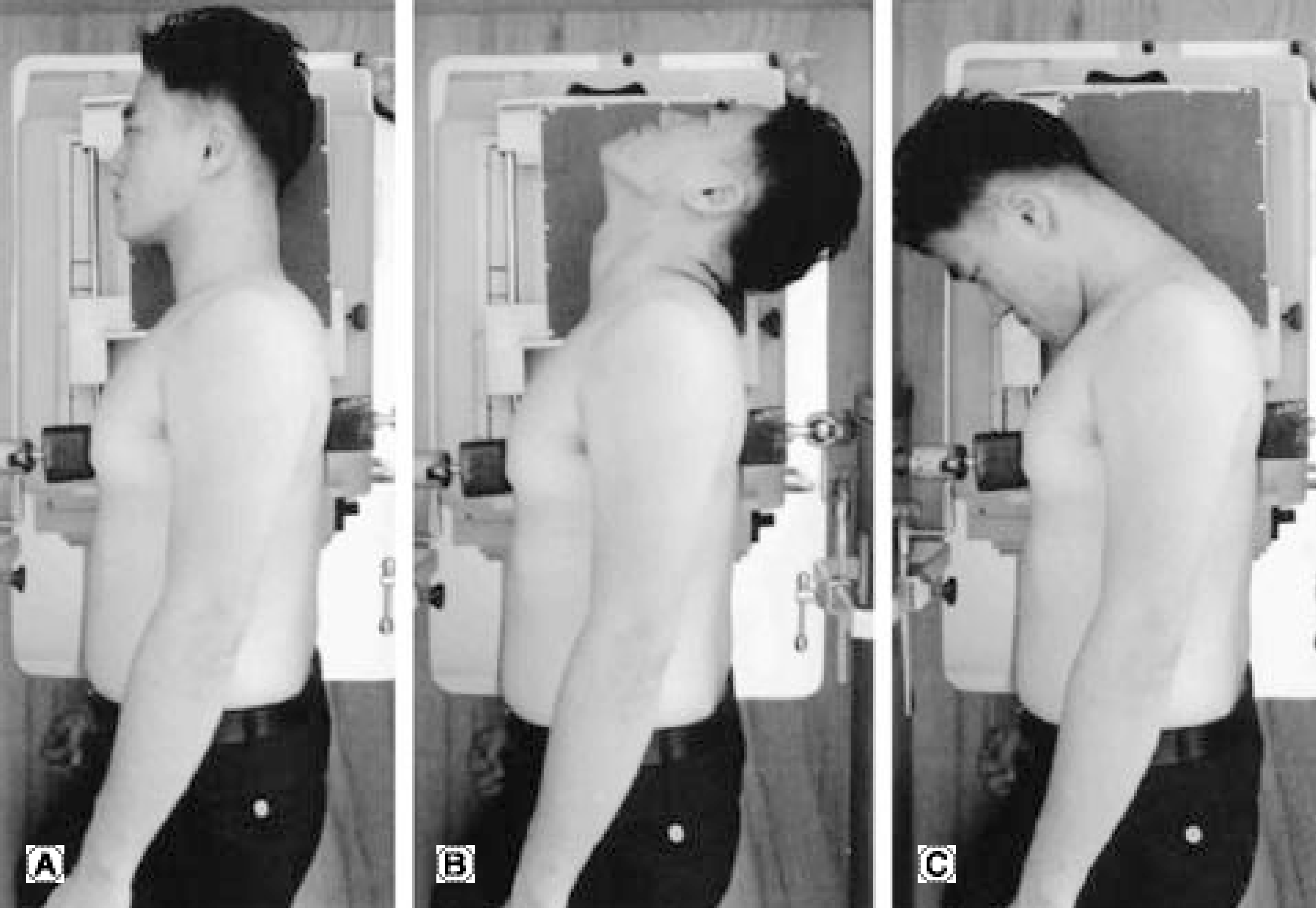 | Fig. 1.(A) Neutral standing position, the thorax fixed by two pellots from ventral and dorsal; (B) Active Extension, (C) Active flexion. |
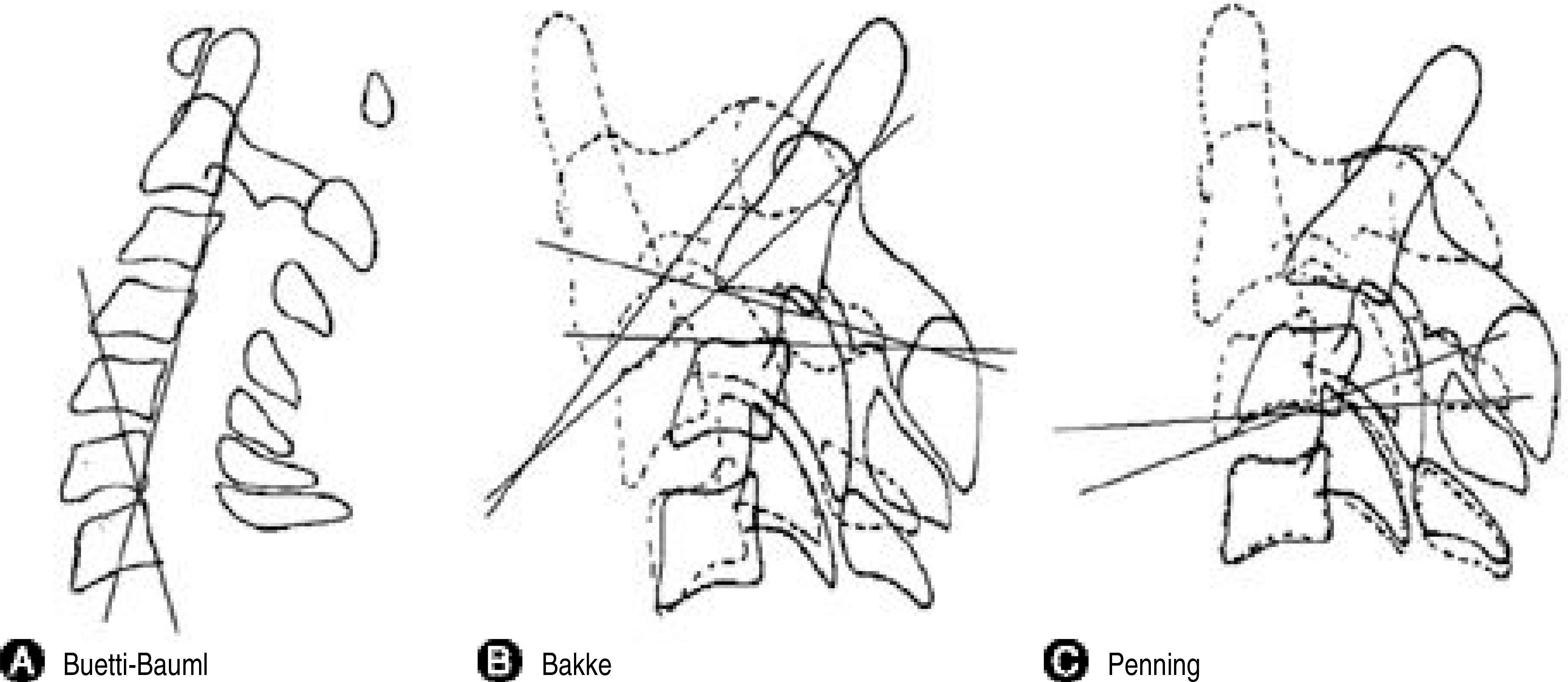 | Fig. 2.Technique of measurement. (A) Buetti-Bauml technique, (B) Bakke technique, (C) Penning technique. |
Table 1.
Summary of the Normal Values Established by Different Authors(°)
Table 2.
Cervical Lordosis(°)
| Age | Male | Female | Mean± SD∗ |
|---|---|---|---|
| 20-29 | 19.3± 9.9 | 22.4± 9.2 | 20.8± 9.7 |
| 30-39 | 21.4± 9.5 | 20.7± 7.6 | 21.1± 8.4 |
| 40-49 | 17.6± 9.4 | 15.4± 5.6 | 16.5± 8.2 |
| 50-59 | 19.6± 9.6 | 19.6± 7.1 | 19.6± 8.0 |
| 60- | 25.7± 6.8 | 16.0± 4.1 | 20.9± 7.4 |
| Mean | 20.7± 9.4 | 18.9± 7.2 | 19.8± 8.4 |
Table 3.
Segmental Motion during Flexion and Extension of Cervical Spine(°)
| Age | C1-2 | C2-3 | C3-4 | C4-5 | C5-6 | C6-7 | Mean |
|---|---|---|---|---|---|---|---|
| 20-29 | 12.7 | 10.3 | 13.6 | 16.0 | 18.8 | 15.5 | 71.4 |
| 30-39 | 10.3 | 9.0 | 13.4 | 15.6 | 17.4 | 14.4 | 66.3 |
| 40-49 | 11.5 | 8.6 | 12.1 | 15.0 | 16.1 | 13.3 | 63.3 |
| 50-59 | 10.7 | 9.8 | 13.3 | 15.3 | 13.0 | 10.8 | 60.0 |
| 60- | 9.6 | 8.5 | 15.1 | 13.3 | 13.7 | 11.5 | 54.9 |
| Mean | 10.8 | 9.2 | 13.5 | 15.1 | 15.6 | 13.0 | 63.0 |
| P value | p=0.01 | NS∗ | NS∗ | NS∗ | p=0.00 | p=0.01 |
Table 4.
Comparision of Segmental Motion between Male and Female(°)
| Male | Female | P value | |
|---|---|---|---|
| C1-C2 | 11.4± 3.2 | 10.6± 2.3 | NS∗ |
| C2-C3 | 8.5± 2.9 | 9.9± 3.0 | NS∗ |
| C3-C4 | 14.0± 9.7 | 13.0± 3.6 | NS∗ |
| C4-C5 | 14.8± 4.0 | 15.4± 4.6 | NS∗ |
| C5-C6 | 15.1± 3.9 | 16.1± 4.8 | NS∗ |
| C6-C7 | 13.3± 4.8 | 12.7± 5.1 | NS∗ |
| Total | 62.8± 9.5 | 63.3± 12.6 | NS∗ |
Table 5.
Comparision of Segmental Motion of the Cervical Spine between Free and Fixed Sternum and Thoracic Spine(°)
| Free | Fixed | P value | |
|---|---|---|---|
| C1-C2 | 10.9± 3.2 | 11.5± 4.3 | NS∗ |
| C2-C3 | 9.9± 2.7 | 12.5± 2.9 | NS∗ |
| C3-C4 | 14.7± 2.4 | 15.5± 3.0 | NS∗ |
| C4-C5 | 18.4± 4.5 | 18.5± 4.3 | NS∗ |
| C5-C6 | 18.3± 4.5 | 19.1± 5.5 | NS∗ |
| C6-C7 | 15.3± 4.3 | 14.9± 3.7 | NS∗ |
| Total | 73.2± 8.9 | 75.4± 8.7 | NS∗ |
 XML Download