Abstract
Epidural tuberculoma without bony involvement was first reported by Rao et al. in 1971; however, extraosseous spinal epidural tuberculoma and tuberculous infection of the cauda equina have never been reported. We experienced a case of epidural tuberculoma without bony involvement, which was diagnosed by decompression and biopsy, and treated with combined antituberculous chemotherapy. It resembled herniated nucleus pulposus at the L4- 5 level, based on its clinical features, a physical exami-nation, myelography and computed tomography. In the course of antituberculous medication, tuberculosis of the cauda equina occurred, which caused paraparesis. Herein, this case is reported in terms of its treatment and clinical course, with a review of the literature.
REFERENCES
1). Arthornthurasook A, Chongpieboonpatana A. Spinal tuberculosis with posterior element involvement. Spine. 1990; 15:191–194.

2). Rao BS, Dinakar I, Rao SK. Extraosseous extradural tuberculous granuloma simulating a herniated lumbar disc. J Neurosurgery. 1971; 35:488–490.
3). Naim-Ur-Rahman. Atypical forms of spinal tuberculosis. J Bone Joint Surg. 1980; 62B:162–165.
4). Reina A. Tuberculous epidural granuloma simulating a herniated lumbar disc. Surg Neurol. 1975; 4(3):336–338.
5). Choksey MS, Powell M, Gibb WR, et al. A conus tuberculoma mimicking an intramedullary tumour. A case report and review of the literature. Br J Neurology. 1989; 3:117–121.

6). Gokalp HZ, Ozkal E. Intradural tuberculoma of the spinal cord. Report of two cases. J Neurosurg. 1981; 55:289–292.
7). Orell RW, King RHM, Bowler JV, Ginsberg L. Periph -eral nerve granuloma in a patient with tuberculosis. J Neurol Neurosurg Psychiatry. 2002; 73:769–772.
8). Nucci F, Mastronardi L, Artico M, Ferrante L, Acqui M. Tuberculoma of the Ulnar Nerve. Case Report. J Neurosurg. 1988; 22:906–907.

9). Griffith DL, Seddon HJ, Roaf R. Potts Paraplegia. Lon -don, Oxford University Press. 1956; 2:4–21. (Cited from Hamada J, Sato K, Seto H and Ushio Y: Epidural tuberculoma of the spine. A case report. J Neurosurgery. 1991; 28:161–163.
10). MacDonell AH, Baird RW, Bronze MS. Intramedullary tuberculomas of the spinal cord. Case report and review. Rev Infect Dis. 1990; 12:432–439.

11). Hamada J, Sato K, Seto H, Ushio Y. Epidural tuberculoma of the spine. A case report. J Neurosurgery. 1991; 28:161–163.
12). Yune SH, Rhee KJ, Rhee JK, Kim HY. Ap pendic eal Tuberculosis of Spine. J Kor Orthop Assoc. 1981; 16(3):675–677.
13). Babhulkar SS, Pande KC. Spinal tuberculosis. Atypical spinal tuberculosis. Clin Orthop. 2002; 398:67–74.
14). Thomas CS, Glenn AM, Mary ADG. Lumbosacral plex -opathy in a patient with pulmonary tuberculosis. CID. 2000; 30:226–227.
15). Citow JS, Ammirati M. Intramedullary Tuberculoma of the Spinal Cord. Case Report. J Neurosurgery. 1994; 35:327–330.
16). Kayaoglu CR, Tuzun Y, Boga Z, Erdogan F, Gorguner M, Aydin IH. Intramedullary spinal tuberculoma. A case report. Spine. 2000; 25:2265–2268.
17). Bertrand I, Guillaume JM, Samson M, Gueguen Y. Tuberculoma intramedullaire dorsal. Rev Neurol. 1958; 98:51–54.
Fig. 1.
(A) Preoperative myelography. Anteroposterior and lateral myelogram showing a complete block of the contrast media at the level of the L4-5 disc due to a space occupying lesion in the epidural space. (B) CT myelography showing a epidural mass, but no destruction of vertebral body, pedicles and transverse processes.
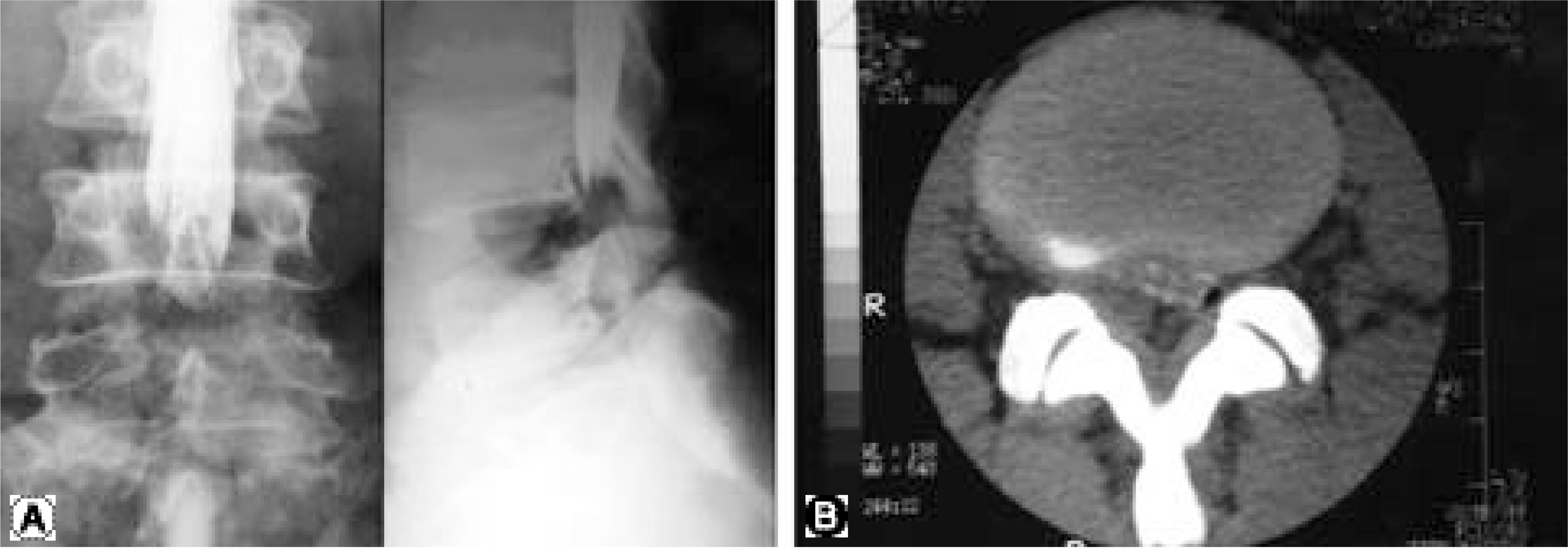
Fig. 2.
(A) Photomicrograph of biopsy specimen showing typical tuberculous granulation tissue. Typical Langhan-type giant cells(arrow), caseation necrosis(open arrow) are seen (Hematoxylin & Eosin staining, × 100). (B) Acid-fast bacilli(small arrow) in granulomatous tissue, taken from same biopsy specimen are also noted (AFB staining × 400).
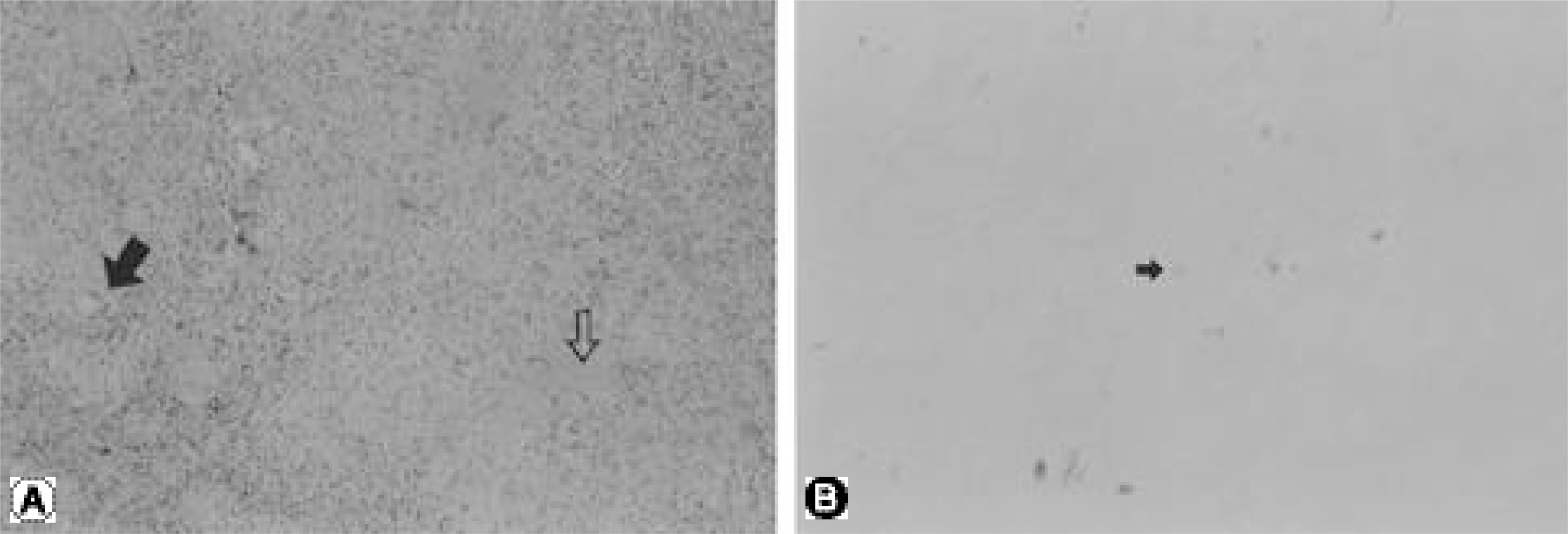
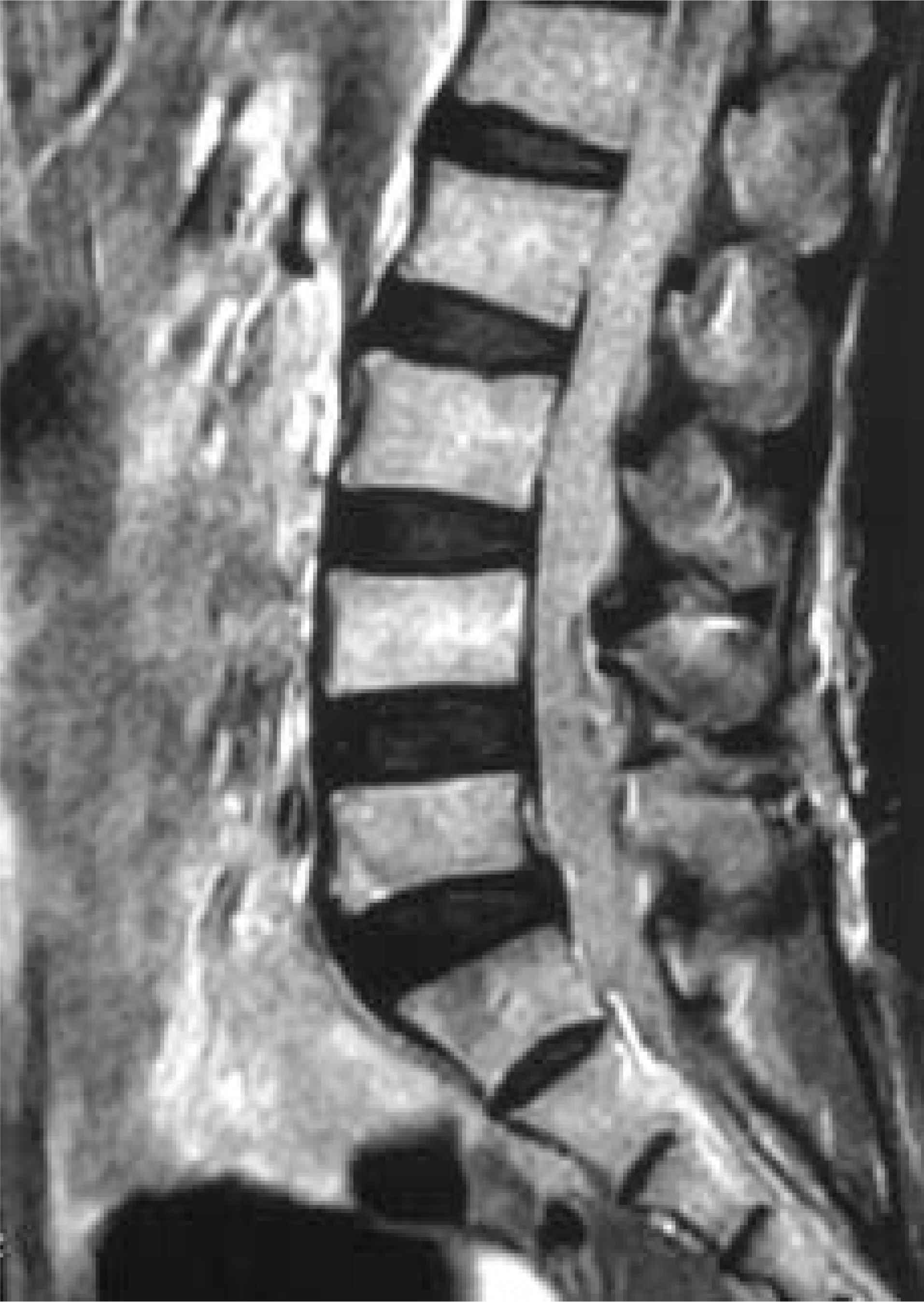
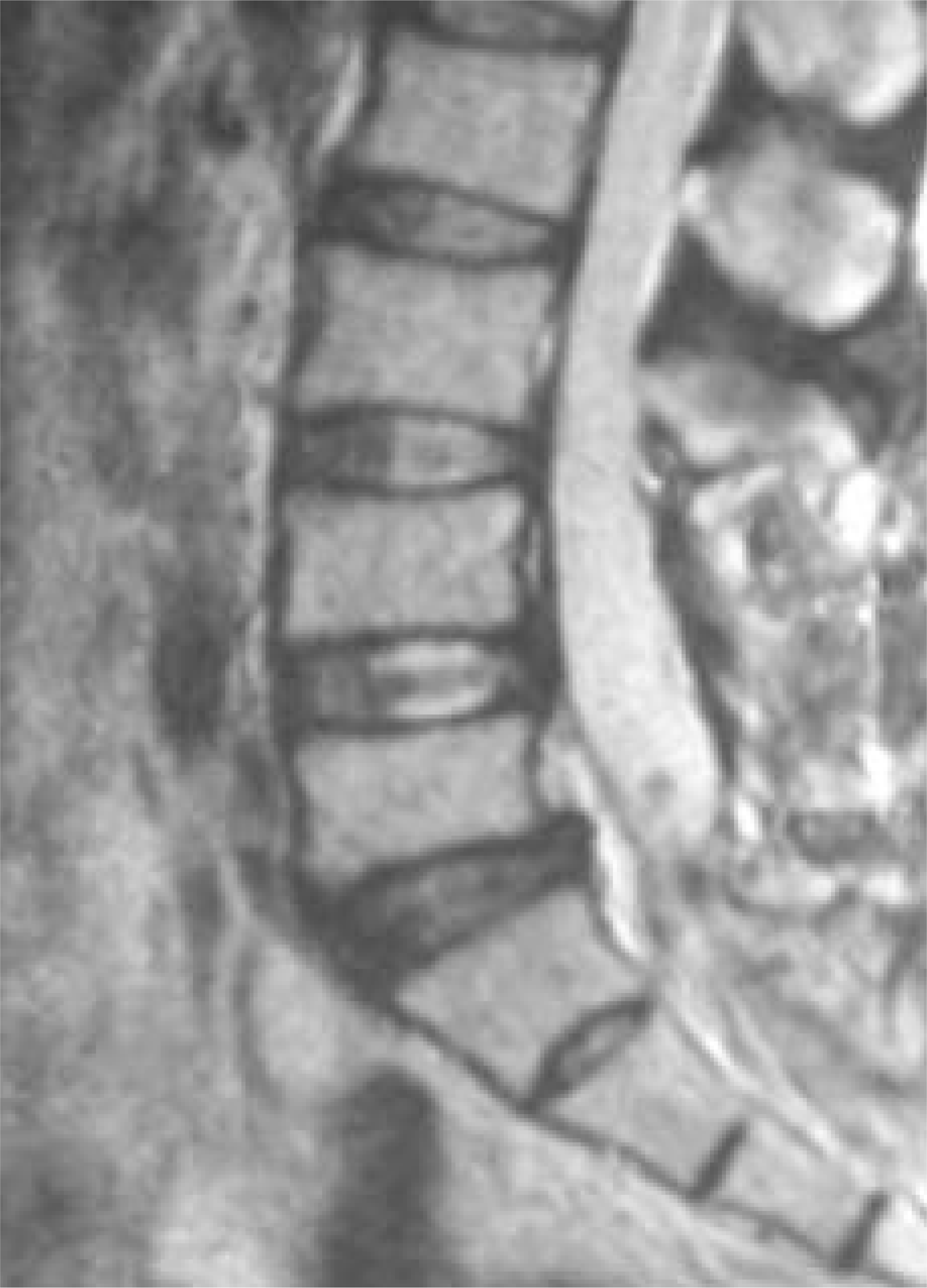
Fig. 3.
T1-weighted MR image which shows indistinguishable cauda equina due to edematous change and epidural col-lection of fluid, considered as cold abscess.


 PDF
PDF ePub
ePub Citation
 Print
Print


 XML Download