

 PDF
PDF ePub
ePub Citation
 Print
Print


Abstract
Objectives
To evaluate the clinical and radiological results of anterior decompression and instrumentation for delayed vertebral body collapse in neurologically compromised osteoporotic compression fractures.
Literature Review Summary
Indications for an operation in delayed vertebral body collapse, following osteoporotic compression fractures, are intractable pain, progressive kyphosis and neurological deficits. The options for an operation are anterior, posterior and combined anterior and posterior approaches. Posterior surgery may need some degree of destruction of intact posterior elements. Combined anterior and posterior surgery increases the morbidity and mortality due to increased operative time and blood loss. Therefore, a one stage anterior surgery is a reasonable choice.
Materials and Methods
Between June 1989 and May 2003, seven cases of delayed vertebral body collapse, with neurological deficit, were treated using anterior decompression and anterior Kaneda instrumentation. All the cases were female, with a mean age of 67, ranging from 57 to 77 years. The average follow up period was 3.4, ranging from 1 to 13 years. One patient had a history of steroid medication. The operation time, intraoperative blood loss and bone mineral density were retrospectively reviewed. The changes in the kyphotic angle, preoperatively, postoperatively and on the last follow- up plain lateral radiograph were measured. The clinical results were evaluated based on a modified Frankel grading and visual analogue scale
Results
The average kyphotic angles preoperatively, postoperatively and at the last follow up were 29。 (25∼47。), 14°(6∼20°) and 19°(10∼27°), respectively. In all cases, the preoperative neurological deficits were improved by more than one degree in the Frankel grading at the final follow up. The mean operation time, blood loss and mean bone mineral density were 3.2 hours, 1514ml and T: - 3.51, respectively. The values from the visual analogue scale preoperatively and at the last follow up were 7.0 and 0.5, respectively.
Go to : 
REFERENCES
1). Kaplan PA, Orton DF, Asleson RJ. Osteoporosis with vertebral compression fractures, retropulsed fragments, and neurologic compromise. Radiology. 1987; 165:533–535.

2). Kempinsky WH. Osteoporotic kyphosis with paraplegia. Neurology. 1958; 8:181–186.
3). Salomon C, Chopin D, Benoist M. Spinal cord compression: an exceptional complication of spinal osteoporosis. Spine. 1988; 13:222–224.
4). Shikata J, Yamamuro T, Iida H, Shimizu K, Yoshikawa J. Surgical treatment for paraplegia resulting from vertebral fractures in senile osteoporosis. Spine. 1990; 15:485–489.
5). Benedek TG, Nicolas JJ, Reece JG. Kummell's disease: a rare cause of post traumatic back pain. Arthritis Rheum. 1980; 23:653.
6). Schmorl G, Junghannas H. The human spine in health and disease. 2nd ed.New York: Grune and Stratton;p. 268–269. 1971.
7). Maldaque BE, Noel HM, Malghem JJ. The intravertebral vacuum cleft: a sign of ischemic vertebral collapse. Radiology. 1978; 129:23–29.
8). Kim KT, Suk KS, Kim JM. Surgical treatment of Kummell disease with neurologic deficits. J Kor Spine Surg. 2001; 8:136–142.
9). Arciero RA, Leung KYK, Pierce JH. Spont a n eous unstable burst fracture of the thoracolumbar spine in osteoporosis. Spine. 1989; 14:114–117.
10). Hammererg KW, DeWald RL. Senile burst fracture: a complication of osteonecrosis. Orthop Trans. 1989; 13:97.
11). Harverson G. Intravertebral vacuum phenomenon: Clin Radiol. 1988; 39:69–72.
12). Kumpan W, Salomonowitz E, Seidt G. The intervertebral vacuum phenomenon. Skeletal Radiol. 1986; 15:444–447.
13). Nicholas J, Benedek T, Reece G. Delayed traumatic vertebral body compression fracture part Ⅰ: clinical features. Semin Arthritis Rheum. 1981; 10:264–270.
14). Hermann G, Goldblatt J, Desnick RJ. Kummell disease: delayed collapse of the traumatised spine in a patient with Gaucher Type 1 disease. J Radiol. 1984; 57:833–835.
15). Kaneda K, Asano S, Hashimoto T, Satoh S, Fujiya M. The treatment of osteoporotic-potstraumatic vertebral collapse using the Kaneda device and bioactive ceramic vertebral prosthesis. Spine. 1992; 17:295–303.
16). Gurr KR, McAfee PC, Shin CM. Biomechanical Analysis of anterior and posterior instumentation system after corpectomy. J Bone Joint Surg. 1988; 70A:1182–1192.
Go to :
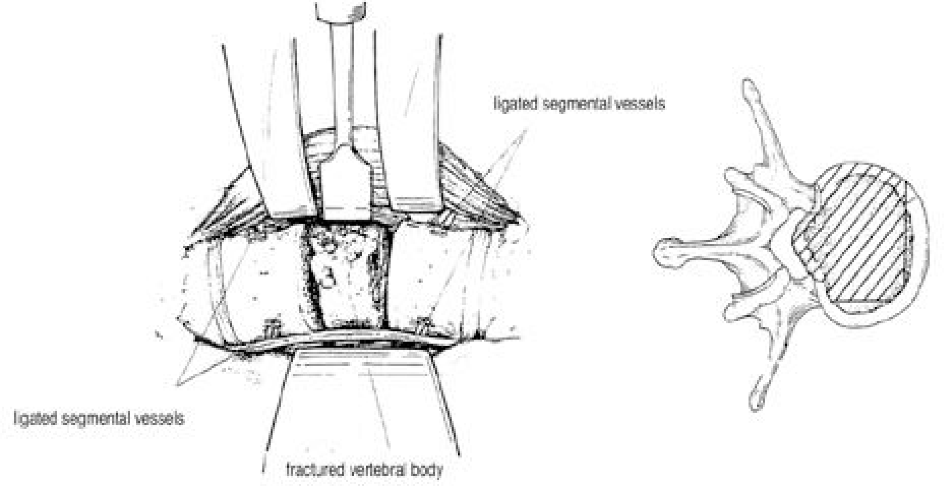 | Fig. 1.Decompression of the spinal canal. Left, A corpectomy is performed using chiesels following resection of the disc above and below the damaged segment. Then, a decompression is carried out by removal of the retropulsed bony fragment into the spinal canal using chiesels, curets, and Kerrison rongeur. Right, Extent of bone resection for anterior decompression is illus-trated (axial plane) |
 | Fig. 2.Case. (A) 69-year-old woman suffering from sustained back pain and voiding difficulty one month after minor fall. She was neurologically compromised and graded as Frankel grade C. Radiograph revealed T12 vertebral collapse with kyphotic deformity of 30。 angulation. (B) Sagittal T2 weighted MR image shows marked hyperintensity in the cleft, with surrounding band of diminished signal intensity. (C) T1 weighted MR image demonstrates hypointensity in the vertebral body, most prominent in the vicinity of the cleft. (D) She was treated by anterior decompression and anterior Kaneda instrumentation. At 6 years postoperatively, radiograph shows complete union with kyphotic deformity of 23。 angulation. |
Table 1.
| case | sex | fracture site | VAS∗ | modified Frankel grade | Kyphotic Angle | BMD† | operation time (Hr) | blood loss (L) | ||||
|---|---|---|---|---|---|---|---|---|---|---|---|---|
| preop | postop | preop | postop | preop | postop | final | ||||||
| 1 | F | L2,L3 | 9 | 1 | C0 | D3 | 47° | 19° | 25° | -3.4 | 3.2 | 2.40 |
| 2 | F | T12 | 7 | 1 | C0 | D3 | 34° | 13° | 27° | -3.7 | 3.1 | 1.25 |
| 3 | F | L2 | 7 | 1 | D2 | D3 | 23° | 16° | 18° | -3.9 | 3.2 | 1.50 |
| 4 | F | T12 | 6 | 0 | D3 | E0 | 25° | 09° | 13° | -3.5 | 3.5 | 1.25 |
| 5 | F | T12 | 7 | 1 | C0 | D2 | 30° | 20° | 23° | -4.2 | 3.2 | 0.70 |
| 6 | F | T12 | 6 | 0 | D3 | D3 | 25° | 06° | 10° | -4.3 | 3.1 | 2.00 |
| 7 | F | T11 | 7 | 0 | D3 | E0 | 25° | 16° | 18° | -1.5 | 3.2 | 1.50 |
| Mean | 7 | 0.5 | 29.8° | 14.1° | 19.1° | -3.5 | 3.2 | 1.51 | ||||
 XML Download