

 PDF
PDF ePub
ePub Citation
 Print
Print


Abstract
Objective
To study the changes in the pain receptor, including the nerve fiber of the disc, immunohistologic studies were performed for S- 100, synaptophysin and CD 31.
Summary of literature review: Degeneration in a human intervertebral disc is one of the most important causes of back pain.
Materials and Methods
This study used disc materials obtained from patients that had undergone an operation. A ccording to the patient's age and diagnoses, the disc materials were subdivided into 3 groups. Group 1; HNP patients aged in their twenties and thirties, with a mean age of 28.3 years. Group 2; HNP patients aged above forty, with a mean age of 43.8 years. Group 3; patients diagnosed with degenerative disc disease through preoperative MRI or discogram, with a mean age of 40.2 years. There were ten in each group. H & E staining, immunohistologic studies for S- 100, synaptophysin and CD 31 staining were investigated to find the relationship between blood vessels and nerve fibers.
Results
From the H & E staining, inflammatory cells were detected in groups 1and 2, and partially detected in group 3, but no significant differences were detected between the groups. The chondrocyte morphologies were similar in groups 1and 2, but the number of chondrocyte in group 3 was significantly decreased, and degeneration was also detected. From the S- 100 staining, 0,10 and 40% positive findings were detected in groups 1, 2 and 3, respectively. For the synaptophysin staining, no positive findings were detected in groups 1 and 2, whereas, 30% positive findings were detected in group 3. No positive results for the CD 31 staining were seen in any of the three groups.
REFERENCES
1). Hirsch C, Ingelmark B, Miller M.The anatomical basis for low back pain: Studies on the presence of sensory nerve endings in ligamentous, capsular, and intervertebral disc structures in the human lumbar spine. Acta Orthop Scand. 1963; 33:1–17.
2). Ashton IH, Walsh DA, Polak JH, Eisenstein SM.Subs -tence P in intervertebral disc, binding sites on vascular endothelium of the human annulus fibrosus. Acta Orthop Scand. 1994; 65:635–639.
3). Copes MH, Marani E, Thomeer RTWN, Oudega M, Groen GJ.Innervation of annulus fibrosus in low back pain(letter). Lancet. 1990; 336:189–190.
4). Cross SA. Pathophysiology of pain. Mayo Clin Proc. 1994; 69:375–383.

5). Kauppila LI.Ingrowth of blood vessels in disc degeneration: Angilgraphic and histological studies of cadaveric spines. J Bone Joint Surg [Am]. 1995; 77-A:26–31.
6). Bogduk N. The innervation of the lumbar spine. Spine. 8:286–293. 1983.
7). Freemont AJ, Peacock TE, Goupille P, Hoyland JA, O’ Brien J, Jason MIV.Nerve ingrowth into diseased intervertebral disc in chronic back pain. Lancet. 1997; 350:178–181.
8). Kojima Y, Maeda T, Arai R, et al. Nerve supply to the posterior longitudinal ligament and the intervertebral disc of the rat vertebral column as steroids by acetyl -cholinesterase histochemistry. J Anat. 1990; 169:237–255.
9). Czervionke LF. Lumbar intervertebral disc disease. Neu -roimaging Clin North Am. 1993; 3:465–486.
10). Doran JF, Jackson P, Kynoch PA, Thompson RJ.Iso -lation of PGP9.5: a new human neuron-specific protein detected by high resolution two-dimensional electrophore -sis. J Neurochem. 1983; 40:1542–1547.
11). Groen GJ, Baljet B, Drukker J.Nerves and plexuses of the human vertebral column. The American Journal of Anatomy. 1990; 188:282–296.
12). Gronblad M, Virri J, Tolonen J, et al. A controlled immunohistochemical study of inflammatory cells in disc herniation tissue. Spine. 1994; 19:2744–2751.
13). Gruber HE, Hanley EN Jr.Analysis of aging and degeneration of the human intervertebral disc. Comparison of surgical specimens with normal controls. Spine. 1998; 23(7):751–757.
Figures and Tables%
Fig. 1.
(A,B) Hematoxylin counter staining A in HNP, B in DDD : Noted differences of chondrocyte and matrix between 2 groups.
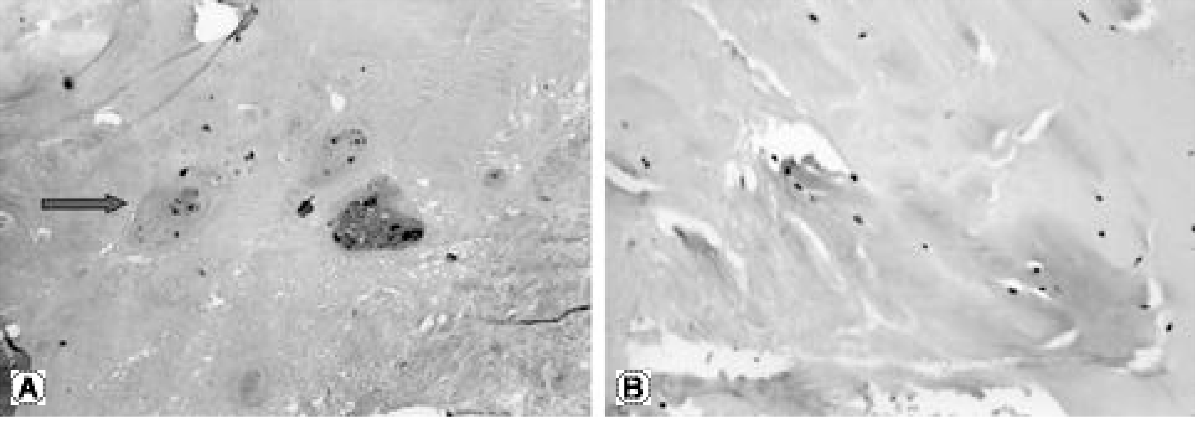
Fig. 2.
S-100 positive finding in group 3(case number 1) : Noted Schwann cell(brown color) and these cells of chondrocyte and matrix look like spindle shape.
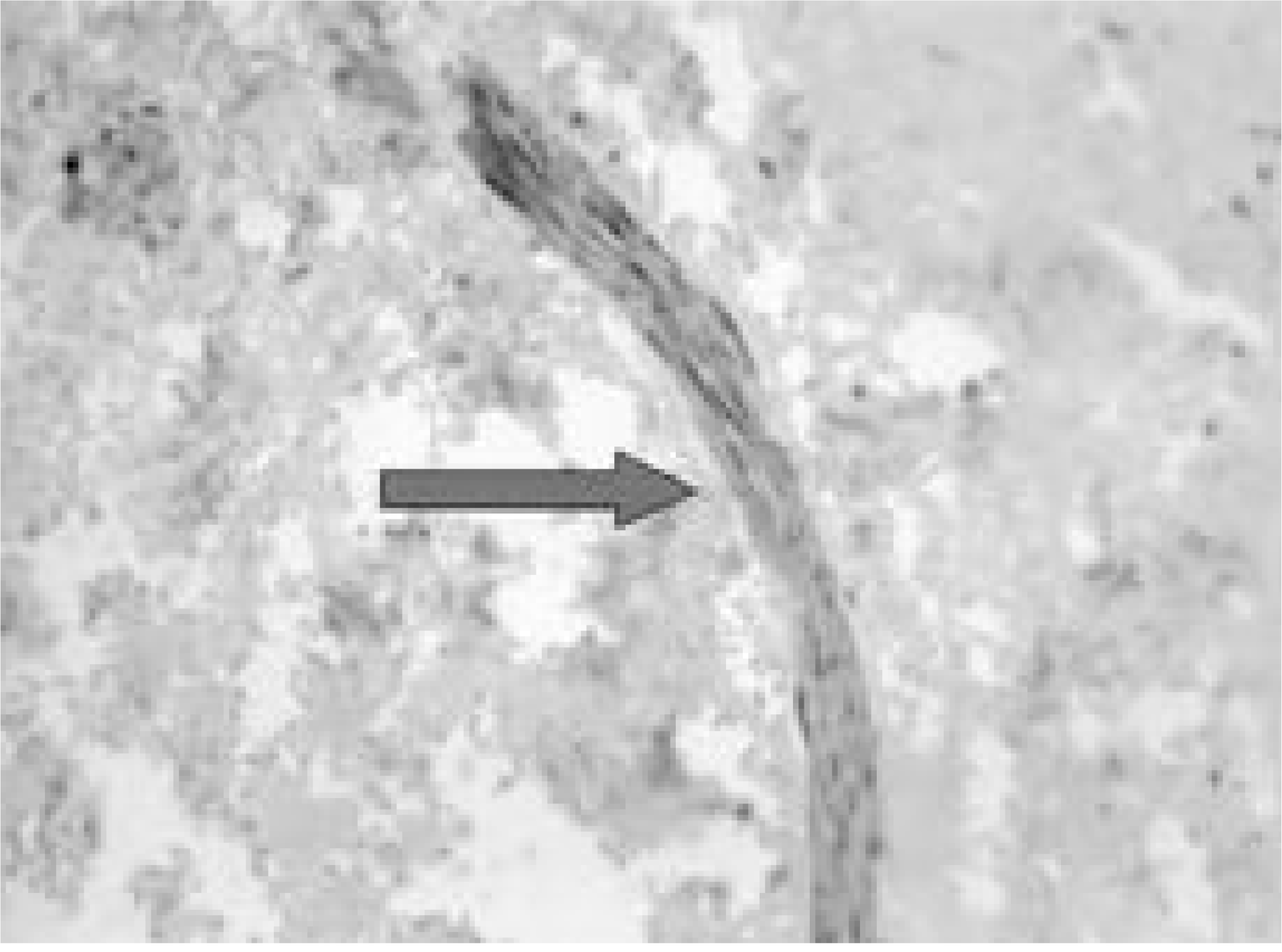
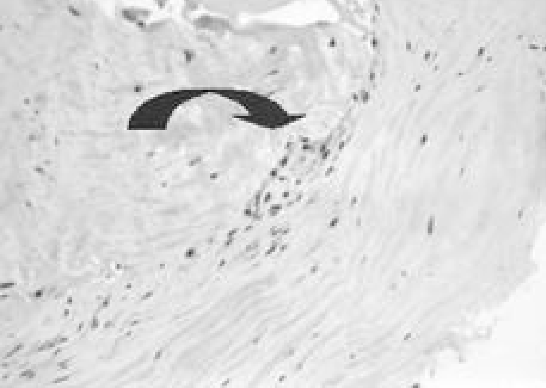
Table 1.
Data of each group
 XML Download