Abstract
Objectives
A n attempt to analyze the degenerative change of an intervertebral disc for adjacent segment degeneration in lumbar degenerative diseases.
Literature Review Summary
A review of the literature failed to uncover any documented study examining the quantitative analysis of the degenerative change of the intervertebral disc for adjacent segment degeneration.
Methods
This study was based on 45 patients, treated operatively or conservatively at this hospital, between A pril 1995 and July 2004. 39 and 6 cases of operative and conservative treatments, respectively, were performed. In the 39 operative treatments, there were 34 cases of fusion and 5 of discectomy. Dynamic X - ray and MRI were performed at the initial evaluation, and again more than 2 years later. In the 34 fusion cases, the upper and lower adjacent segments of the fused level were studied, and in the 11 nonfusion cases (conservative treatment or discectomy), the L3- 4, L4- 5 and L5- S1 level were studied. The instability of the dynamic X - ray and Thompson grade changes of the disc on MRI were also evaluated. Statistical analysis was carried out using the Wilcoxon signed rank test.
Results
Adjacent segment degeneration was found in 10 of the 34 cases (29.4%) on plain X - ray. The average Thompson grades of the 33 upper segment cases were 2.6 and 3.4 preoperatively and postoperatively (P=0.000), and for the 24 of the lower segment cases were 2.9and 3.2 (P=0.033), respectively. No statistical increase in the Thompson grade was found in the nonfusion group.
Go to : 
REFERENCES
1). Hambly MF, Wiltse LL, Raghavan N, Schneiderman G. The transition zone above a lumbosacral fusion. Spine. 1998; 23(16):1785–1792.

2). Aota Y, Kumano K, Hirabayashi S. Postfusion instability at the adjacent segments after rigid pedicle screw fixation for degenerative lumbar spinal disorders. J spinal disord. 1995; 8(6):464–473.

3). Lehmann TR, Spratt KF, Tozzi JE, et al. Long-term follow-up of lower lumbar fusion patients. Spine. 1987; 12:97–104.

4). Penta M, Sandhu A, Fraser RD. Magnetic resonance imaging assessment of disc degeneration 10 years after anterior lumbar interbody fusion. Spine. 1995; 20:743–747.

5). Shahin E, David WC. Risk factors for adjacent segment failure following lumbar fixation with rigid instrumentation for degenerative instability. J Neurosurg. 1999; 90:163–169.
6). Rahm MD, Hall BB. Adjacent segment degeneration after lumbar fusion with instrumentation: A retrospective study. J spinal disord. 1996; 9(5):392–400.
7). Brodsky AE. Post-laminectomy and post-fusion stenosis on the lumbar spine. Clin Orthop. 1976; 115:130–139.
8). Kumar MN, Baklanov A, Chopin D. C o r r e l a t i o n between sagittal plane changes and adjacent segment degeneration following lumbar spine fusion. Eur Spine J. 2001; 10:314–319.
9). Nagata A, Schendel MJ, Transfeldt EE, et al. The effects of immobilization of long segments of the spine on the adjacent and distal facet force and lumbosacral motion. Spine. 1993; 18:2471–2479.

10). Wimmer C, Gluch H, Krismer M, et al. AP-translation in the proximal disc adjacent to lumbar spine fusion: a retrospective comparison of mono- and polysegemental fusion in 120 pateients. Acta Orthop Scand. 1997; 68:269–272.
11). Wiltse LL, Radecki SE, Biel HM, et al. Comparative study of the incidence and severity of degenerative change in the transition zones after instrumented versus noninstrumented fusions of the lumbar spine. J Spinal Disord. 1999; 12:27–33.

12). Frymoyer JW, Hanley E, Howe J, Kuhlmann D, Matteri R. Disc excision and spine fusion in the management of lumbar disc disease: A minimum ten-year follow-up. Spine. 1978; 3:1–6.
13). Dekutoski MB, Schendel M, Ogilvie JW, OLsewski JM, Wallace LJ, Lewis JL. Comparison of in vivo and in vitro adjacent segment motion after lumbar fusion. Spine. 1994; 19:1745–1751.

15). Chung JY, Seo HY, Jung JW. Surgical treatment of adjacent degenerative segment after lumbar fusion-pre -liminary report. J Kor Spine Surg. 2000; 7(2):264–270.
16). Ha KY, Kim YH, Kang KS. Surgery for adjacent segment changes after lumbosacral fusion. J Kor Spine Surg. 2002; 9(4):332–340.

17). Kim SS, Michelsen CB. Revision surgery for failed back surgery syndrome. Spine. 1992; 17:957–960.

18). Whitecloud TS, David JM, Olive PM. Operative treatment of the degenerated segment adjacent to a lumbar fusion. Spine. 1994; 19:531–536.

19). Gary G, Jeffrey CW, Nitin NB, Wellington KH, Edgar GD. Adjacent segment degeneration in the lumbar spine. J Bone Joint Surg. 2004; 86-A:1497–1503.
20). White III AA, Panjabi MM. Clinical biomechanics of the spine. 2nd ed.Philadelphia: JB Lippincott;p. 192–193. 1978.
21). Thompson JP, Pearce RH, Schechter MT, Adams ME, Tsang IK, Bishop PB. Preliminary evaluation of a scheme for grading the gross morphology of the human intervertebral disc. Spine. 1990; 15:411–415.

23). Albee FH. Transplantation of a portion to the tibia into the spine for Pott’s disease. JAMA. 1911; 57:885.
24). Brunet JA, Wiley JJ. Acquired spondylolysis after spinall fusion. J Bone Joint Surg(Br). 1984; 66:720–724.
26). Weinhoffer SL, Guyer RD, Herbert M, et al. Intradiscal pressure measurements above an instrumented fusion. A cadeveric study. Spine. 1995; 20:526–531.
27). Cunningham BW, Kotani Y, McNulty PS, et al. The effect of spinal destabilization and instrumentation on intradiscal pressure: an in vitro biomechanical analysis. Spine. 1997; 22:2655–2663.
28). Thompson JP, Lewis JL. Comparison of in vivo and in vitro adjacent segment motion after lumbar fusion. J Rheumatol Suppl. 1991; 27:1745–1751.
Go to : 
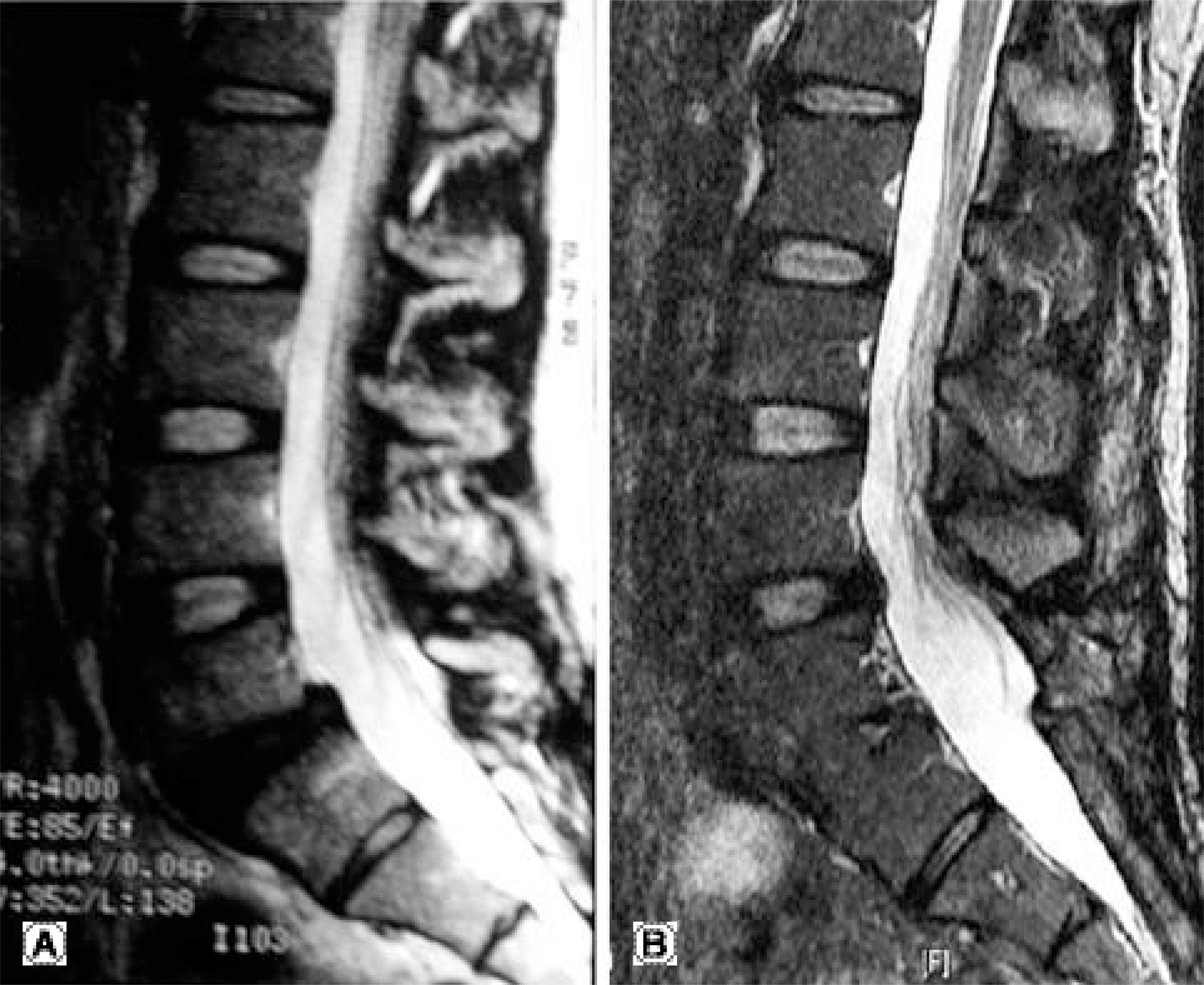
 | Fig. 1.(A) A 53-year-old woman with isthmic spondylolisthesis L4. The disc signal of L3-4 and L5-S1 was Thmpson grade I. Anterior interbody fusion and posterior fixation was done. (B) 9 years later, there was no remarkable change of adjacent disc. |
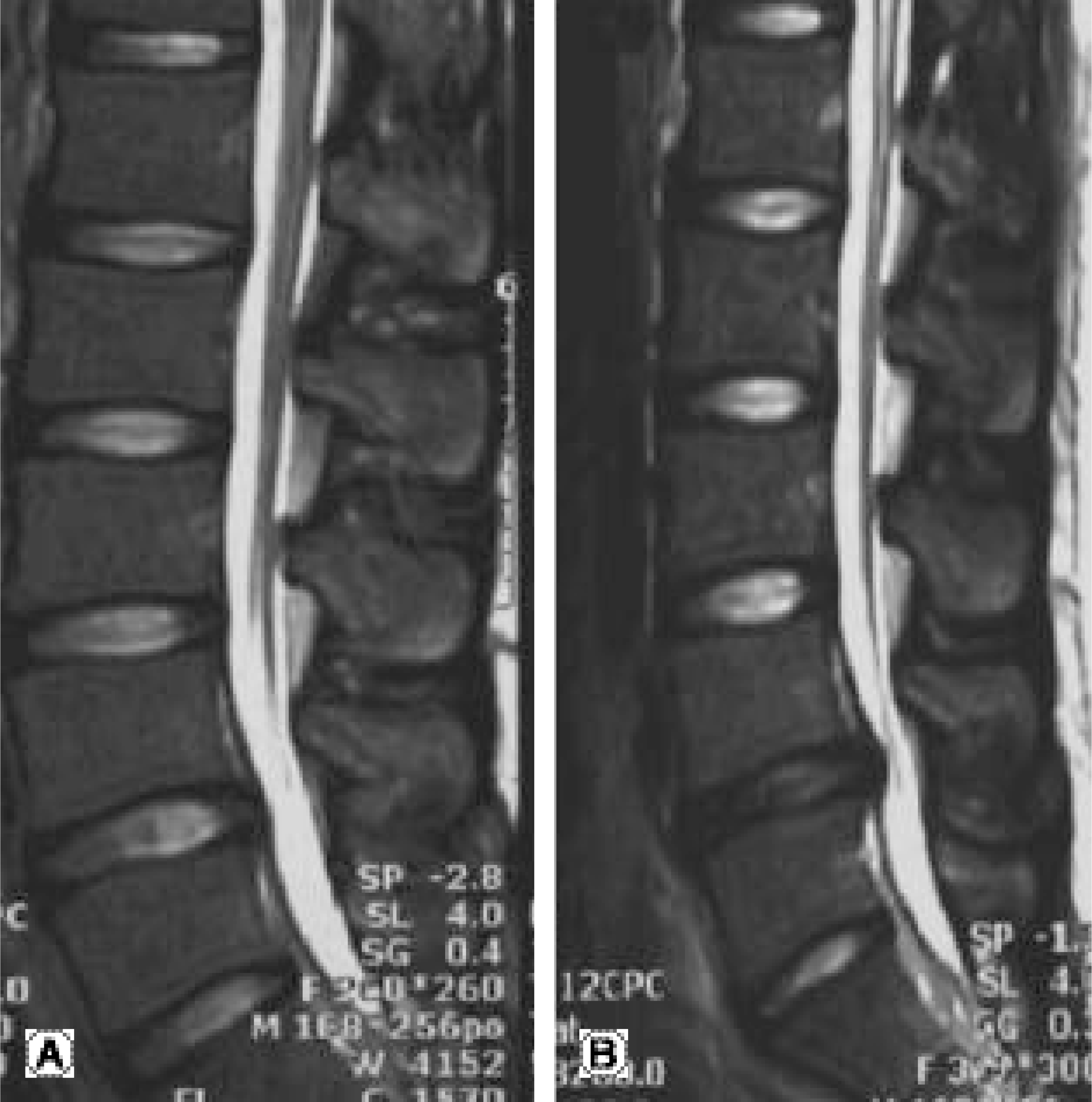
 | Fig. 2.(A) A 22-year-old man with mild bulging of L4-5 and Thompson grade was grade II. Conservative treatment was done. (B) 2 years later, L4-5 herniation was severed and disc signal has changed to grade IV. |
Table 1.
Patients data


 PDF
PDF ePub
ePub Citation
 Print
Print


 XML Download