

 PDF
PDF ePub
ePub Citation
 Print
Print


Abstract
Objectives
The optimal trajectories for C1lateral mass screws and C2 parspedicle screws were sought, and their accuracy evaluated.
Literature Review Summary
There have been a few suggestions for the trajectories of the screws listed above, but these are somewhat vague or impractical.
Materials and Methods
Using 1 mm- sliced CT scan images of 128 patients, and a V - works spine surgery simulator 4.0(Cybermed, Inc., Korea), the optimal trajectories with which 4.0 mm screws can be inserted without breaching bone cortices were determined. The anatomical characteristics of the cases having a cortical perforation were analyzed.
Results
The insertion point suggested for a C1 screw was 1 mm lateral to the middle of the junction of the posterior arch and posterior inferior part of the lateral mass. The screw was directed 15 degrees medially and toward the junction of the superior 2/3 and inferior 1/3 of the anterior tubercle in the lateral fluoroscopic view. The C2 screw was directed 30 degrees medially, and toward the anterior end of the superior articular process, in the lateral fluoroscopic view. The insertion point was one where the screw was inserted close to the superomedial border of the pedicle. Using these trajectories, all (256/256) of the C1 screws were inserted safely. However, 6.3% (16/256) of the C2 screws breached the inferolateral cortices of the pedicles, due to the pedicles being either too narrow or too medially angulated.
Conclusions
Herein, more practical and safe screw trajectories have been suggested. Using these trajectories, all the C1 and most of the C2 screws were able to be inserted safely. However, there were some cases in which the C2 screws could not be inserted without breaching the vertebral artery groove. Therefore, preoperative thin- slice CT scanning, with Three-dimensional reconstruction and/or Three-dimensional CT- angiography, is recommended for these cases.
REFERENCES
1). Abumi K, Kaneda K. Pedicle screw fixation for nontrau -matic lesions of the cervical spine. Spine. 1997; 22:1853–1863.
2). Ebraheim NA, Mission JR, Xu R, Yeasting RA. The optimal transarticular C1-2 screw length and the location of the hypoglossal nerve. Surg Neurol. 2000; 53:208–210.

3). Ebraheim N, Rollins JR Jr, Xu R, Jackson WT. Anatomic consideration of C2 pedicle screw placement. Spine. 1996; 21:691–695.
4). Goel A, Desai KI, Muzumdar DP. Atlantoaxial fixation using plate and screw method: A report of 160 treated patients. Neurosurgery. 2002; 51:1351–1357.
5). Harms J, Melcher RP. Posterior C1-C2 fusion with polyaxial screw and rod fixation. Spine. 2001; 26:2467–2471.
6). Hong X, Dong Y, Yunbing C, Qingshui Y, Shizheng Z, Jingfa L. Posterior screw placement on the lateral mass of atlas.: An anatomic study. Spine. 2004; 29:500–503.
7). Howington JU, Kruse JJ, Awasthi D. Surgical anatomy of the C-2 pedicle. J Neurosurg Spine. 2001; 95:88–92.
8). Resnick DK, Lapsiwala S, Trost GR. Anatomic suitability of the C1-C2 complex for pedicle screw fixation. Spine. 2002; 27:1494–1498.
9). Wang MY, Samudrala S. Cadaveric morphometric analysis for atlantal lateral mass screw placement. Neurosurgery. 2004; 54:1436–40.
10). Xu R, Ebraheim NA, Missoon JR, Yeasting RA. The reliability of the lateral radiograph in determination of the optimal transarticular C1-C2 screw length. Spine. 1998; 23:2190–2194.
11). Xu R, Nadaud MC, Ebraheim NA, Yeasting RA. Morphology of the Second cervical vertebra and the posterior projection or the C2 pedicle axis. Spine. 1995; 20:259–263.
Fig. 1.
Trajectories of lateral mass screws of C1 are shown. (A) Traditional multiplanar reformatting (MPR) images and a posterior view of a three-dimensional image are shown. (B) Trajectory MPR images and a lateral view of a three-dimensional image are shown. (C) Traditional MPR images and an anterior view of a three-dimension-al image are shown.
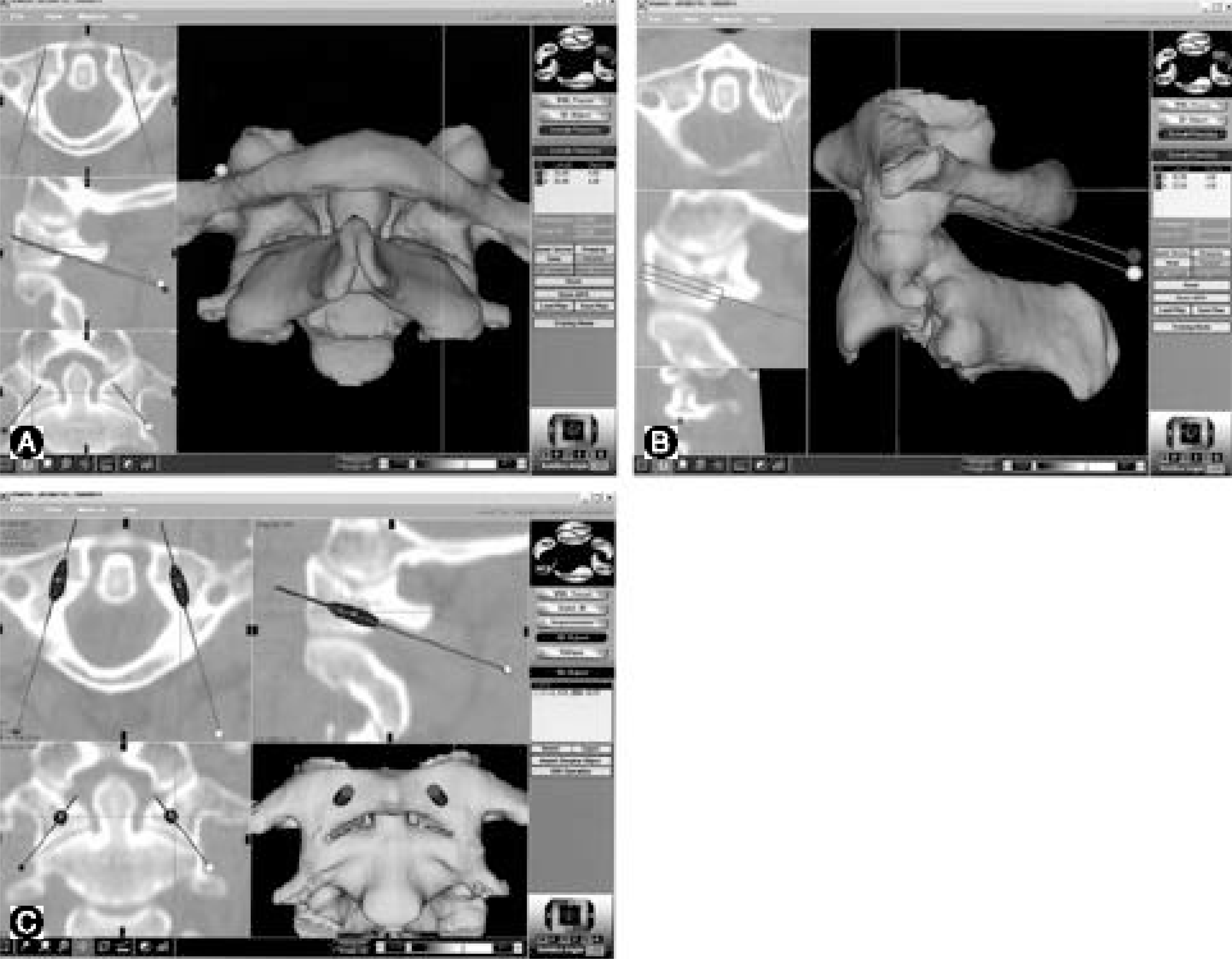
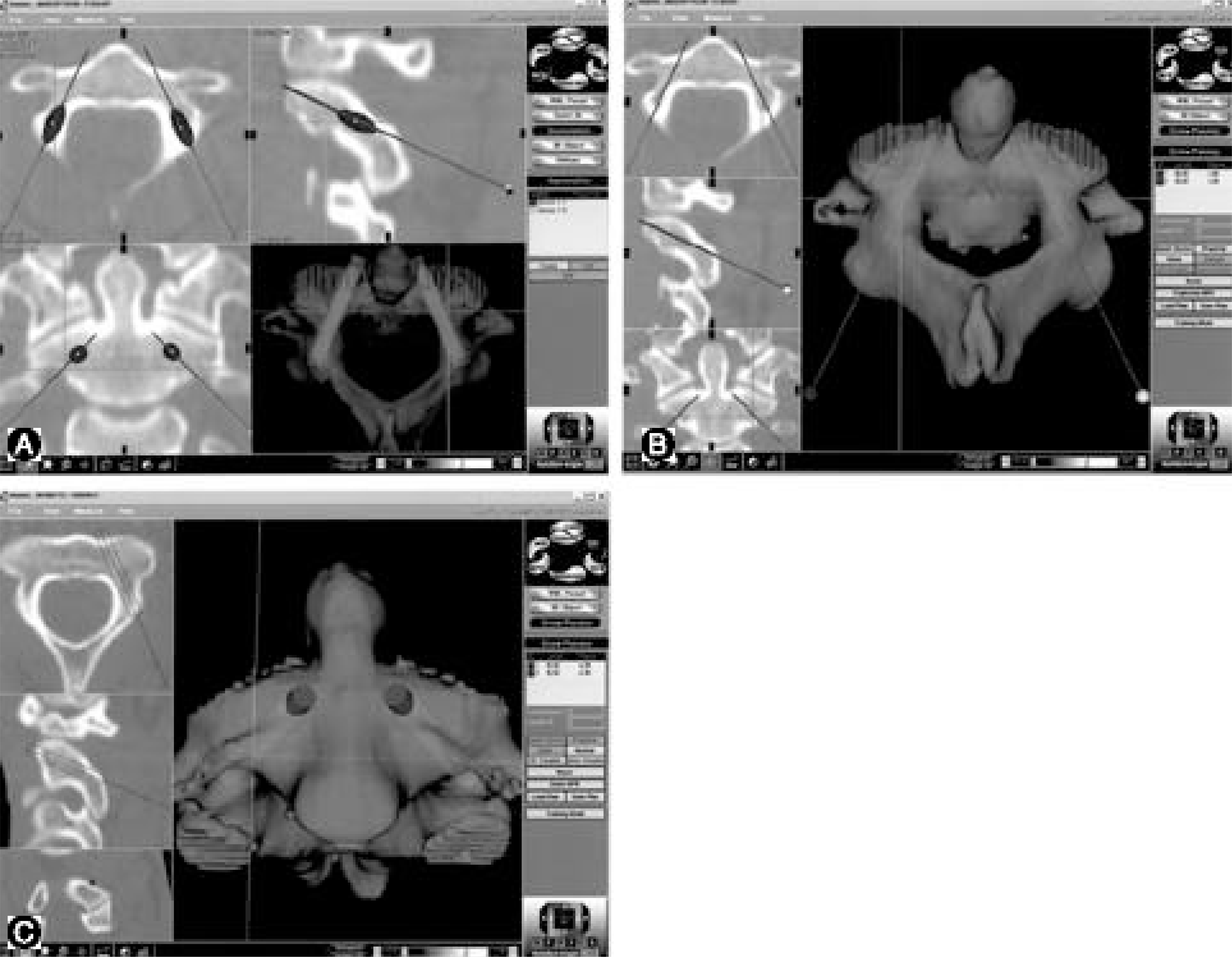
Fig. 2.
Trajectories of parspedicle screws of C2 are shown.(A) Traditional MPR images and a superoposterior view of a three-dimensional image are shown. (B) Traditional MPR images and a superoposterior view of a three-dimensional image are shown. (C) Trajectory MPR images and an anterior view of a three-dimensional image are shown.
 XML Download