Abstract
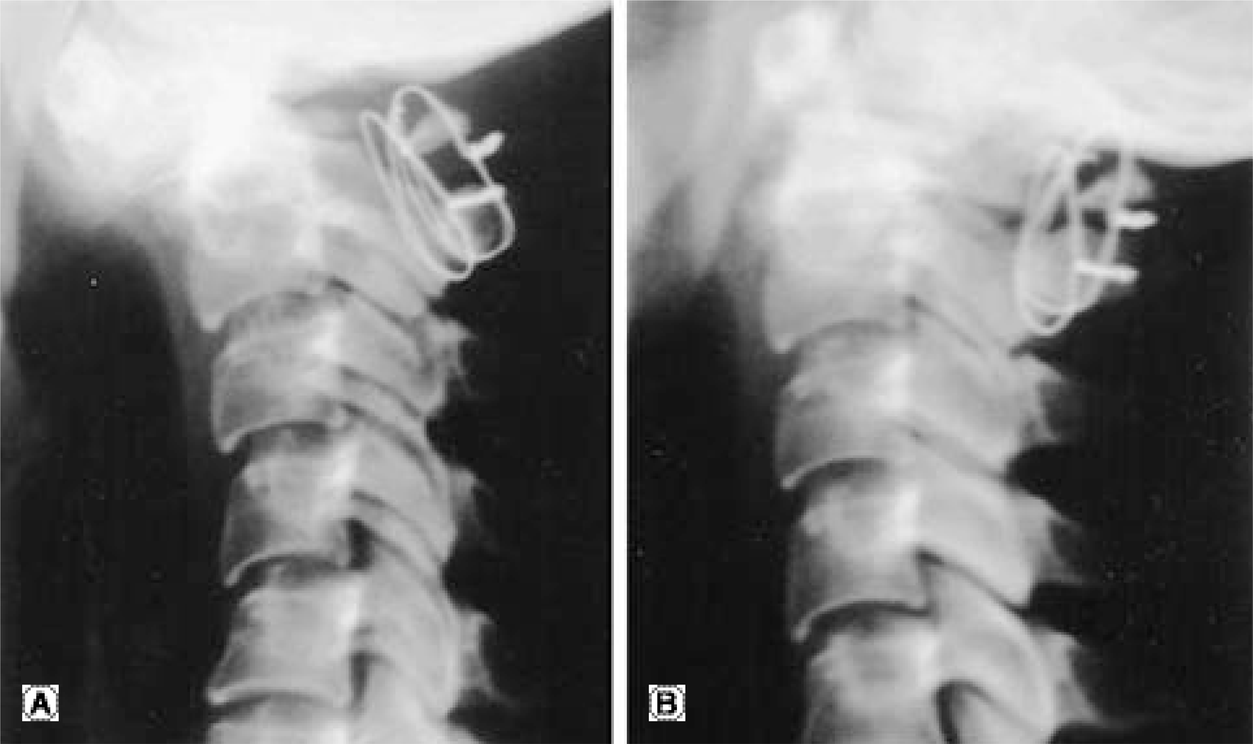
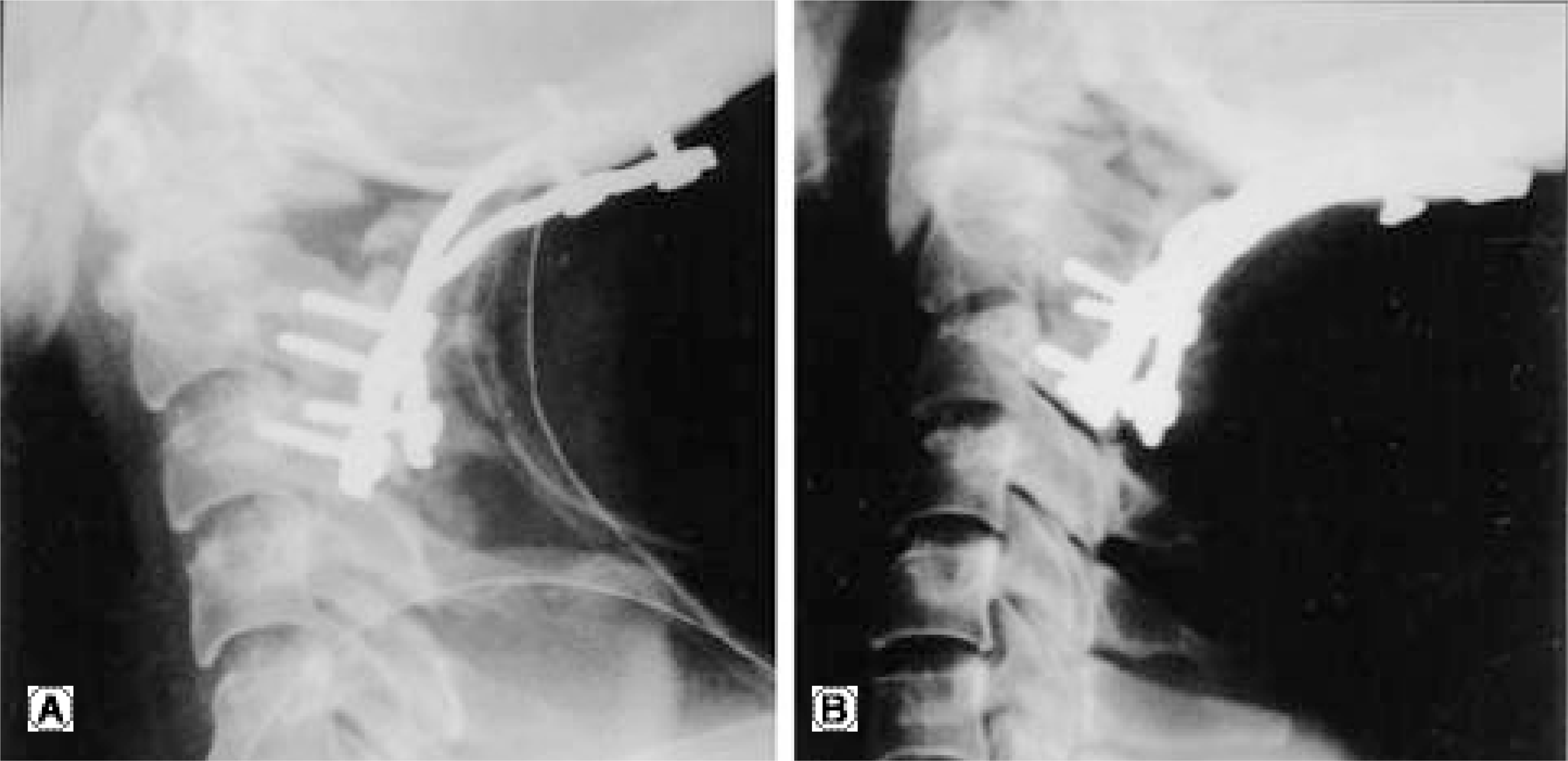
A 31- year-old female complained of neck pain and limitation in neck motion. She had a 3 month history of treatment with Halovest at another hospital for a fracture of the odontoid process due to a car accident. The patient complained of persistent pain and limitation in neck motion following the cessation of Halovest. A dynamic radiograph demonstrated instability on C1- 2 and she underwent a posterior cervical fusion with wiring. A wound infection developed, and loosening of the wire and lysis of the posterior arch at C1- 2 were seen on a follow up plain radiograph 2 months postoperatively. She was transferred to our hospital where she underwent occipitocervical fusion with a double plate after control of the infection. There were rigid fixations of the plate and bone union on a follow up radiograph 24 months postoperatively.
Go to : 
REFERENCES
1). Carlson GD, Bohlman HH. Surgical techniques for occiput to C2 arthrodesis. dillin WH, Simeone FA, editors. Posterior cervical spine surgery. 3rd ed.Philadelphia: p. 41. 1998.
2). Sasso RC, Jeanneret B, Fischer K, Magerl F. Occipitocervical fusion with posterior plate and screw instrumentation. Spine. 1994; 19:2264–2268.
3). Smith MD, Anderson P, Grady MS. Occipit oce rvical arthrodesis using contoured plate fixation. Spine. 1993; 18:1984–1990.
4). Elia M, Mazzara JT, Fielding JW. Onlay technique for occipitocervical fusion. Clin Orthop. 1992; 280:170–174.

5). Newman P, Sweetman R. Occipitocervical fusion: an operative technique and indications. J Bone Joint Surg. 1969; 51B:423–431.
6). Wertheim SB, Bohlman HH. Occipitocervical fusion: indications, techniques, and long-term results in 13 patients. J Bone Joint Surg. 1987; 69A:833–836.
7). Clark CR, Goetz DD, Menezes AH. Arthrodesis of the cervical spine in rheumatoid arthritis. J Bone Joint Surg. 1989; 71A:381–392.

8). Clark CR, Keggi KJ, Panajabi MM. Methylmethacry -late stabilization of the cervical spine. J Bone Joint Surg. 1984; 66A:40–46.
9). MaKenzie AI, Uttley D, Marsh HT, Bell BA. Craniocervical stabilization using Luque/Hartshill rectangles. Neu-rosergery. 1990; 26:32–36.
10). Malcolm GP, Ransford JR, Crockard HA. Treatment of non-rheumatoid occipitocervical instability. internal fixation with the Hartshill-Ransford loop. J Bone Joint Surg. 1994; 76B:357–366.

11). Takada T, Shono T, et al. Posterior occipitocervical reconstruction using cervical pedicle screws and plate-rod systems. Spine. 1999; 24:1425–1434.

12). Cahill DG. Posterior occipitocervical reconstruction using cervical pedicle screws and plate-rod systems. Spine. 2000; 25:1318.

13). Heller JG, Sillcox III DH, Sutterlin III CE. Complication of posterior cervical plating. Spine. 1995; 20:2442–2448.
14). Lieberman IH, Webb JK. Occipitocervical fusion using posterior titanium plate. Eur Spine J. 1998; 7:308–312.
15). Bradford LC, Panayiotis JP, Patricia GN, et al. Biomechanical evalution of new posterior occipitocervical instrumentation system. Clin Orthop. 2003; 411:103–115.
Go to : 


 PDF
PDF ePub
ePub Citation
 Print
Print


 XML Download