

 PDF
PDF ePub
ePub Citation
 Print
Print


Abstract
Study Design
A retrospective analysis of the distribution and patterns of posterior column injury in flexiondistraction injuries of the thoracolumbar spine.
Objectives
To recognize the various types of posterior column injury in terms of the path of the distraction force in Flexion-distraction injuries of the thoracolumbar spine.
Summary of Literature Review
As posterior column injuries are associated with instability of the spine, many authors have described and classified posterior column injuries. However, there are no descriptions or classifications in terms of the path of the distraction force in the literature.
Materials and Method
The preoperative plain X - rays, axial CT, MRI (in 5 patients) and operation records of 34 patients were reviewed in relation to the patterns of posterior column injury.
Results
Posterior column injuries can be classified into two main types. In TypeⅠ (30/34), the distraction failure started from the spinous process one level above the fractured body (TypeⅠA) or the posterior ligament complex between the spinous processes of the fractured and the level above (TypeⅠB). In TypeⅡ (4/34), the distraction failure started from the spinous process of the fractured vertebra and from the interspinous ligament between the fractured level and the level below.
Go to : 
REFERENCES
1). Whitesides TE Jr. Traumatic kyphosis of the thoracolumbar spine. Clin Orthop. 1977; 128:78–91.

2). Liu YJ, Chang MC, Wang ST, Yu WK, Liu CL, Chen T H. Flexion-distraction injury of the thoracolumbar spine. Injury. 2003; 34:920–923.
3). Holdsworth FW. Fractures, dislocations, and fracture-dislocations of the spine. J Bone Joint Surg. 1970; 52A:1534–1551.
4). Denis F. Spinal instability as defined by the three-column spine concept in acute spinal trauma. Clin Orthop. 1984; 189:65–76.
5). Denis F. The three column spine and its significance in the classification of acute thoracolumbar spinal injuries. Spine. 1983; 8:817–831.
6). Kaufer H, Hayes JT. Lumbar fracture dislocation. a study of twenty-one cases. J Bone Joint Surg. 1966; 48A:712–730.
7). McAfee PC, Yuan HA, Lasda NA. The unstable burst fracture. Spine. 1982; 7:365–373.
8). Shin BJ, Kim SK, Lee BI, Kim YI, Rah SK, Choi CU. Posterior column injuries in thoracolumbar and lumbar burst fracture. J Kor Spine Surg. 1997; 4:67–73.
9). Shin BJ, Kim BW, Kim YI, Rah SK. Difference of fracture patterns by the level of the thoracolumbar burst fracture. J Kor Spine Surg. 1998; 5:47–52.
10). Berry JL, Moran JM, Berg WS, et al. A morphometic study of human lumbar and selected thoracic vertebrae. Spine. 1987; 12:362–366.
11). Daffner RH, Deeb ZL, Goldberg AL, Kanada barow A, Rothfus WE. The radiologic assessment of post-traumatic vertebral stability. Skeletal radiol. 1990; 19:103–108.
12). Abe E, Sato K, Shimada Y, Mizutani Y, Chiba M, Ok uyama K. Thoracolumbar burst fracture with horizontal fracture of the posterior column. Spine. 1997; 22:83–87.
13). Gumley G, Taylor TKA, Ryan MD. Distraction fractures of the lumbar spine. J Bone Joint Surg. 1982; 64B:520–525.
14). Gertzbein SD, Court-Brown CM. Fle xi on-distraction injuries of the lumbar spine. Clin Orthop. 1988; 227:52–60.
15). Terk MR, Hume-Neal M, Fraipont M, Ahmadi J, Col-letti PM. Injury of the posterior ligament complex in patients with acute spinal trauma: evaulation by MR imaging. Am J Roentgenol. 1997; 168:1481–1486.
16). Haba H, Taneichi H, Kotani Y, et al. Diagnostic accura -cy of magnetic resonance imaging for detecting posterior ligamentous complex injury associated with thoracic and lumbar fracture. J Neurosurg. 2003; 99:20–26.
17). Kliewer MA, Gray L, Paver J. Acute spinal ligament disruption: MR imaging with anatomical correlation. J Magn Reson Imaging. 1993; 3:855–861.
18). Saifuddin A, Noordeen H, Taylor BA, Bayley I. The Role of imaging in the diagnosis and management of thoracolumbar burst fractures: current concepts and a review of the literature. Skeletal Radiol. 1996; 25:603–613.
19). Moon SH, Park MS, Suk KS, et al. Feasibility of ultra -sound examination in posterior ligament complex injury of thoracolumbar spine fracture. Spine. 2002; 27:2154–2158.
Go to :
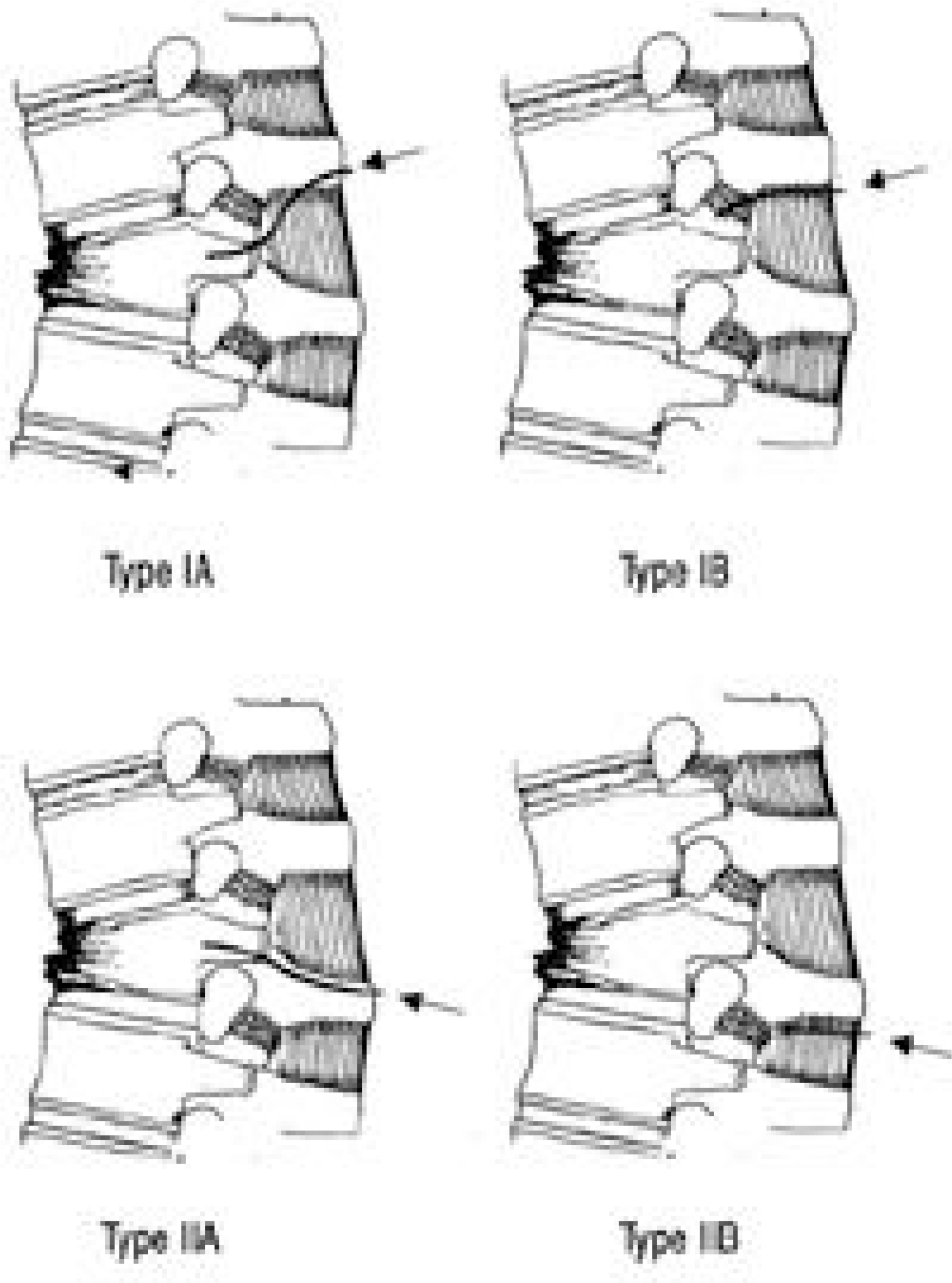 | Fig. 1.Classification of posterior column injury according to the path of distraction force TypeⅠⅠ A: Distraction failure started from the spinous process of one level above. ⅠB : Distraction failure started from the posterior ligament complex between the spinous processes of fractured and one level above. TypeⅡⅡ A : Distraction failure started from the spinous process of the fracture vertebra. ⅡB : Distraction failure started from the posterior ligament complex between the spinous processes of the fractured and one level below. |
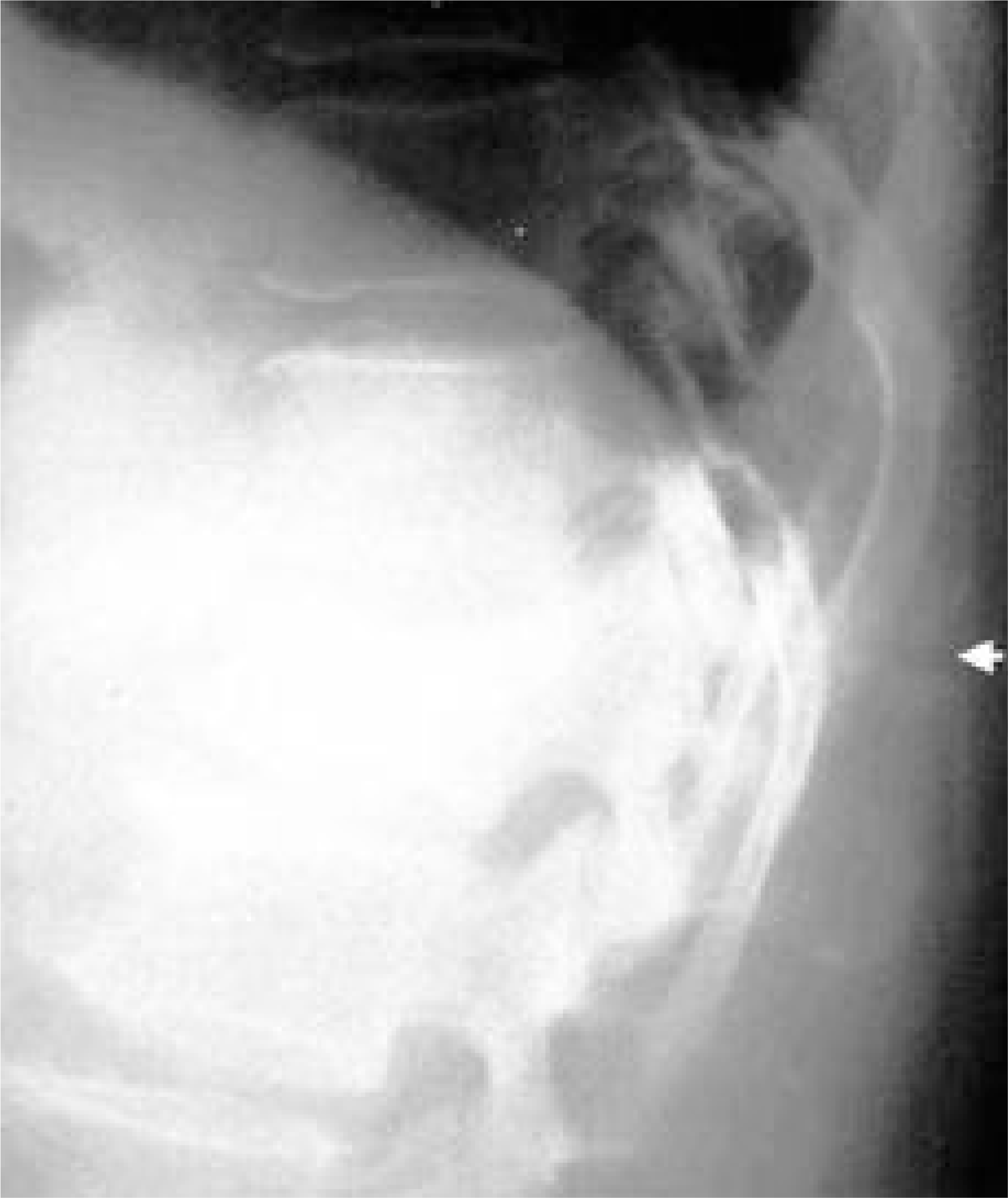 | Fig. 2.An example of Type Ⅰ A-2 T-L spine lateral X-ray shows wedge compression of L1 body and spinous process fracture of T12(arrow). |
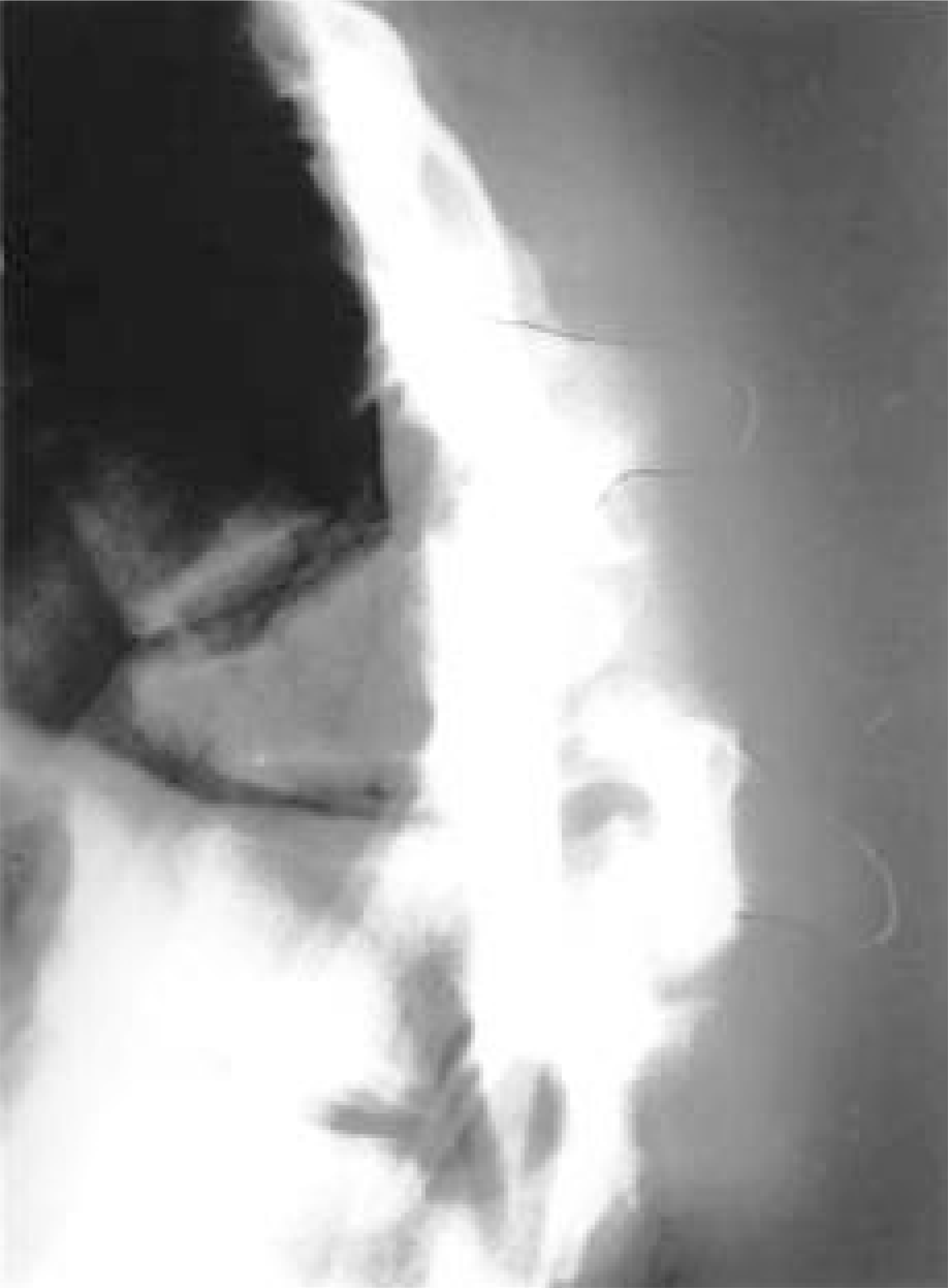 | Fig. 3.An example of Type Ⅰ B-3 T-L lateral X-ray shows severe wedge compression of T11 and widened interspinous space between T10 and T11. |
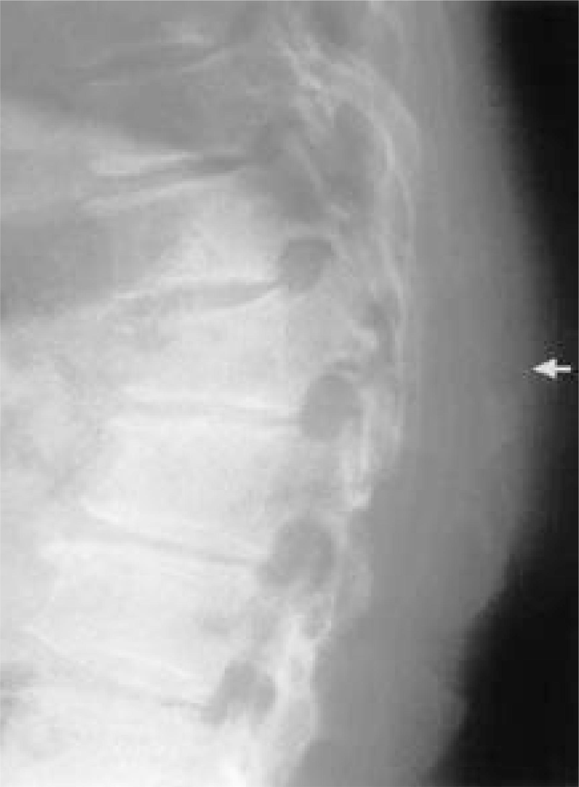 | Fig. 4.An example of Type Ⅱ A T-L spine lateral X-ray shows wedge compression of L1 body and horizontal fracture of spinous process of the fractured vertebra(arrow). |
 | Fig. 5.
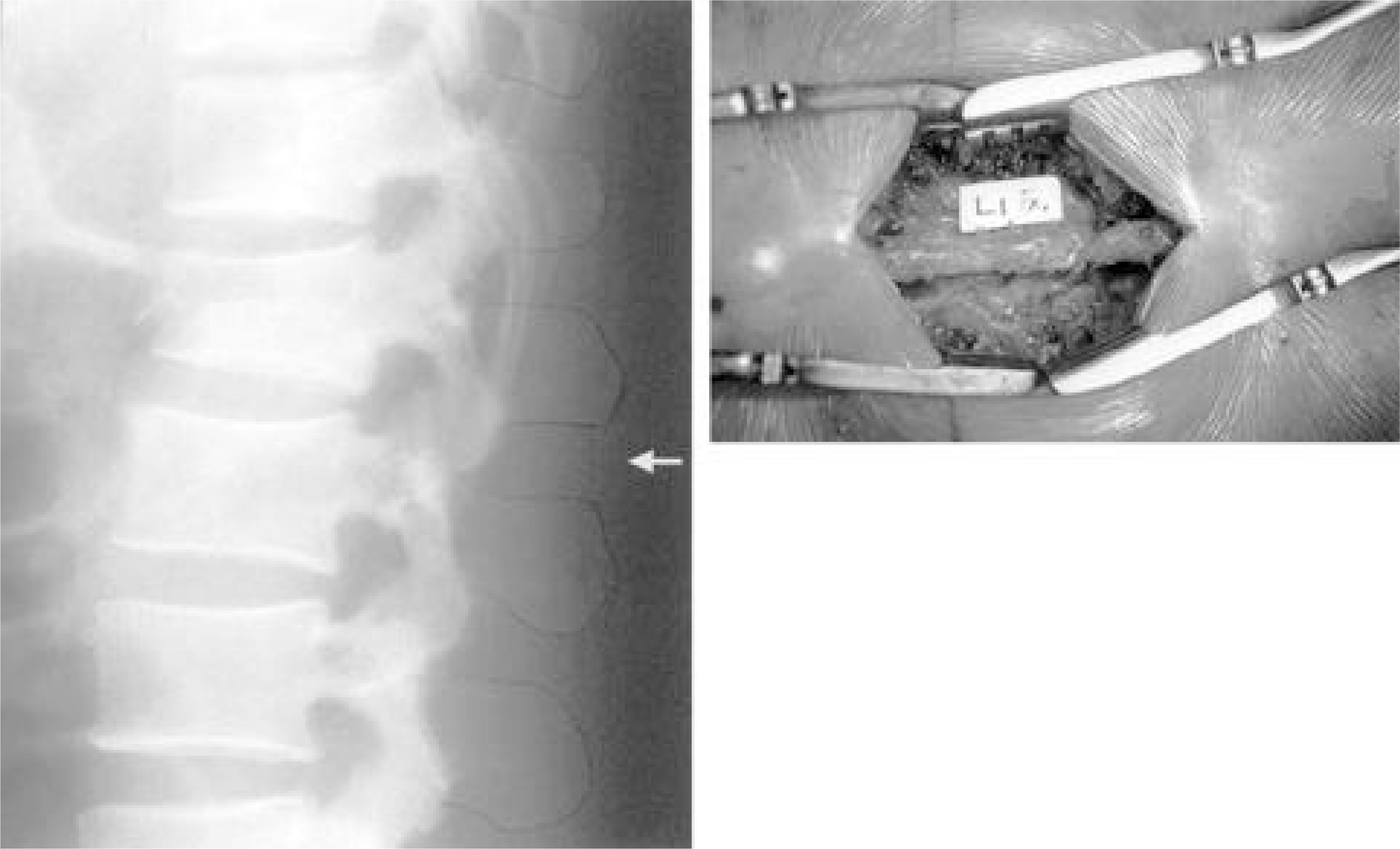
(A) An example of Type Ⅱ B T-L lateral X-ray shows wedge compression of L1 body and minimal widening of interspinous space between L1 and L2(arrow).(B) Operative finding shows disruption of posterior ligament complex between L1 and L2. |
Table 1.
Distribution of fractured vertebra according to posterior column injury
| LEVEL | PCI∗ | Ⅰ A | Ⅰ B | Ⅱ A | Ⅱ B | No. of casew |
|---|---|---|---|---|---|---|
| T11 | 2 | 2 | ||||
| T12 | 1 | 7 | 1 | 9 | ||
| L1 | 8 | 8 | 2 | 1 | 19 | |
| L2 | 2 | 2 | 4 | |||
| No. of case | 11 | 19 | 3 | 1 | 34 |
Table 2.
Detection rate of posterior column injury according to the radiologic examination
| PCI† | DM∗ | Simple AP | Simple lat. | Soft tissue. lat. | CT | |
|---|---|---|---|---|---|---|
| TypeⅠ A | 7/11(64%) | 8/11(72%) | 10/11(91%) | 8/11(73%) | ||
| TypeⅠ B‡ | 15/19(79%) | 15/19(79%) | 17/19(89%) | 9/19(47%) | ||
| TypeⅡ A | 2/3(67%) | 3/3(100%) | 3/3(100%) | 1/3(34%) | ||
| TypeⅡ B | 1/1(100%) | 1/1(100%) | 1/1(100%) | 0/1(0%) |
 XML Download