

 PDF
PDF ePub
ePub Citation
 Print
Print


Abstract
Objective
To evaluate the concordance, or discordance, of the osteoporotic diagnosis between femur neck and lumbar spine, using DEX A T- scores and the WHO classification.
Materials and Methods
The BMD (Define?) on both hips and of the lumbar spine of 718 Korean females were measured. The mean age of the subjects was 55.5 years (31- 91). The BMD data were obtained from 3 hip regions and from the lumbar spine, anteroposteriorly, using dual- energy x- ray absorptiometry (Lunar). The BMDs of femur neck and the L2- 4 vertebrae were classified into normal (a T- score >- 1), osteopenia (- 1 ≤ T- score <- 2.5) and osteoporosis (- 2.5≤T- score) using the WHO definitions.
Results
There was significant correlation between the femur neck and lumbar BMDs (r=0.772). However, the discordance rate was 33% for all the cases, but this was 20% in the subjects below 50 of age, 31% in the subjects in their 50's, 47% in their 60's and 42% when 70 or above.
The discordance rates of the normal, osteopenic and osteoporotic groups were 21, 54 and 17% respectively, with the highest discordance rate in the osteopenia group.
A mong the 649 persons in the normal or osteopenia groups, in relation to the femur neck BMD, there were 67 (10.3%) in the osteoporotic group with L2- 4 BMD. But the reverse situation was only 12 persons from 594 (2.0%). (Eds note: this whole section makes little sense? What were the BMDs? The last sentence is completely meaningless.)
Go to : 
REFERENCES
1). Feyerabend Angela J.., James L., Lear MD. Regional variations in Bone Mineral Density as Assessed with Dual-Energy Photon Absorptiometry and Dual X-Ray Absorptiometry. Radiology,. 186:467–469. 1993.

2). Blake GM., Fogelman I. Interpretation of bone den -sitometry studies. Semin Nucl Med,. 27:248–60. 1997.
3). Cameron JR., Sorenson J. Measurement of bone mineral in vivo: an improved method. Science. 142:230–232. 1964.
4). Compston JE. Osteoporosis. Clin Endocrinol. 33:653–682. 1990.
5). Douglas OH., Toyokawa S., Masaru U., Takahashi H., Katsumi K. Timing of menopause, reproductive years, and bone mineral density. a cross-sectional study of post -menopausal Japanese women. Am. J. of Epid.,. 148:1055–1061. 1998.
6). Kroger H., Tuppurainen M., Honkaneu R. Bone mineral density and risk factors for osteoporosis-a population based study of 1600 perimenopausal women. Calif Tissue int. 55:1–7. 1994.
7). Kanis JA., Melton LJ., Christiansen C., Johnston CC., Khaltaev N. The diagnosis of osteoporosis. J bone mineral research,. 9:1137–1141. 1994.
8). Kao. DH., Chen CC., Wang SJ. Normal data for lum -bar spine bone mineral content in healthy elderly Chinese. influences of sex, age, obesity and ethinicity. Nuclear Medicine Communications,. 15:916–920. 1994.
9). Koh SK., Cho SH., Hwang YY. Spinal bone mineral den -sity of normal and osteoporotic 재 men in Korea. Jounal of Korean Medical Science,. 7(2):136–140. 1992.
10). Kokai K., Kazuhiro K., Kaoru Y., Shozo O., Tetsuo I. Bone mineral density of the spine in normal Japanese sub -jects using Dual-Energy X-ray Absorptiometry. effect of obesity and menopausal status. Calcif Tissue,. 49:101–106. 1991.
11). Kroger H., Heikkinen J., Laitinen K., Kotaniemi A. Dual-Energy X-ray absorptiometry in normal women: A cross-sectional study of 717 Finnish volunteers. Osteo -porosis Int,. 2:135–140. 1992.
12). Laura K. Bachrach, Trevor Hastie, May-Choo Wang, Balasubramanian Narasimhan, and Robert Marcus. Bone Mineral acquisition in healthy Asian, Hispanic, Black, and Caucasian youth. a longitudinal study. The J Clinical Endocrinology & Metabolism,. 84(12):4702–4712. 1999.
13). Lee SJ., Koo JW., Suh JS., An JC. Measure of Bone Min -eral Density for Korean Adult with Dual Energy X-ray Absortiometry. J Korean Bone Metabolism. 1(2):201–208. 1994.
14). Meltzer M., Lesing HJ., Siegel JA. Bone mineral density and fracture in postmenopausal women. Calcif Tissue Int. 45(3):142–5. 1989.
15). Moon WN., Oh HJ., Suh SW. Differences in Bone Mineral Density by using Different Densitometers or by Measuring Different Sites of Spine in Osteoporotic Vertebral Fractures. J Korean Orthop Assoc. 34(6):1153–1157. 1999.
16). Riggs GL., Wahner HW., Seeman E. Change in bone mineral density of the proximal femur and spine with aging: Difference between the postmenopausal and senile osteoporosis syndrome. J clin invest. 70:716–723. 1982.
17). Rowe SM., Jung ST., Lee JY. Epidemiology of Osteo -porosis in Korea. Osteoporos Int,. 7(s3):S88–S90. 1997.
18). Saburo Ide, Yoshio Hirota, Takao Hotokebuchi, Shin-ichiro Takasugi, Yoichi Sugioka and Hitomi Hayabuchi. Osteoporosis and years since menopause. European jour -nal of Epidemiology,. 15:739–745. 1999.
19). Toshitsugu Sugimoto, Masaharu Tsutsumi, Yoshio Fjujh, Mitsuru Kawakatsu, Hirokuni Negishi, Lee MC, Keh-Ssung Tsai, Masaaki Fukase, and Takuo Fujita. Comparison of bone mineral content among Japanese, Koreans, and Taiwanese assessed by Dual-Photon Absorptiometry. J Bone and Mineral Research. 7(2):153–159. 1992.
20). Sugimoto T.., Kanbara Y.., Shirashi H.., Kawakatsu M.Femoral and spinal bone mineral density in Japanese osteoporosis with hip fracture. Osteoporosis Int. 4:144–148. 1994.
21). Wood AJJ. Treatment of Postmenopausal osteoporosis. New England J Medicine,. 338:736–746. 1998.
22). Woodson Grattan C.DXA T-Score concordance and discordance between the PA spine and total hip sites. Am. Society Bone Min Res. abstract book,. 1999.
Go to :
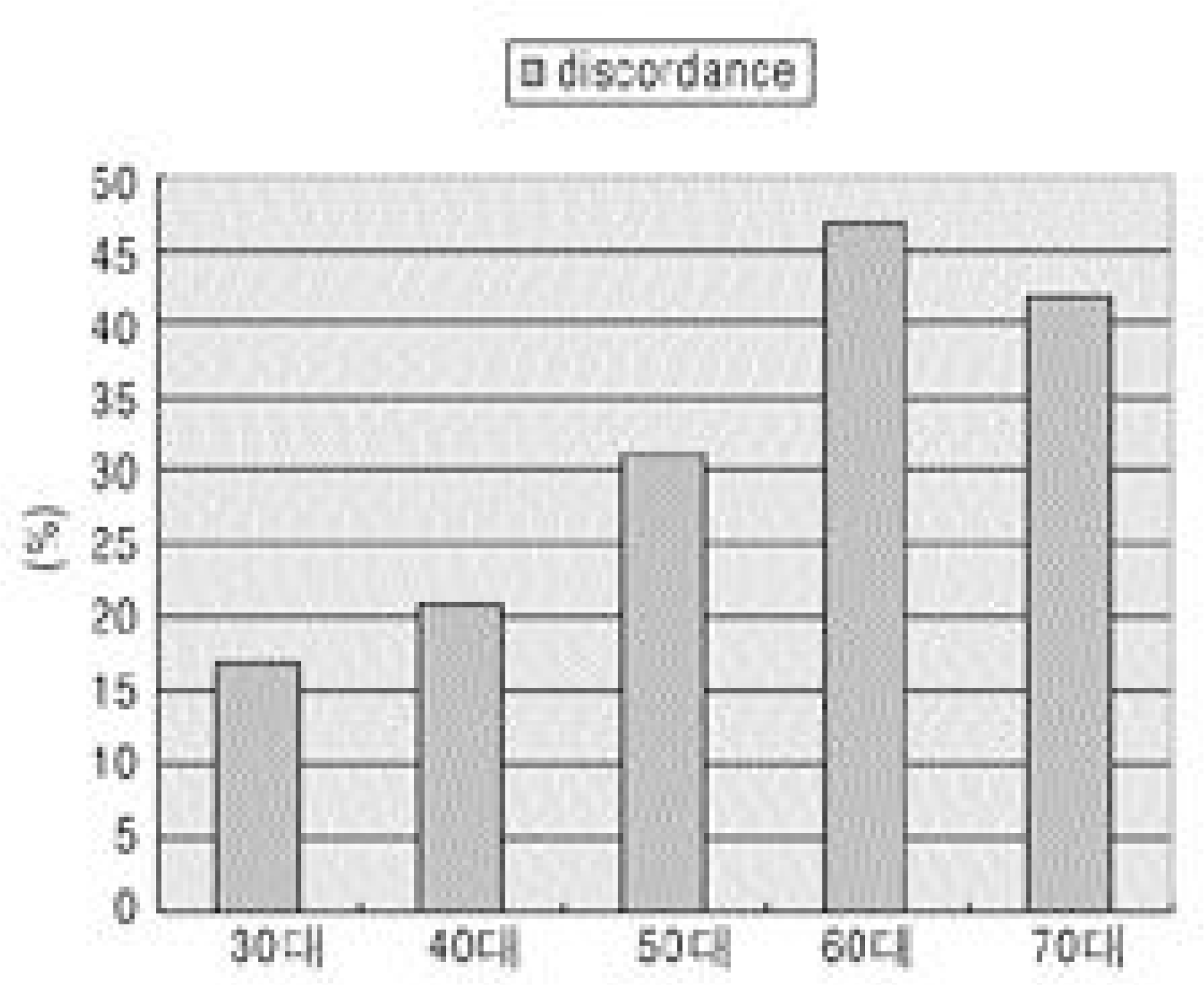 | Fig. 1.The diagnostic discordances of bone loss according to ages. It was increased with ages and showed highest val-ues in 60's. |
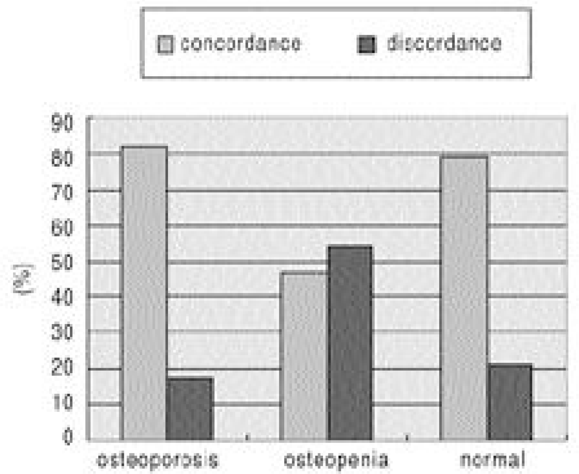 | Fig. 2.The ratio of discordance and concordance according to WHO classification of femur neck site. The discordance ratio in osteopenia group is highest. |
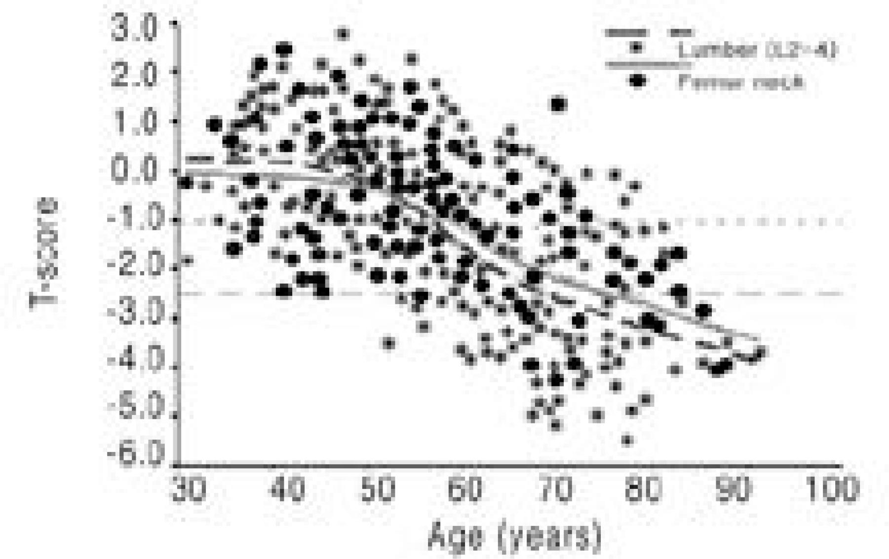 | Fig.3.Scattered plots of BMD, T-scores according to ages checked at lumbar spines (star) and femur neck (circle) |
Table 1.
The details of concordance and discordance between lumbar and femur neck sites
 XML Download