

 PDF
PDF ePub
ePub Citation
 Print
Print


Abstract
Objectives
To measure the first and second cervical spine, using MRI for a C1- 2 transarticular screw fixation, and find the safe trajectory for the screw.
Summary of Literature Review
Posterior atlantoaxial transarticular screw fixation is an excellent procedure that is associated with high fusion rates. However, there is a potential risk of vertebral artery injury.
Materials and Methods
Sagittal MR images, of the cervical spine transecting mid portion of the C1- 2 facet joints, were obtained in 24 patients. The mean age of the patients was 45.5 years. The male to female ratio of the patients was 15:9. From the sagittal images the ideal screw trajectory was made, and 48 oblique axial MR images, depending on the ideal screw trajectory in the sagittal plane, were obtained. On the oblique sagittal images, the width of the isthmic portion of the C2, the ideal length of the transarticular screw, the ideal insertion angle of the screw and the ideal entry point were measured using a PA CS digital measuring instrument. The location of the vertebral artery was also evaluated.
Results
The mean width of the isthmic portion of the C2 was 6.2 mm, ranging from 2.3 to 7.6 mm. The mean ideal screw length was 40.5 mm, ranging from 34.0 to 46.8 mm. The mean ideal insertion angle was 1.1°, ranging from - 2.4 to 4.7°, medi-ally. There were no significant differences in the width or the angle in relation to the sex of the patients. However, the length of the screw was significantly longer in the male (42.1mm) than the female patients (38.0 mm). Three of 24 patients (3 of 48 C1- 2 facet joints) had a narrow isthmus due to a high riding vertebral artery.
Conclusions
A C1- 2 transarticular screw fixation has a risk of injury to the vertebral artery. Therefore, the preoperative mea-surement of the C1- 2 region and an evaluation of the vertebral artery are recommended in each patient. A magnetic resonance image is a useful method for easily evaluating the anatomic structure of the C1- 2 region, with no additional study.
Go to : 
REFERENCES
1). Coric D., Branch CL., Wilson JA., Robinson JC. Arteriovenous fistula as a complication of C1-2 transartic -ular screw fixation. Case report and review of the literature. J Neurosurg,. 85:340–3. 1996.
2). Grob D., Crisco J., Panjabi MM., Dvorak J. Biome -chanical evaluation of four different posterior atlantoaxial fixation technique. Spine,. 17:480–90. 1991.
3). Grob D., Jeanneret B., Aebi M., Markwalder T. Atlantoaxial fusion with transarticular screw-fixation. J Bone Joint Surg,. 73-B:972–6. 1991.
4). Grob D., Magerl F. Surgical stabilization of C1 and C2 fractures. Orthopade,. 16:46–54. 1987.
5). Hurlbert RJ., Crawford NR., Choi WG., Dickman CA. A biomechanical evaluation of occipitocervical instrumentation: screw compared with wire fixation. J Neurosurg,. 90(1 Suppl):84–90. 1999.

6). Jeanneret B., Magerl F. Primary posterior fusion C1/2 in odontoid fracture: indications, technique, and results of transarticular screw fixation. J Spinal Disord,. 5:464–75. 1992.
7). Madawi AA., Casey AT., Solanki GA., Tuite G., Veres R., Crockard HA. Radiological and anatomical evaluation of the atlantoaxial transarticular screw fixation technique. J Neurosurg,. 86:961–8. 1997.
8). Mizuno J., Nakagawa H. Spinal instrumentation for unstable C1-2 injury. Neurol Med Chir,. 39:434–9. 1999.
9). Neo M., Matsushita M., Iwashita Y., Yasuda T., Sakamo-to T., Nakamura T. Atlantoaxial transarticular screw fixation for a high-riding vertebral artery. Spine,. 28:666–70. 2003.
10). Paramore CG., Dickman CA., Sonntag VK. Th e anatomical suitability of the C1-2 complex for transarticu -lar screw fixation. J Neurosurg,. 85:221–4. 1996.
11). Prabhu VC., France JC., Voelker JL., Zoarski GH. Vertebral artery pseudoaneurysm complicating posterior C1-2 transarticular screw fixation: case report. Surg Neu -rol,. 55:29–34. 2001.
12). Song GS., Theodore N., Dickman CA., Sonntag VK. Unilateral posterior atlantoaxial transarticular screw fix -ation. J Neurosurg,. 87:851–5. 1997.
13). Suk KS., Kim KT., Lee SH. C1-2 transarticular screw fix -ation as a revision surgery for failed C1-2 fusion. J Kor Spine Surg,. 9:251–6. 2002.
14). Wright NM., Lauryssen C. Vertebral artery injury in C1-2 transarticular screw fixation: results of a survey of the AANS/CNS section on disorders of the spine and peripheral nerves. American Association of Neurological Surgeons/Congress of Neurological Surgeons. J Neuro -surg. 88:636–40. 1998.
Go to :
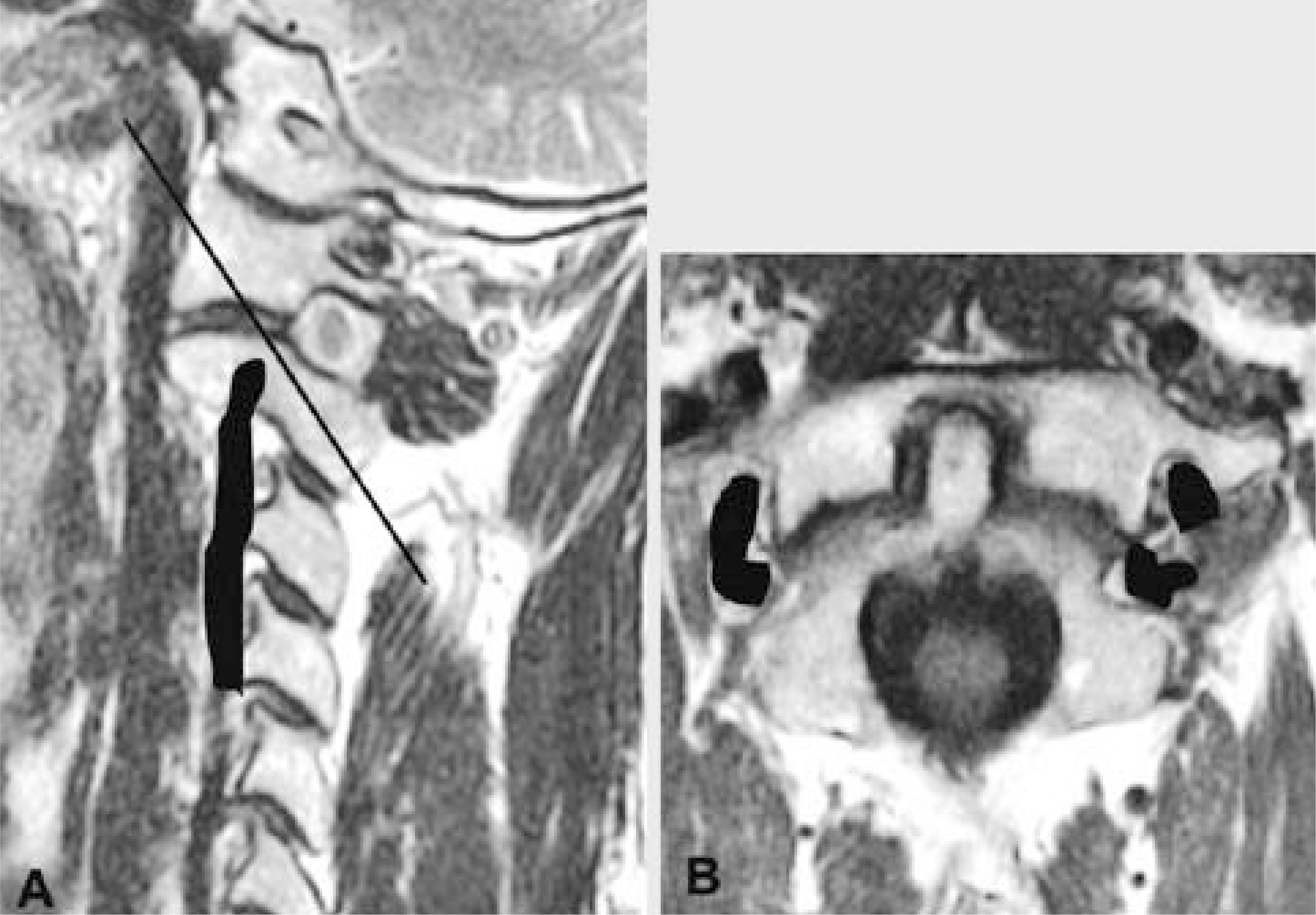 | Fig. 1.A sagittal MR image of the cervical spine transecting center of the C1-2 joint. Black line is an ideal trajectory of the C1-2 transarticular screw. Vertebral artery is marked as black color (A). An oblique axial image of the C1-2 joint obtained along the ideal trajectory line on the sagittal MR image. Vertebral artery is marked as black color (B). |
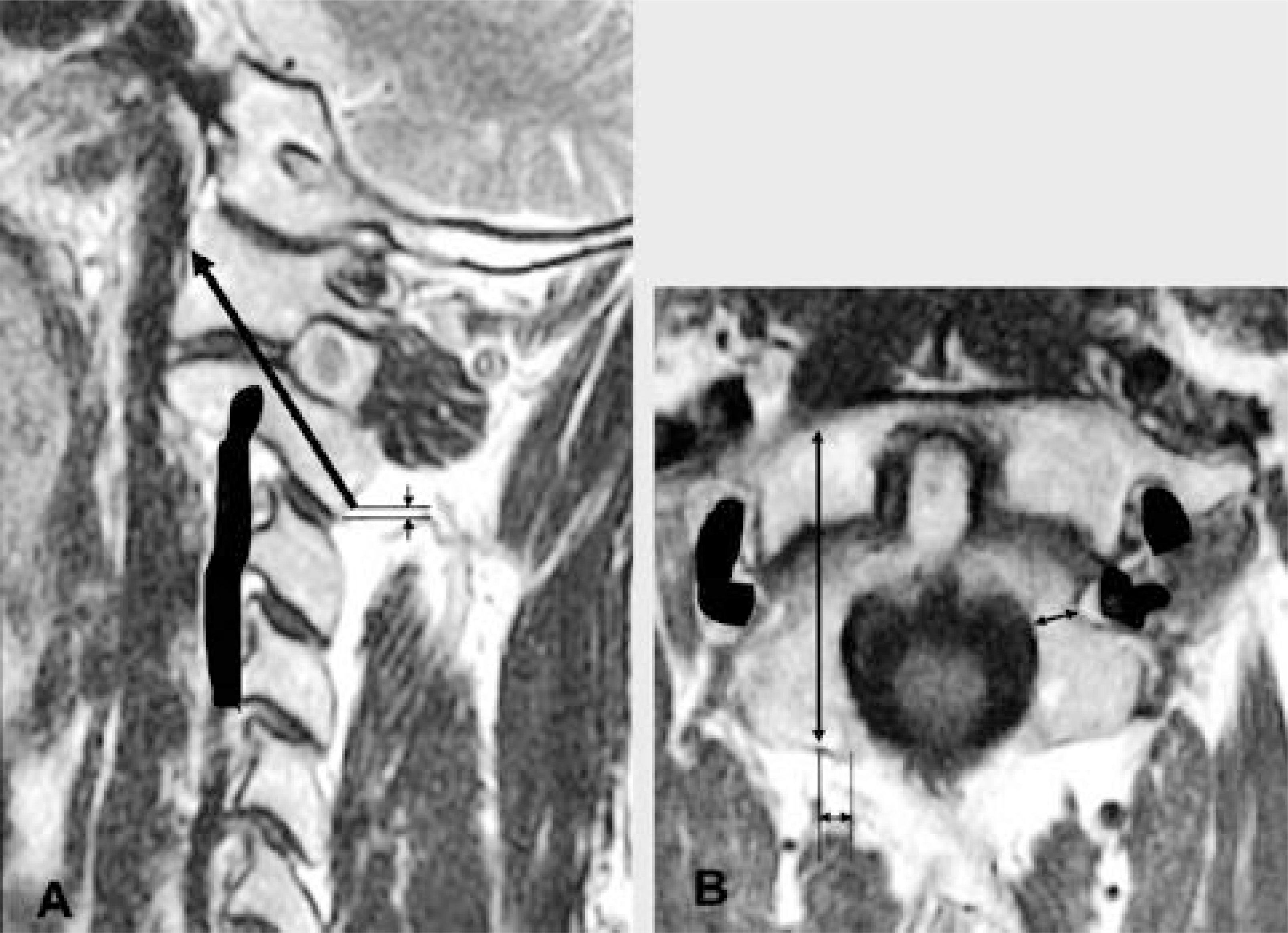 | Fig. 2.Ideal entry points, insertion angle and length of the C1-2 transarticular screw were on the sagittal and oblique axial MR images using PACS digital measuring instrument. |
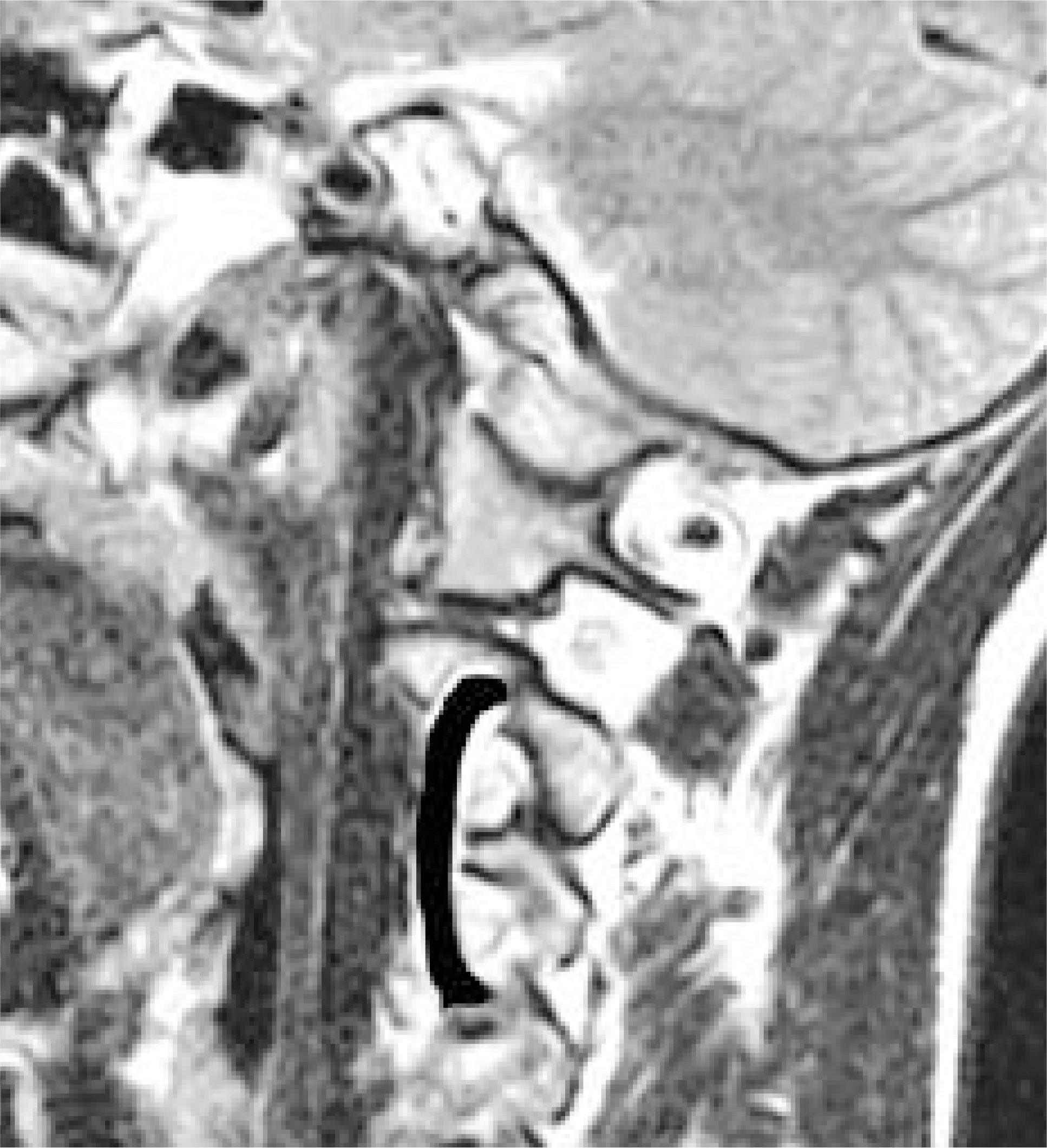
Table 1.
Measurements of C1-2 region for transarticular screw fixation
 XML Download