

 PDF
PDF ePub
ePub Citation
 Print
Print


Abstract
Study design
To examine the factors considered in the selection of therapeutic methods, and the methods for accessing postop-erative clinical outcomes, in degenerative lumbar spondylolisthesis.
Objectives
In this retrospective study, patients who had taken only posterolateral fusion, and with a posterior lumbar interbody fusion, were evaluated. The analyses of the pre- and post- operative factors associated with the clinical outcomes of the surgery for degenerative lumbar spondylolisthesis were also performed.
Materials and Method
Of the patients who had received the surgery for degenerative lumbar spondylolisthesis, between Janu-ary 1995 and December 2000, there were 59 for whom follow- up observations were possible, and these were selected for the present study. The patients were comprised of 19 males and 40 females, with ages ranging from 42 to 74 years (58.4± 8.4 years old). Of the 59 patients, 39, and 20, received a posterolateral fusion, or both a posterolateral fusion and a posterior lumbar inter-body fusion, respectively. In the present study, the pre- operative factors considered were the surgical method, sex, age, L1 axis S1 distance (LA SD), lordosis angle and the degree and duration of spondylolisthesis, with the degree of fusion, the lordosis angle of the fused body, the lordosis angle at the final follow- up and the lordosis angle of the fused body at the final follow- up, used as the post- operative factors. Each factor was statistically tested to see if it had a significant correlation with clinical outcomes (Recovery rate by Hirabayashi's method). A value of P <0.05 was considered as being statistically significant.
Result
The posterolateral fusion group showed a significantly lower recovery rate with an LSAD over 35 mm, a degree of spondylolisthesis over 10 mm and a preoperative lordosis angle under 20°, indicating that an additional posterior lumbar interbody fusion would provide a good clinical outcome. At the final follow-up, both groups showed significantly lower recovery rates with a lumbar lordosis angle under 20°, and the posterolateral fusion group showed significantly lower recovery rates when the post-operative lordosis angle of the fused segment was under 18°, and with a lordosis angle of the fused segment was under 18° at the final follow-up. These post-operative factors showed significant correlations with the clinical outcomes.
Conclusions
It is considered that an additional posterior lumbar interbody fusion is indicated in patients with a LSAD over 35 mm, an anterior slippage over 10 mm and a lumbar lordosis angle over 20°. It is also considered that the lordosis angles of the fused segment, and the post-operative lumbar lordosis, are important factors that require peri-operative correction and maintenance.
Go to : 
REFERENCES
01). Ahn JS., Lee JK., Yang JY., Kim YM., Kim SB., Lee MJ. Relationship between union of grafted autologous bone and clinical results of operative treatment of degen -erative spondylolisthesis by posterolateral fusion. J of Korean Orthop,. 34:95–101. 1999.
02). Anker P., Steffee AD. Interbody fusion and instru -mentation. Clin Orthop,. 300:90–101. 1994.
03). Briggs H., Milligan PR. Chip fusion of the low back following exploration of the spinal canal. J Bone Joint Surg,. 26:125–130. 1944.
04). Chung JY., Seo HY., Kim JS. The results and affecting factors of posterior lumbar interbody fusion with TPM cages in spondylolisthesis. J of Korean Spine Surg,. 7(4):586–596. 2000.
05). Fujiya M., Saita M., Kaneda K., Abumi K. Clinical study on stability of combined distraction and compression rod instrumentation with posterolateral fusion for unstable degenerative spondylolisthesis. Spine,. 15:1216–1222. 1990.

06). Gill K., Blumenthal SL. Posterior lumbar interbody fusion-A 2-year follow-up of 238 patients. Acta Orthop Scand Suppl,. 251:108–110. 1993.
07). Heim SE. Transpedicle instrumentation in the degenerative spine. Clin Orthop,. 337:97–110. 1997.
08). Hirabayashi K., Watanabe K., Wakano K, et al. Expan -sive open-door laminoplasty for cervical spinal stenotic myelopathy. Spine,. 8:693–699. 1983.
09). Inoue S., Watanabe T., Sumio G., Takahashi K., Takada K., Sho E. Degenerative spondylolisthesis. Pathophys -iology and results of anterior interbody fusion. Clin Orthop,. 227:90–98. 1988.
10). Kaneda K., Kazama H., Satoh S., Fujiya M. Follow-up study of medial facetectomies and posterolateral fusion with instrumentation in unstable degenerative spondylolis -thesis. Cli Orthop,. 203:159–167. 1986.
11). Kawakami M., Tamaki T., Ando M., Yamada H., Hashizume H., Yoshida M. Lumbar saggital balance influences the clinical outcome after decompression and posterolateral spinal fusion for degenerative lumbar spondylolisthesis. Spine,. 27(1):59–64. 2002.
12). Kim EH., Song IS. Additional posterior lumbar inter -body fusion using threaded cage in spondylolisthesis with instability. J of Korean Spine Surg,. 7(4):544–551. 2000.
13). Lee NH., Lee JW. Anterior interbody fusion versus pos -terolateral fusion with transpedicular fixation for isthmic spondylolisthesis in adults. Spine,. 24(8):812–816. 1999.
14). Kim YT., Bin SI., Kang JS., Na HY. Treatment of spinal stenosis with spondylolisthesis using transpedicular screw system. J of Korean Orthop,. 27(7):1772–1784. 1992.
15). Lang P., Genant HK., Chafetz , et al. Three-dimensional computed tomography and multiplanar reformation in the assessment of pseudoarthrosis in posterior lumbar inter -body fusion patients. Spine,. 13:69–75. 1988.
16). Lee SU., Kim KT., Ahn OK., Ok JC. Posterolateral fusion in spondylolisthesis. J of Korean Orthop,. 31:695–701. 1996.
17). Lenke LG., Birdwell KH., Bullis D., Betz RR., Baldus C., Schoenecker PL. Results of in situ fusion for isthmic spondylolisthesis. J Spinal Disorder,. 5:433–441. 1992.
18). Lin PM., Cautilli RA., Joyce MF. Posterior lumbar interbody fusion. Clin Orthop,. 180:154–168. 1983.
19). Lombardi JS., Wiltse LL., Reynolds J., Widell EH., Spencer C III. Treatment of degenerative spondylolisthe -sis. Spine,. 10:821–827. 1985.
20). Matsunaga S., Sakou T., Morizona Y., Masuda A., Demirtas AM. Degenerative spondylolisthesis pathogene -sis and natural course of slippage. Spine,. 15:1204–1210. 1990.
21). Norman J., Rosenberg NJ. Degenerative spondylolis -thesis. J bone Joint Surg,. 57-A:467–474. 1975.
22). Rosenberg NJ. Degenerative spondylolisthesis: Predis -posing factors. J Bone Joint Surg,. 57-A:467–474. 1975.
23). Suk KS., Jeon CH., Lee HM., Kim NH., Kim HC. Comparison between posterolateral fusion with pedicle screw fixation and antrior interbody fusion with pedicle screw fixation in spondylolytic spondylolisthesis of the lum -bar spine. J of Korean Spine Surg,. 6(3):397–406. 1999.
24). Suk SI., Lee CK., Kim WJ., Kim HG. Adding posteri -or lumbar interbody fusion to pedicle screw fixation and posterolateral fusion after decompression in spondylolytic spondylolisthesis. J of Korean Orthop,. 32:1638–1646. 1995.
25). Tokuhashi Y., Shimamura T, et al. Application of pedicle screw fixation for degenerative lumbar spondylolisthesis. Sekitui Sekizui,. 9:777–782. 1996.
26). Watts C., Kington R., Roulhac G. Cha ymopa pain treatment of intervertebral disc disease. J Neurosurg,. 42:374–378. 1975.
27). Wiltse LL., Newman PH., Macnab I. Classification of spondylolysis and spondylolisthesis. Clin Orthop,. 117:23–29. 1976.
28). Yone K., Sakou T., Kawauchi Y, et al. Indication of fusion for lumbar spinal stenosis in elderly patients and its signifi -cance. Spine,. 21:242–248. 1996.
Go to :
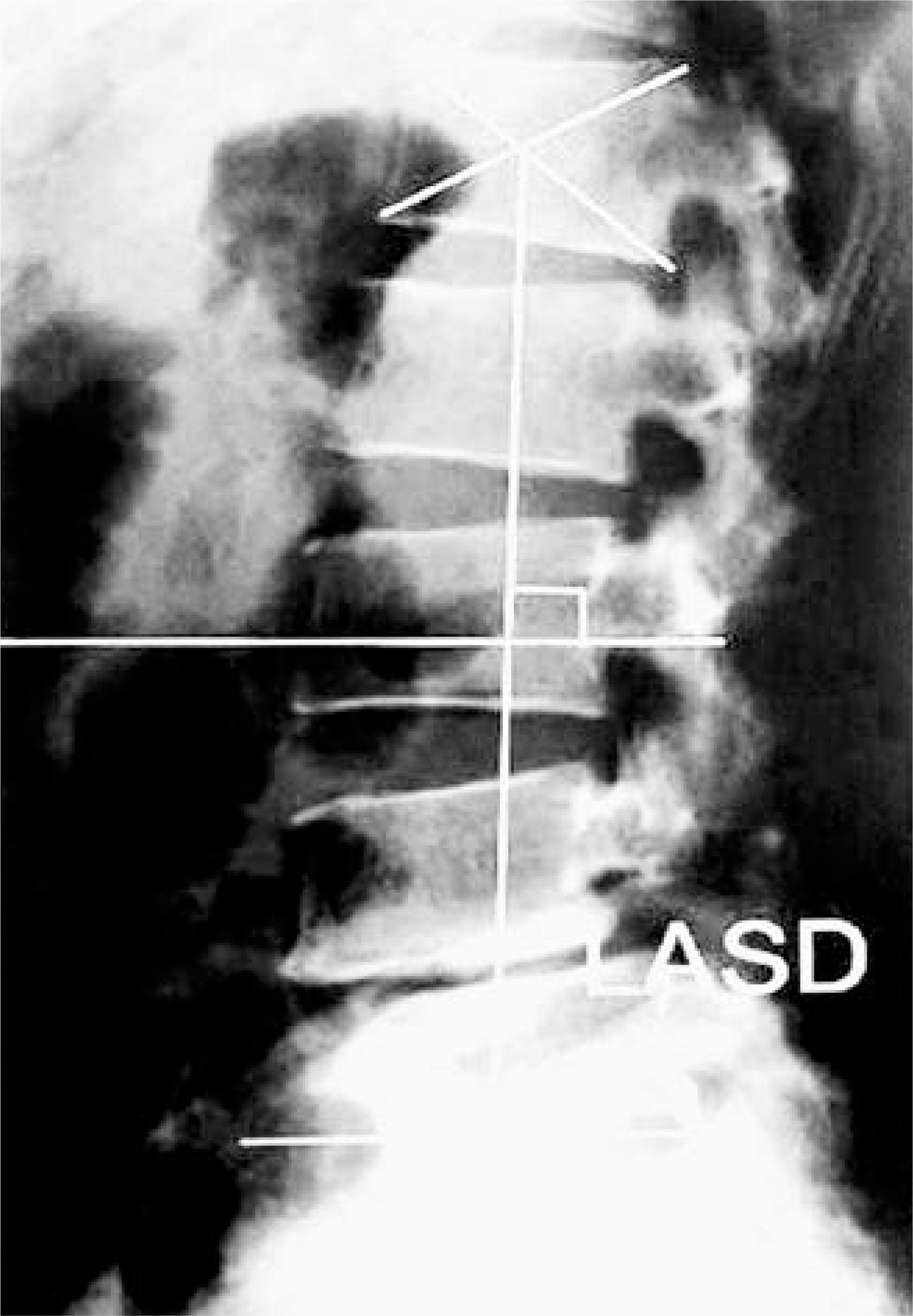 | Fig. 1.LASD(L1 axis S1 distance) is horizontal distance between L1 axis line that is perpendicular to L3 verte-bral body and the back corner of the S1 body. |
 | Fig. 2.
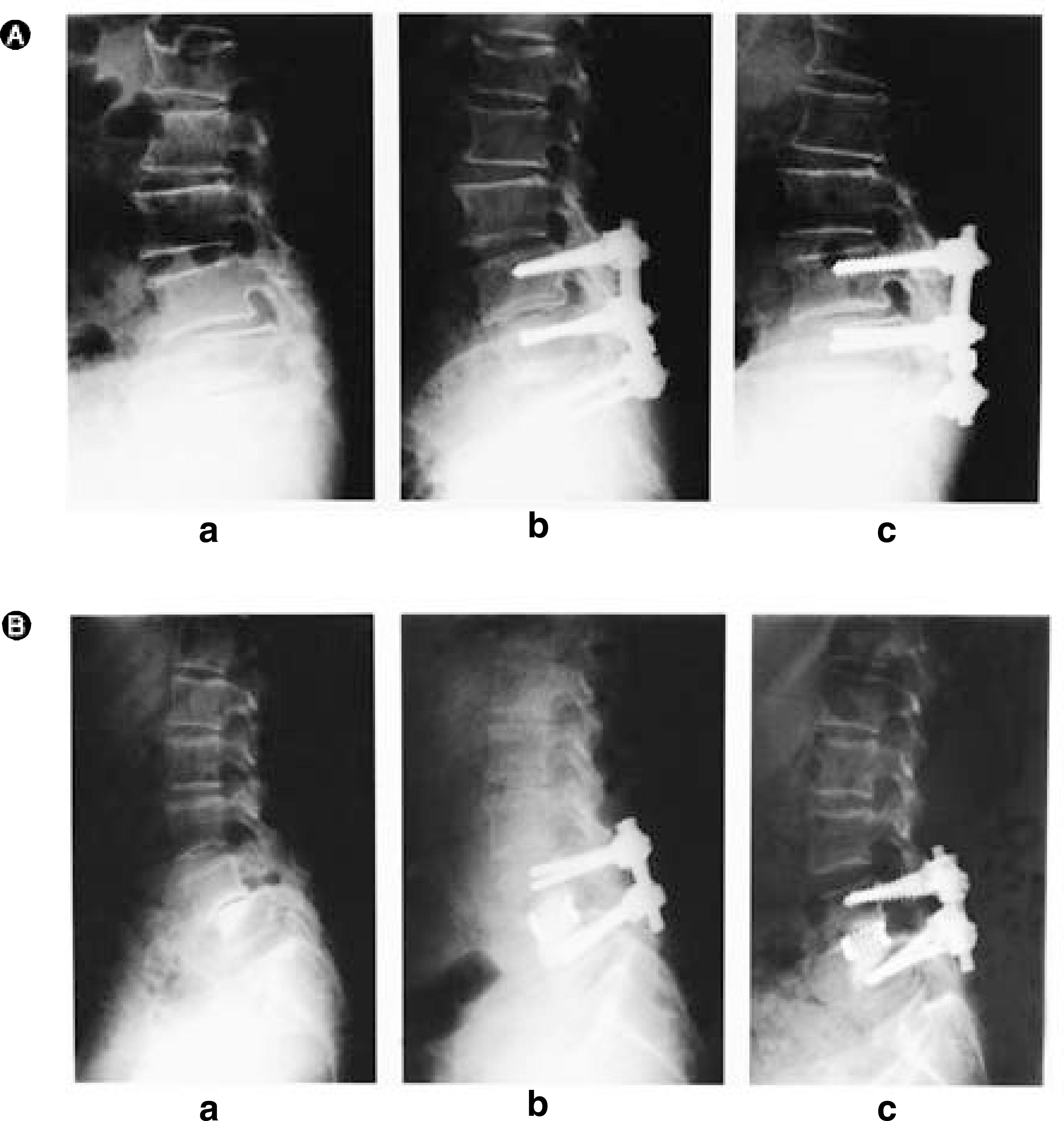
A. 65-year-old female patient with degenerative lumbar spondylolisthesis, L4 on L5. (a) Lateral view of preoperative plain x-ray. LASD is 35 mm, preoperative lumbar lordosis is 15 degree and anterior slippage of L4 is 10 mm. (b) Immediate lateral view of postoperative plain x-ray, treated with posterior decompression and instrumented postero lateral fusion of L3-4-5. Postoperative fused segment lordosis is 10 degree. (c) Twelve months follow up lateral view of plain x-ray, shows fol-low-up lumbar lordosis is 16 degree and follow-up fused segment lordosis is 10 degree. At this point, recovery rate of the patient was 31.5 %. B. 62-year-old female patient with degenerative lumbar spondylolisthesis, L4 on L5. (a) Lateral view of preoperative plain x-ray. LASD is 52 mm, preoperative lumbar lordosis is 30 degree and anterior slippage of L4 is 12 mm. (b) Immediate lateral view of postoperative plain x-ray, treated with posterior decompression, instrumented posterolateral fusion of L4-5 and posterior lumbar interbody fusion of L4 and L5. Postoperative fused segment lordosis is 5 degree. (c) Fourteen months follow up lateral view of plain x-ray, shows follow-up lumbar lordosis is 18 degree and follow-up fused segment lordosis is 5 degree. At this point, recovery rate of the patient was 70.6 %. |
Table 1.
Japanese Orthopaedic Association Scoring System for Treatment of lower Back Pain(JOA Score)
Table 2.
Recovery rate according to preoperative factors
| Factors | Op methods(RR: %) | ||
|---|---|---|---|
| PLF | PLF + PLIF | ||
| Sex | male | 51.3± 11.8(n=12) | 45.0± 11.1(n=7) |
| female | 45.0± 11.1(n=27) | 60.7± 13.1(n=13) | |
| Age | < 60 | 49.8± 13.0(n=19) | ∗67.5± 8.7(n=11) |
| Age | ≥ 60 | 43.6± 8.8(n=21) | ∗49.0± 10.4(n=9) |
| LASD | < 35 mm | ∗50.6± 11.3(n=28) | 59.9± 15.4(n=9) |
| ≥ 35 mm | ∗37.6± 5.0(n=11) | 61.9± 10.9(n=11) | |
| † Pre-L | < 20° | ∗40.1± 9.3(n=11) | 54.2± 11.5(n=7) |
| Pre-L | ≥ 20° | ∗49.6± 11.3(n=28) | 64.7± 12.4(n=13) |
| Slippage | < 10 mm | ∗50.8± 11.7(n=21) | 47(n=1) |
| Slippage | ≥ 10 mm | ∗42.4± 9.8(n=18) | 61.8± 12.7(n=19) |
| Duration | < 30 mo | 49.7± 12.8(n=22) | ∗67.2± 8.8(n=12) |
| Duration | ≥ 30 mo | 43.4± 8.7(n=17) | ∗51.8± 2.7(n=8) |
Table 3.
Recovery rate according to postoperative factors
Table 4.
Correlation of factors and recovery rate by Pearson analysis
 XML Download