Abstract
Diffuse idiopathic skeletal hyperostosis (DISH), also known as Forestier’s disease and ankylosing hyperostosis, is a relatively common disease that predominantly affects middle- aged and elderly men. It is often asymptomatic; especially dysphagia is a rather frequent and prominent symptom, particularly when the cervical spine is involved with the hyperostotic state. A case of DISH, with dysphagia, was experienced, which was treated by excision of the bony spur, with an anterior approach.
Go to : 
REFERENCES
1). McCafferty R, Harrison M, Tamas L, Larkins M. Ossification of the anterior longitudinal ligament and Forestier's disease: an analysis of seven cases. J Neurosurg. 1995; 83:13–17.

2). Resnick D, Niwayama G. Radiographic and pathologic features of spinal involvement in diffuse idiopathic skeletal hypeostosis (DISH). Radiology. 1976; 119:559–568.
3). Resnick D, Shaul S, Robins J. Diffuse idiopathic skeletal hyperostosis (DISH): Forestier's disease with extraspinal manifestations. Radiology. 1975; 115:513–524.

4). Resnick D, Shapiro R, Wiesner K, Niwayama G. Diffuse idiopathic skeletal hyperostosis (DISH). Semin Arthritis Rheum. 1978; 7:153–187.
5). Boachie-Adjei O, Bullough P. Incidence of ankylosing hyperostosis of the spine (Forestier's disease) at autopsy. Spine. 1987; 12:739–743.

6). Tsukamoto Y, Onitsuka H, Lee K. Radiologic aspects of diffuse idiopathic skeletal hyperostosis in the spine. AJR. 1977; 129:913–918.

7). Scutellari P, Orzincolo C, Princivalle M, Franceschini F. Diffuse idiopathic skeletal hyperostosis. Review of diagnostic criteria and analysis of 915 cases. Radiol Med. 1992; 83:729–736.
8). Resnick D. Diagnosis of bone and joint disorders, 3rd. ed. Philadelphia: WB Saunders Co;1463-1495. 1995.
9). Mata S, Fortin P, Fitzcharles M, et al. A controlled study of diffuse idiopathic skeletal hyperostosis: clinical features and functional status. Medicine. 1997; 76:104–117.

10). Weinfeld RM, Olson PN, Maki DD, Griffiths HJ. The prevalance of diffuse idiopathic skeletal hyperostosis (DISH) in two large American Midwest metropolitan hospital populations. Skeletal Radiol. 1997; 26:222–225.
11). Littlejohn G, Smythe H. Marked hyperinsulinemia after glucose challenge in patients with diffuse idiopathic skeletal hyperostosis. J Rheumatol. 1981; 8:965–968.
12). Littlejohn G. Insulin and new bone formation in diffuse idiopathic skeletal hyperostosis. Clin Rheumatol. 1985; 4:294–300.

13). Eviatar E, Harell M. Diffuse idiopathic skeletal hyperostosis with dysphagia. J Laryngol Otol. 1987; 101:627–632.

14). McGarrah P, Teller D. Posttraumatic cervical osteophytosis causing progressive dysphagia. South Med J. 1997; 90:858–860.

15). Akhtar S, O’ Flynn P, Kelly A, Valentine P. The management of dysphagia in skeletal hyperostosis. Spine. 1991; 16:235–237.
Go to : 
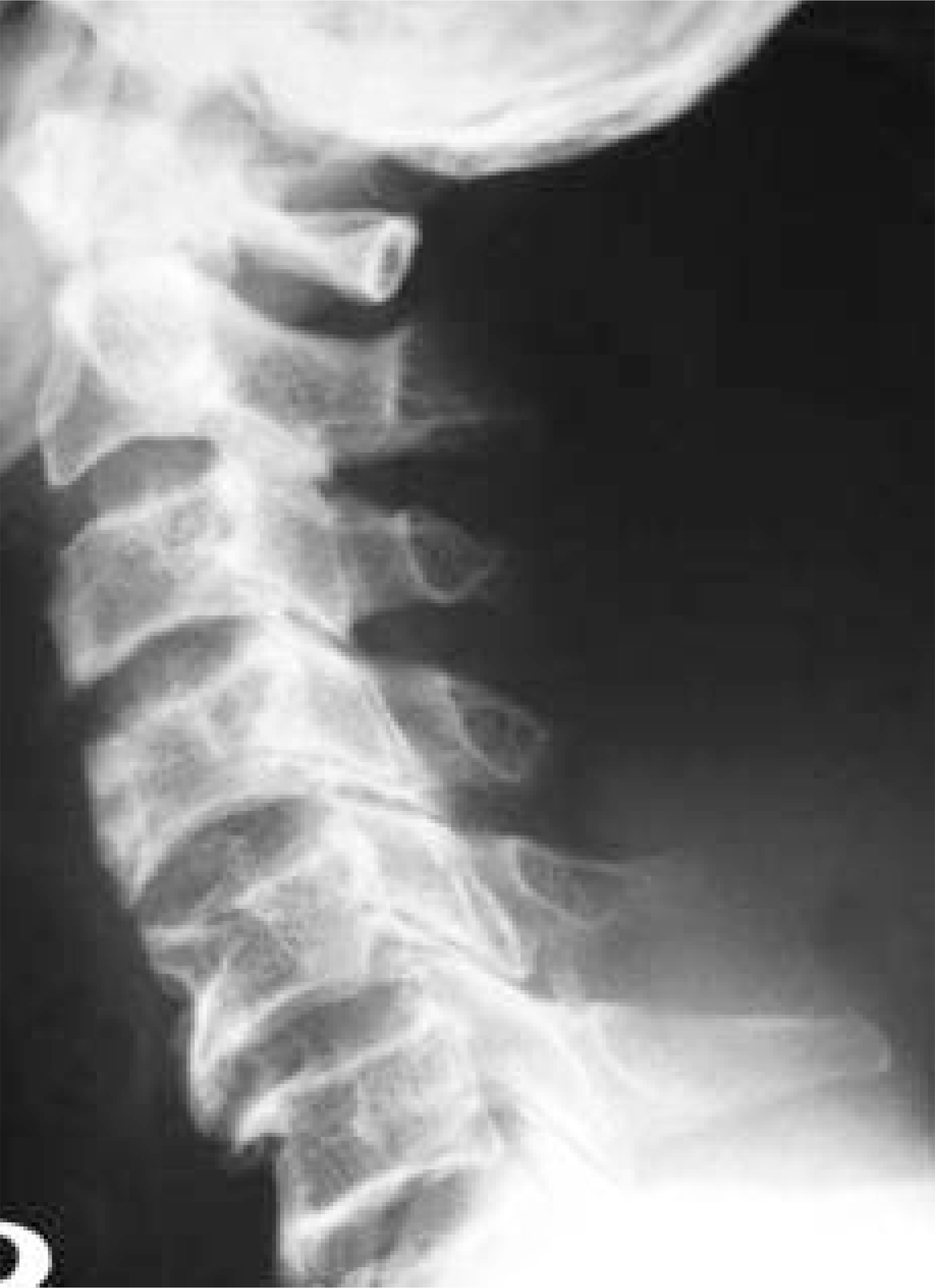
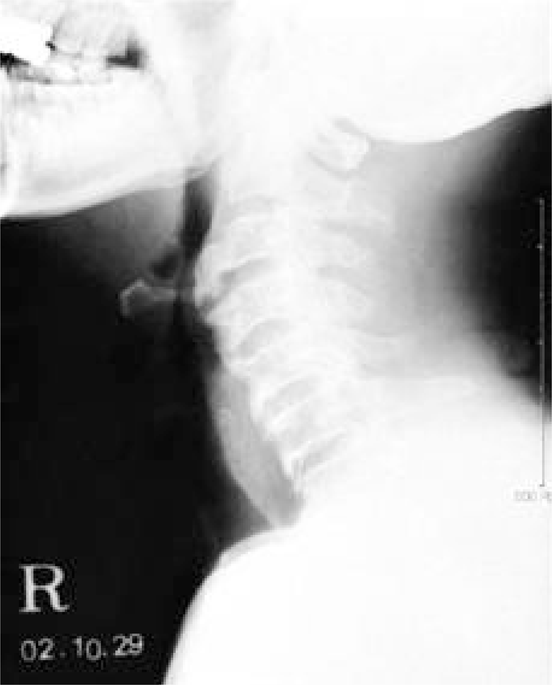 | Fig. 1.Preoperative cervical spine lateral radiograph. Note anteriorly displacing laryngeal air shadow with large osteophyte at C3-4. |
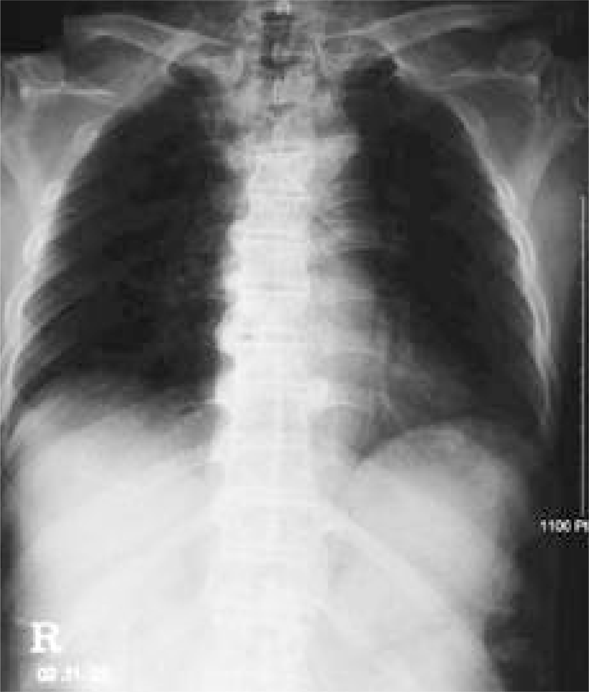 | Fig. 2.A anteroposterior view of thoracic spine, demonstrate flowing hyperostosis at right side. |
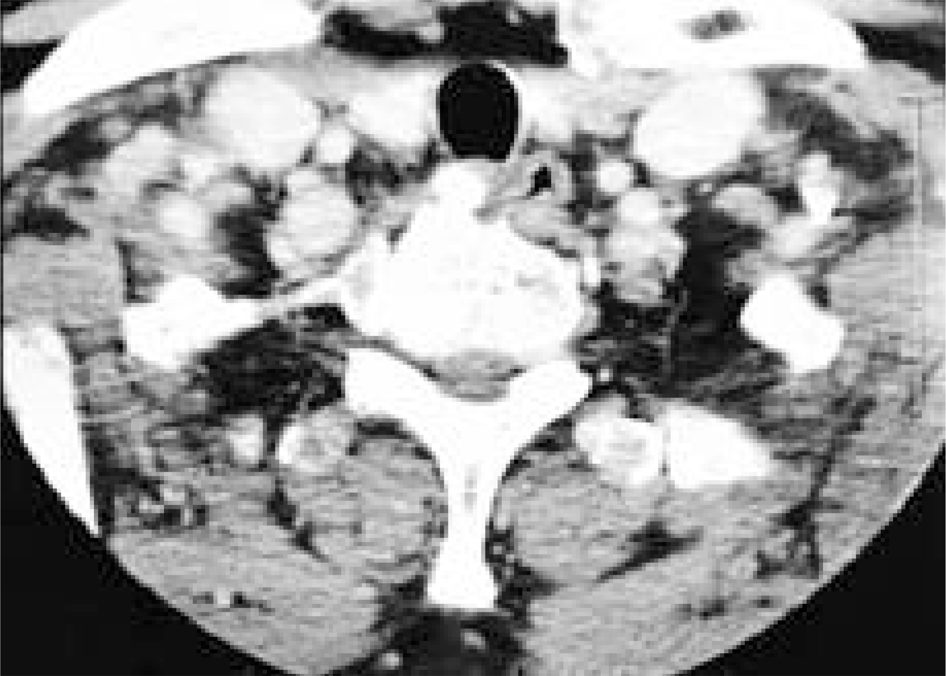 | Fig. 3.Preoperative cervical spine CT demonstrating compression of esophagus by anterior bony spur. |
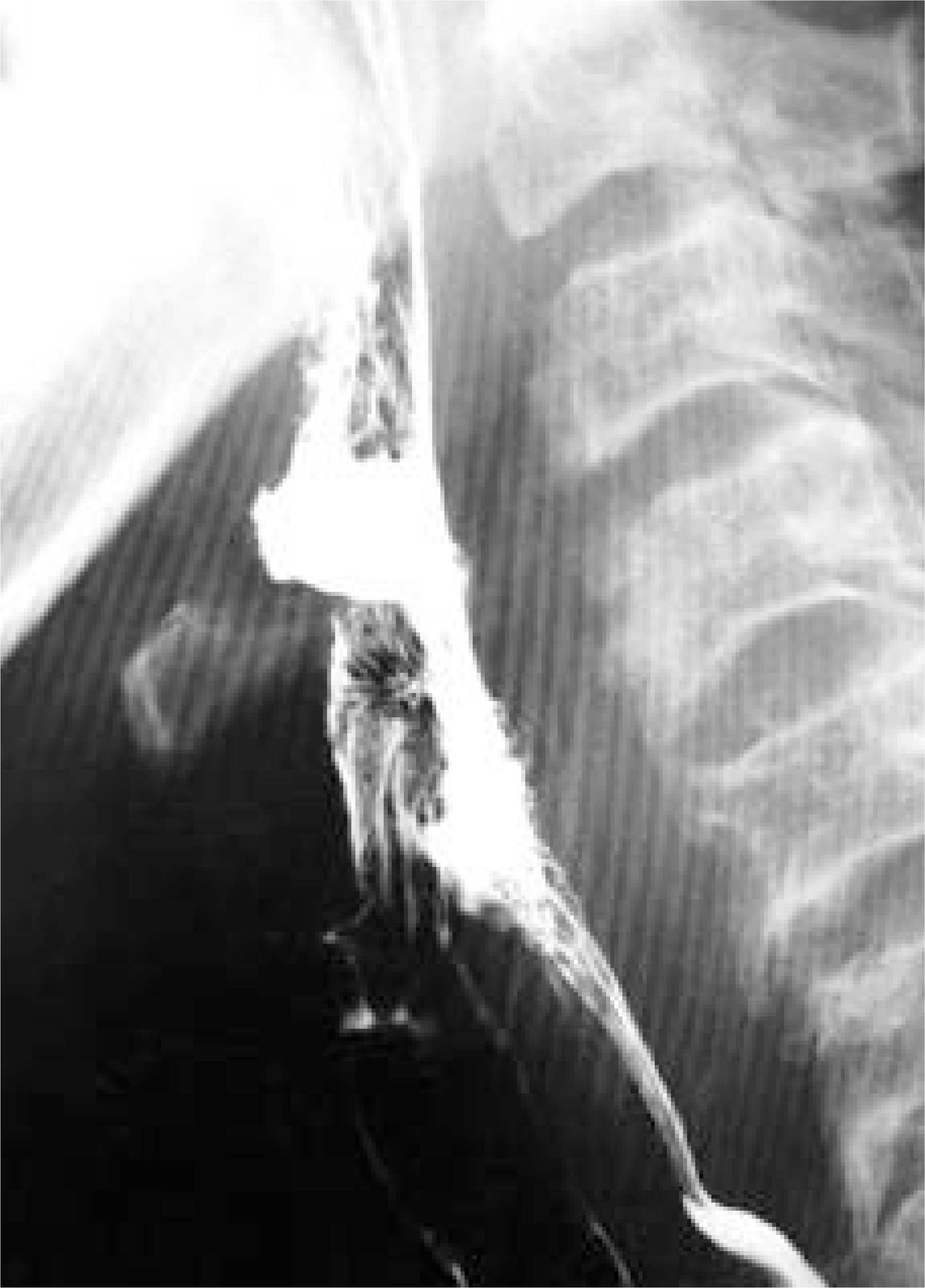
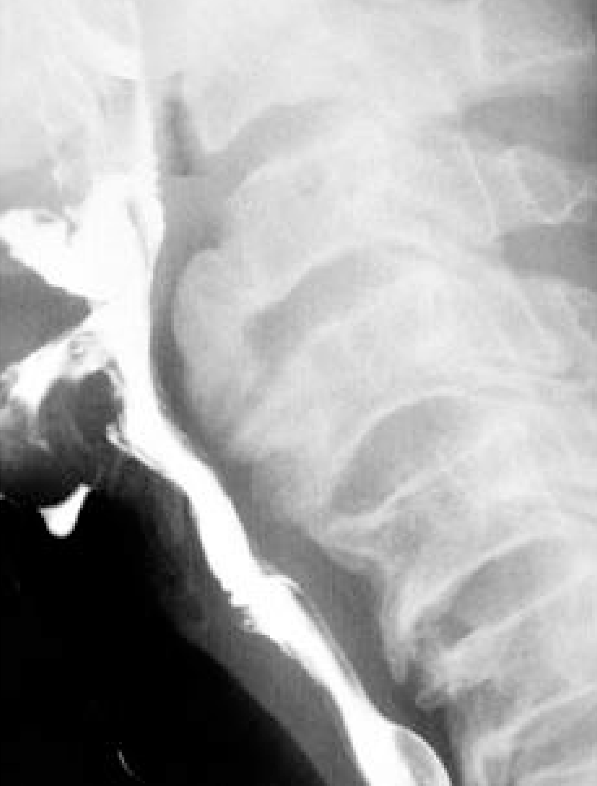 | Fig. 4.Preoperative esophagogram. Note extrinsic compression of esophageal posterior wall by protruding bony spur of the anterior aspect of C3-4. |


 PDF
PDF ePub
ePub Citation
 Print
Print


 XML Download