

 PDF
PDF ePub
ePub Citation
 Print
Print


Abstract
Objectives
To determine the normal range of the sagittal spinal alignment, and define significant spinopelvic compensations over the hip axis for the sagittal balance with aging.
Summary of Literature Review
Normative data of the sagittal spinal alignment has wide variation and limited clinical useful-ness. In addition, the extent to which the “ normal” sagittal spinal contour changes with aging remains unknown.
Materials and Methods
Inclusion criteria were an age between 20 and 29 years (n=50), group A, and between 55 and 65 years (n=50), group B, for the asymptomatic subjects. Measurements made on the standing lateral radiographs included the following: thoracic kyphosis, lumbar lordosis and sagittal vertical axis. In addition, measurements of the sacropelvic translation, spinopelvic balance, pelvic incidence, pelvic tilting and sacral slope were made.
Results
The average thoracic kyphosis was 24°, ranging from 3 to 42°, in group A, and 33°, ranging from 9 to 53°, in group B (p<0.001). The average lumbar lordosis was - 47°, ranging from - 65 to - 23°, and - 51°, ranging from - 69 to - 33°, in groups A and B, respectively (p>0.05). The C7 plumb line, on average, fell 15.4 mm more anteriorly to the posterosuperior corner of S1 in group B than in group A (p<0.05). The anterior positioning of the C7 was also positively correlated with decreasing lordosis (p<0.001). The average sacropelvic translation was - 41mm, ranging from - 76 to 20 mm, and - 48 mm, ranging from - 76 to - 17 mm, in groups A and B, respectively (p<0.05). The average spinopelvic balance was - 57 mm, ranging from - 104 to - 4 mm, and - 49 mm, ranging from - 101 to - 3 mm, in groups A and B, respectively. The C7 plumb line fell posterior to the hip axis in all cases. The average pelvic incidence was 46°, ranging from 30 to 61°, and 54°, ranging from 28 to 76°, in groups A and B, respectively (p<0.05). The average pelvic tilt was 14°, ranging from 4 to 33°, and 19°, ranging from 3 to 37°, in groups A and B, respectively (p<0.05). The average sacral slope was 32°, ranging from 17 to 47°, and 35°, ranging from 25 to 50°, in groups A and B, respectively (p<0.05). There was significant correlation between pelvic incidence and lumbar lordosis (p<0.001).
REFERENCES
1). Bradford DS, Moe JH, Montalvo FJ, et al. Scheu er-mann's kyphosis and roundback deformity: results of Mil -waukee brace treatment. J Bone Joint Surg. 1974; 56A:740–58.
2). Jackson RP, McManus AC. Radiographic analysis of sagittal plane alignment and balance in standing volunteers and patients with low back pain matched for age, sex and size. Spine. 1994; 19:1611–8.

3). Jackson RP, Peterson MD, McManus AC, et al. Com -pensatory spinopelvic balance over the hip axis and better reliability in measuring lordosis to the pelvic radius on standing lateral radiographs of adult volunteers and patients. Spine. 1998; 23:1750–67.
4). Roaf R. Vertebral growth and its mechanical control. J Bone Joint Surg. 1960; 42B:40–59.
5). Vendatam R, Lenke LG, Keeney JA, et al. Comparison of standing sagittal spinal alignment in asymptomatic adolescents and adults. Spine. 1998; 23:211–5.
6). Hammerberg EM, Wood KB. Sagittal Profile of The Elderly. Journal of Spinal Disorders & Techniques. 2003; 16:44–50.
7). Gelb DE, Lenke LG, Bridwell KH, Blanke K, McEnery KW. An analysis of sagittal spinal alignment in 100 asymptomatic middle and older aged volunteers. Spine. 1995; 20:1351–8.
8). Vendatam R, Lenke LG, Bridwell KH, et al. The Effect of Variation in Arm Position on Sagittal Spinal Alignment. Spine. 2000; 25(17):2204–9.
9). Stagnara P, De Mauroy JC, Dran G, et al. Reciprocal angulation of vertebral bodies in a sagittal plane: approach to references for the evaluation of kyphosis and lordosis. Spine. 1982; 7:335–42.
10). Jackson RP. The sagittal plane-From birth to the grave: Spinopelvic alignment and balance. CME course, SRS meeting. 1997.
11). Legaye J, Duval-Beauper G, Hecquet J, et al. Pelvic incedence: a fundamental pelvic parameter for three-dimensional regulation of spinal sagittal curves. Eur Spine J. 1998; 7:99–103.
12). Vaz G, Roussouly P, Berthonnaud E, Dimnet J. Sagittal morphology and equilibrium of pelvis and spine. Eur Spine J. 2002; 11:80–87.
13). Wilies P. Postural deformities of the A-P curves of the spine, Lancet. I:1937; 911–919.
14). Peterson MD, Janckson , McManus AC. Standing sagittal spinal balance, alignment and lumbopelvic relationship. Presented at the 30th annual meeting of the Scoliosis Research Society, Ashiville, NC, September. 1995. 13–16.
15). Lee CS, Oh WH, Chung SS, Lee SG, Lee JY. Analysis of the sagittal alignment of normal spines. J Korean Orthop Assoc. 1999; 34:949–954.
16). During J, Goudfrooij H, Keessen W, Beeker TW, Crowe A. Toward standards for posture. Postural characteristics of the lower back system in normal and pathologic conditions. Spine. 1985; 10:83–87.
17). Duval-Beaupere G, Schmidt C, Cosson P. A bary-centremetric study of the sagittal shape of spine and pelvis: The conditions required for an economic standing position. Ann. Biomed. Eng. 1992; 20:451–462.
18). Jackson RP, Kanemura T, Kawakami N, Hales C. Lumbopelvic lordosis and pelvic balance on repeated standing lateral radiographs of adult volunteers and untreated patients with constant low back pain. Spine. 2000; 25:575–586.
Figures and Tables%
Fig. 1.
(A) Photograph of arms raised horizontally forward at 60° flexion at the shoulder. (B) Photograph of ar arms raised horizontally forward at 90° flexion at the shoulder.

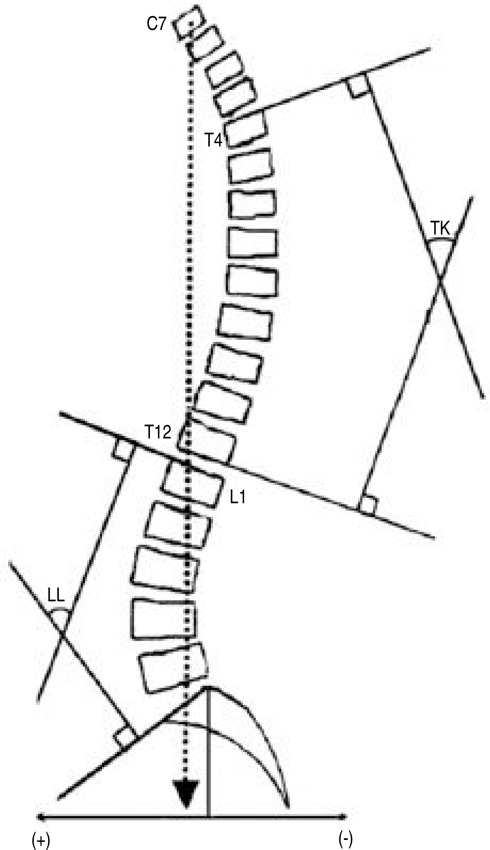
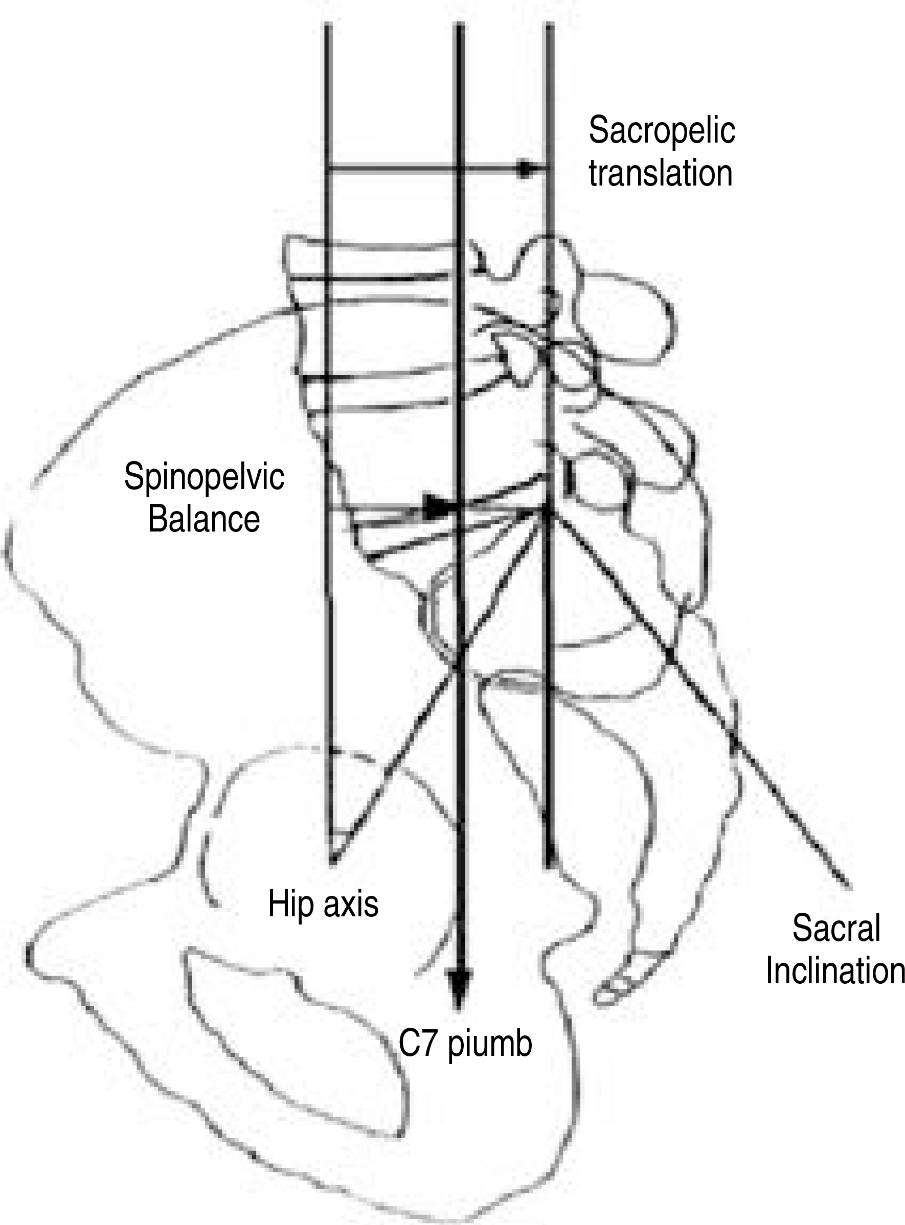
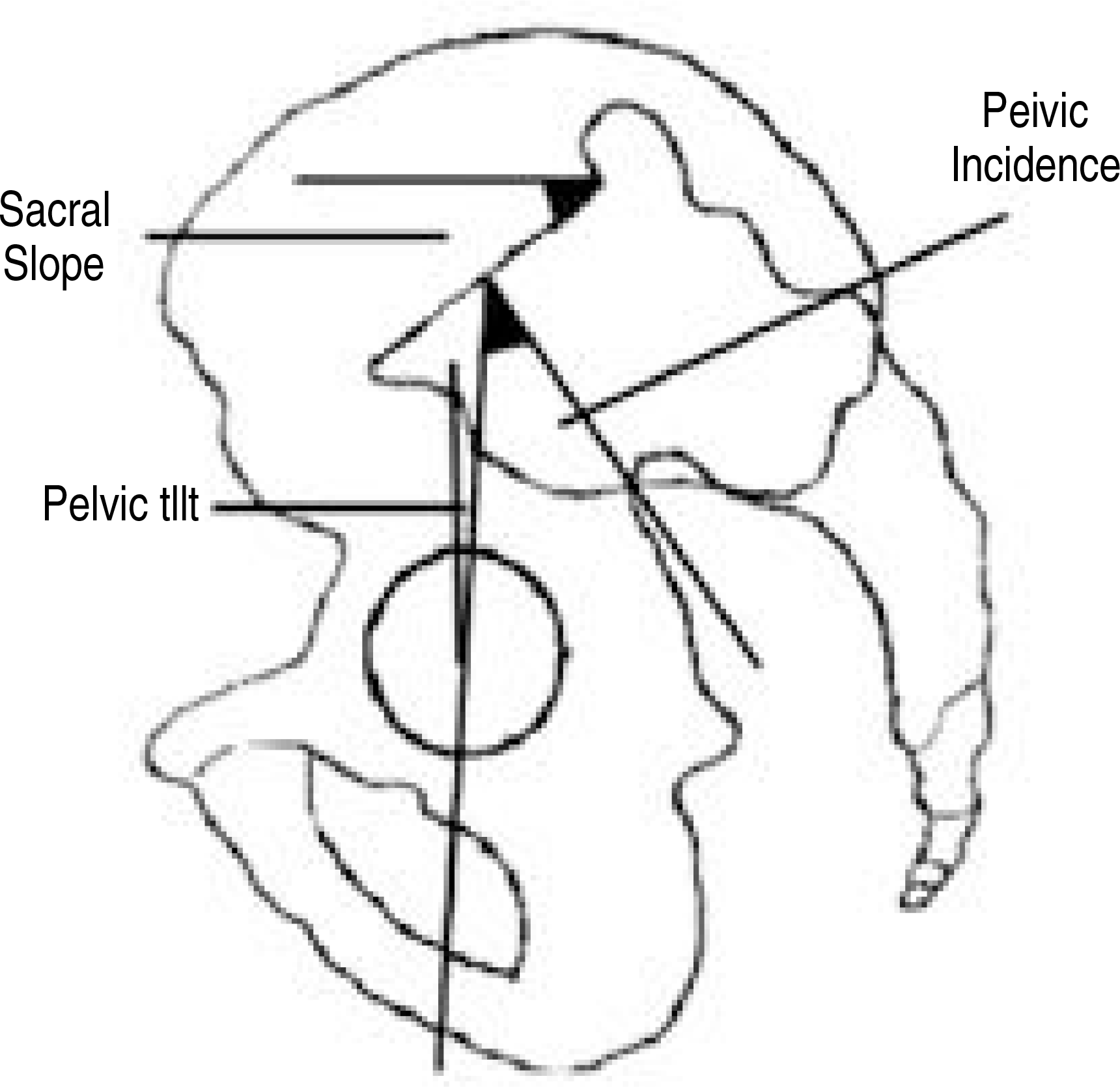
Table 1.
Average values of thoracic kyphosis, lumbar lordosis and sagittal vertical axis in each group.
| A 군 | B 군 | p value | |
|---|---|---|---|
| T-kyphosis (˚) | 24± 7.72 | 33± 9.60 | p=0.000 |
| L-lordosis (˚) | -47± 10.07 | -51± 9.28 | p=0.170 |
| Sagittal vertical axis (mm) | -16± 21.18 | -0.6± 22.94 | p=0.002 |
Table 2.
Average values of spinopelvic parameters in each group.
Table 3.
Statistical analysis between the parameters.
| p value | |
|---|---|
| lordosis - SVA | ∗∗∗ |
| lordosis - pelvic incidence | ∗∗∗ |
| sacral inclination - SVA | NS |
| lordosis - kyphosis | ∗∗∗ |
| sacral slope-lordosis | ∗∗∗ |
| pelvic tilt-lordosis | ∗∗∗ |
Table 4.
Average values of each parameters according to arms raised horizontally forward at 60° and 90° flexion at the shoulder.
 XML Download