Abstract
Obj ectives
To analyze the clinical and radiological results of different surgical methods in osteoporotic vertebral fracture patients, with neurologic deficits in the thoracolumbar junction.
Summary of Literature review
Various surgical methods have been reported for osteoporotic vertebral fractures, with neurologic deficits, in the thoracolumbar junction. These are: anterior decompression, anterior decompression and anterior or posterior reconstruction, and Egg shell procedure. However, it is controversial as to which method is better.
Materials and Methods
13 patients that had undergone surgical treatment for osteoporotic vertebral fractures, with neurologic deficits, With a mean age of 68± 8.4, ranging from 51 to 79 years. Six of the cases were male and seven were female. The mean follow up period was 18 months. The patients were divided into two groups.
· Group A (n=8): A nterior decompression, anterior interbody fusion, with cage or autologous strut iliac bone block, and instrumentation (anterior or posterior).
· Group B (n=5): Posterior decompression and posterior reconstruction (egg shell procedure). The kyphotic angles, neurologic improvements and complications in each group were analyzed preoperatively, postoperatively and at final follow up.
Results
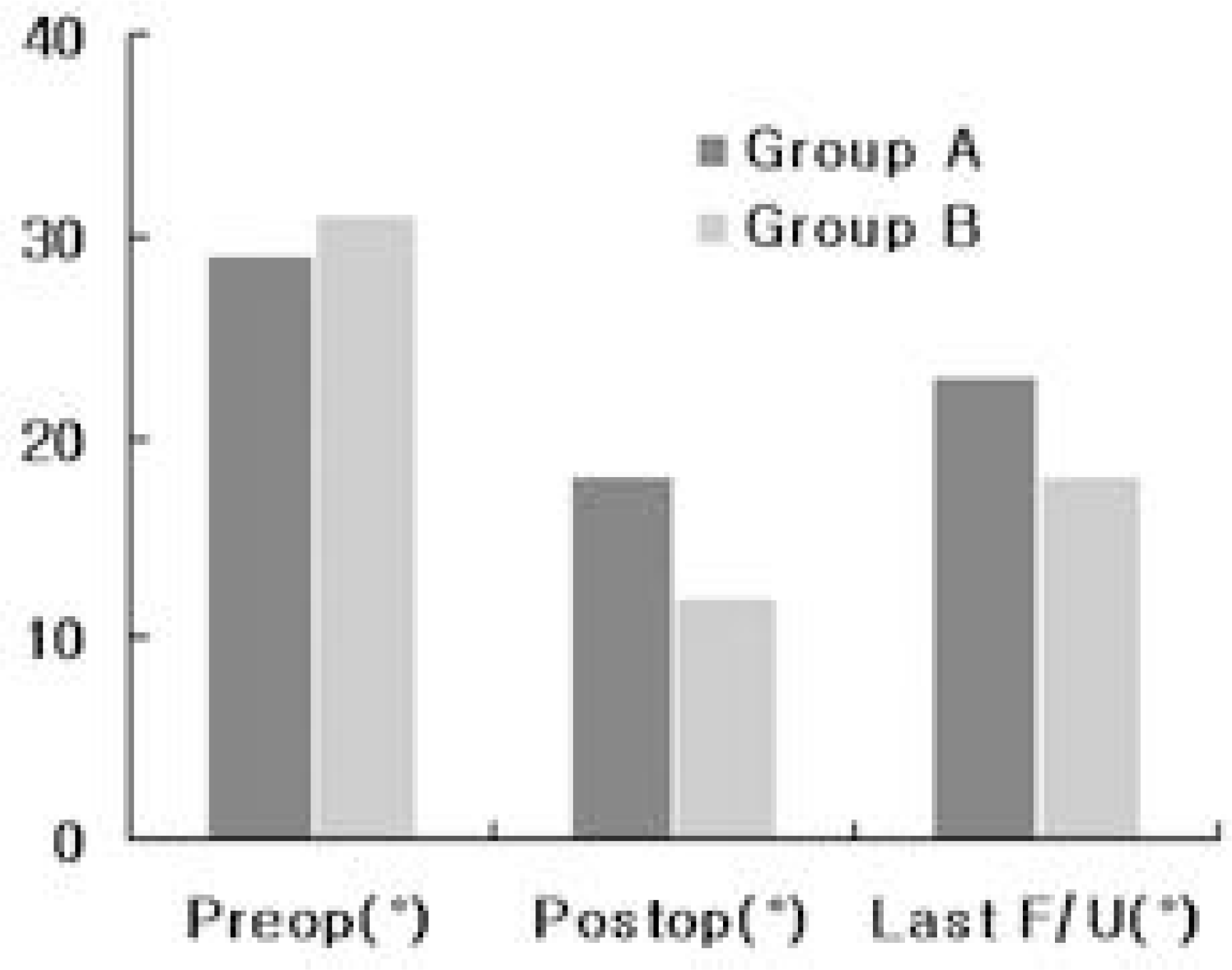
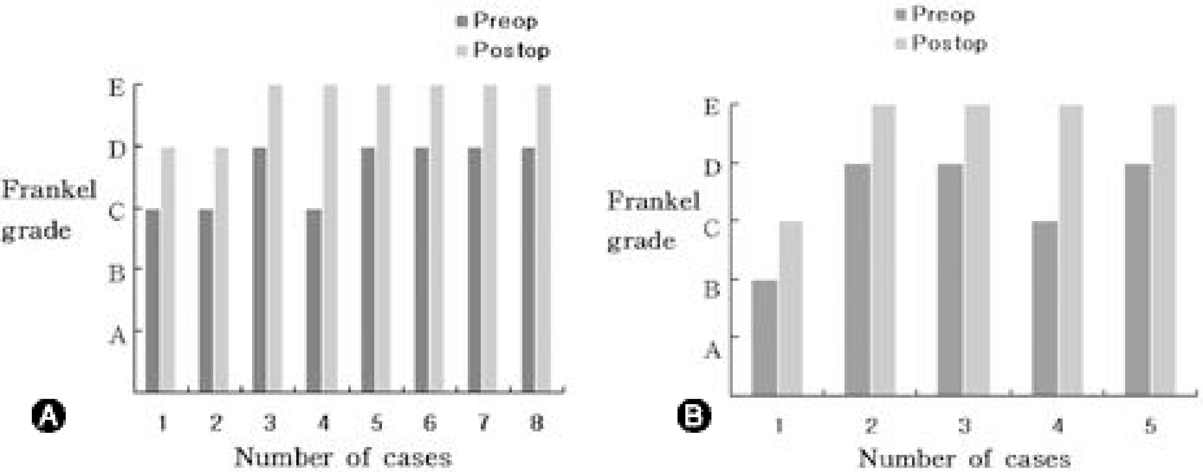
In group A, the mean kyphotic angles were 29± 5.9°, 18± 6.7° and 23± 7.7° preoperatively, postoperatively and at the final follow up, respectively. In group B, the mean kyphotic angles were 31± 1.1°, 12± 6.3° and 18± 5.5° preoperatively, postoperatively and at the final follow up, respectively. In group A, 3 and 5 patients were graded as Frankel grades C and D, respectively. In group B, 1, 1 and 3 patients were graded as Frankel grades B, C and D, respectively. The neurological status improved in all the patients, by mean 1.1grades in group A and 1.2 grades in group B. In group A, postoperative transient dyspnea and screw loosening occurred in one and two patients, respectively. In group B, postoperative paralytic ileus and screw loosening occurred in one two patients, respectively.
Go to : 
REFERENCES
1). Adam PH. Osteoporosis. Clin Rheum Dis. 1981; 7:557–593.
2). Johnston CC, Epstein S. Clinical, Biochemical, Radiographic, Epidemiologic and economic features of Osteoporosis. Orthop Clin North Am. 1981; 12:559–569.

3). Mazess RB. Aging bone loss. Clin Orthop. 1982; 323:239–252.
5). Kaneda KS, Ascano S, Hashimoto T, Satoh S, Fujiya M. The treatment of osteoporotic vertebral collapse using the Kaneda device and a bioactive ceramic vertebral prosthesis. Spine. 1992; 17:S295–303.
6). Salomon C, Chopin D, Benoist M. Spinal cord compression: An exceptional complication of spinal osteoporosis. Spine. 1988; 13:222–224.
7). Suk SI, Kim WJ, Lee SM, Chung ER, Nah KH. Posterior vertebral column resection for wevere spianl deformities. Spine. 2002; 27:2374–2382.
8). Kim KT, Suk KS, Kim JM, Lee SH. Delayed vertebral collapse with neurological deficits secondary to osteoporosis. International Orthopaedics(SICOT). 2003; 27:65–69.

9). Shikata J, Yamamuro T, Ida H, Shimizu K, Yoshikawa J. Surgical treatment for paraplegia resulting from vertebral fractures in senile osteoporosis. Spine. 1990; 5:485–489.

10). Hammerberg KW, DeWald RL. Senile burst fracture: a complication of osteoporosis. Orthop Trans. 1989; 13:97.
11). Soshi S, Shiba R, Kondo H, Murota K. An experimental study on transpedicular screw fixation in relation to osteoporosis of the lumbar spine. Spine. 1991; 16:1335–1341.

12). Cook SD, Barbera J, Rubi M, Salkeld SL, Whitecloud TS III. Lumbosacral fixation using expandable pedicle screws: An alternative in reoperation and osteoporosis. Spine. 2001; 1:109–114.
13). Bai B, Kummer FJ, Spivak J. Augmentation of anterior vertebral body screw fixation by an injectable, biodegradable calcuium phosphate bone substitute. Spine. 2001; 26:2679–2683.
Go to : 


 PDF
PDF ePub
ePub Citation
 Print
Print


 XML Download