Abstract
Objective
To evaluate the results after an anterior decompression and fusion, with anterior instrumentation, using a Z- plate in osteoporotic vertebral fractures.
Summary of Literature review
Despite conservative treatment, continuous severe pain, progressive neurological impairments and deformity may need surgical treatment in osteoporotic vertebral fractures accompanied with neurological deficit.
Materials and Methods
Fourteen patients that had undergone anterior decompression and an autogenous iliac bone graft, with anterior internal fixation, between 1997 and 2001, under the diagnosis of an osteoporotic vertebral fracture, were reviewed. The chief complaints, severity of pain measured, using the Denis pain scale, fracture patterns, fracture level, changes of kyphotic angle (revised with sagittal index) and complications were analyzed.
Results
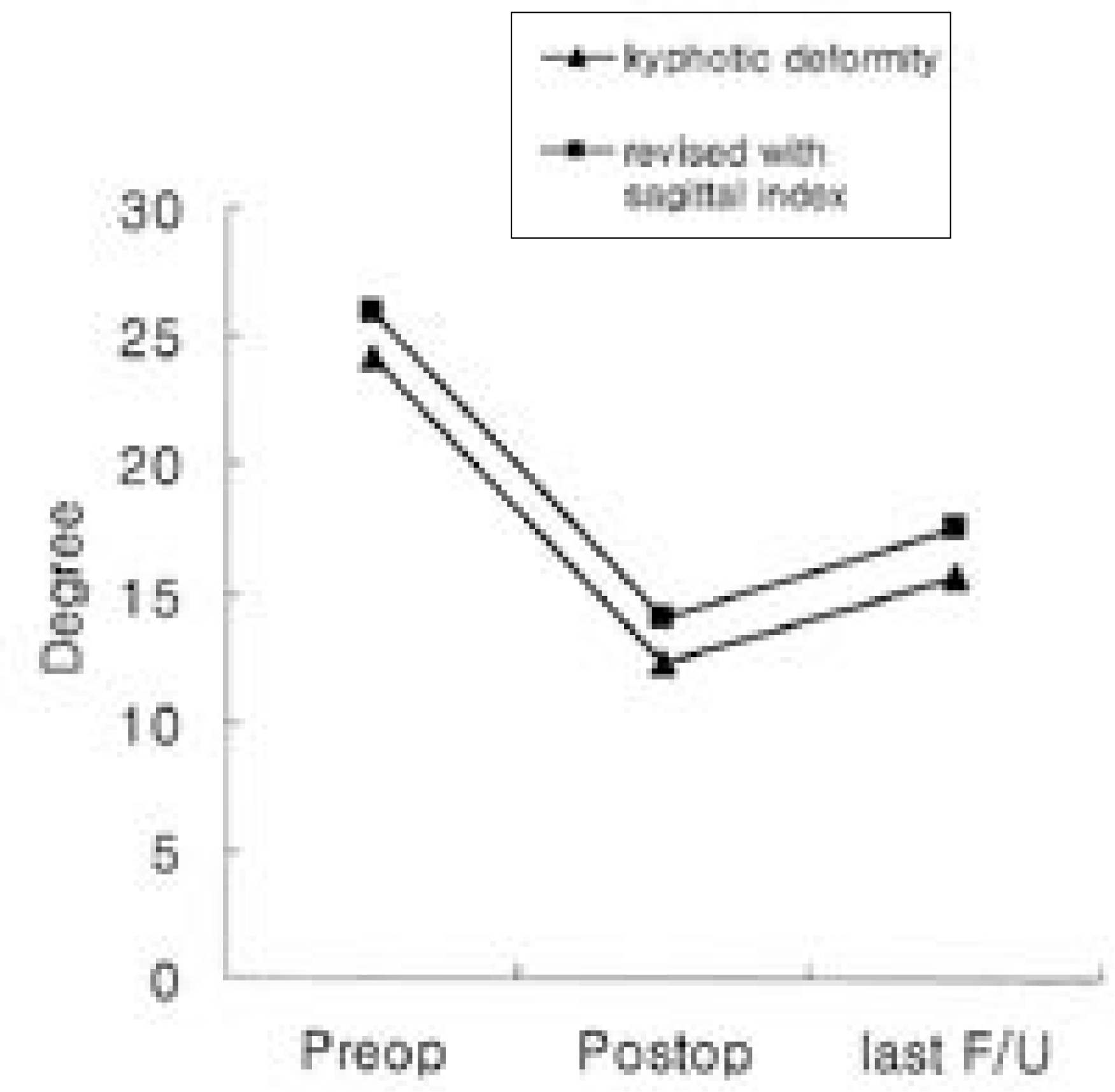
Symptoms subsided completely in 5 patients, one case showed no definite improvement and 8 showed improved symptoms. The fracture levels included: 1 and 2 cases at the 11th and 12th thoracic spine, and 8, 1 and 2 in the 1st, 2nd and 3rd lumbar spine, respectively. 10 patients showed wedge type fractures, three a compression type and one a biconcave type. The average kyphotic deformity decreased 49.0% (50.9% when revised with sagittal index) after surgery, but the average loss of correction angle was 28.8% (26.0% when revised with sagittal index), compared with the immediate postoperative correction angle.
The complications included
screw loosening and adjacent vertebral fractures in 3 and 4 patients, respectively. Two patients had the combined problem of screw loosening and an adjacent vertebral fracture.
Conclusion
In anterior decompression and fusion, with instrumentation, for osteoporotic vertebral fracture treatment, the complications were primarily related, directly or indirectly, to the underlying osteoporosis. Complete neurological recovery occurred 9 of the 11 patients, but residual pain was common.
Go to : 
REFERENCES
1). Ha KY, Kim KW, Park SJ, Paek DH, Ha JH. The surgical treatment of osteoporotic vertebral collapse caused by minor trauma. J Kor Orthop Assoc. 1998; 33:105–111.

2). Mochida J, Toh E, Chiba M, Nishimura K. Treatment of osteoporotic late collapse of a vertebral body of thoracic and lumbar spine. J Spinal Disord. 2001; 14:393–398.

3). Eastell R, Cedel SL, Wahner HW, Riggs BL, Milton LJ I II. Classification of vertebral fractures. J Bone Miner Res. 1991; 6:207–215.

4). Denis F. Spinel stability as defined by the three-column spine concepts in acute spinal trauma. Clin Orthop. 1984; 189:65–76.
5). Frankel HL, Hancock DO, Hyslop G. The value of postural reduction in the initial management of closed injuries of the spine with paraplegia and tetraplegia. Part I. Paraplegia. 1969; 7:179–192.
6). Farcy JPC, Weidenbaum M. Sagittal index in management of thoracolumbar burst fracture. Spine. 1990; 15:958–965.
8). Soshi S, Shiba R, Kondo H, Murota K. An experimental study on transpedicular screw fixation in relation to osteoporosis of the lumbar spine. Spine. 1991; 16:1335–1341.

9). Kaneda KS, Ascano S, Hashimoto T, Satoh S, Fjiya M. The treatment of osteoporotic-posttraumatic vertebral collapse using the Kaneda device and a bioactive ceramic vertebral prosthesis. Spine. 1992; 17:S295–303.

10). Kirkpatrick JS, Wilber RG, Likavec M, Emery SE. Anterior stabilization of thoracolumbar burst fractures using the Kaneda device: a preliminary report. Orthopae-dis. 1995; 18:673–678.

11). Hoan- VN, Steven L, Daniel G. Osteoporotic Vertebral Burst fractures with Neurologic Compromise. J Spinal Disord. 2003; 16:10–19.
12). Suk SI, Kim JH, Lee SM, Chung ER. Anterior-posterior surgery versus posterior closing wedge osteotomy in post - traumatic kyphosis with neurologic compromised osteo -.
Go to : 
Figures and Tables%
 | Fig. 1.Classification of osteoporotic vertebral fractures by type (A) Wedge deformity, (B) Biconcavity deformity, (C) Compression deformity |
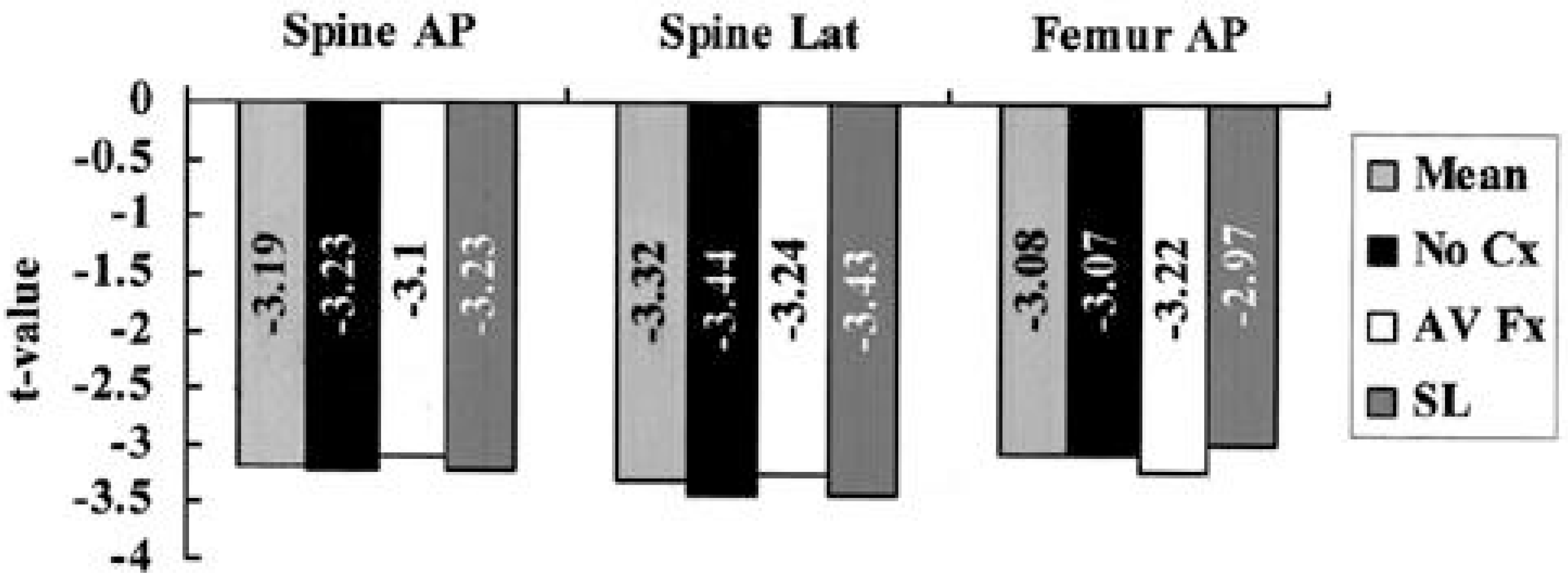 | Fig. 4.Comparison of t-value of osteoporosis complication and non-complication case: there is no statistical significance (p > 0.05) (Mean: average t-value of whole cases No Cx: average t-value of cases without complications AV Fx: average t-value of cases which show adjacent vertebral fracture SL: average t-value of cases which show screw loosening) |
Table 1.
Summary of 14 Cases
Table 2.
Modified Frankel Grade
| Grade | Motor Function | Function of bladder & bowel |
|---|---|---|
| A | 0 | paralysis |
| B | 0-1 | paralysis |
| C | 2 | paralysis or dysfunction |
| D1 | 3 | paralysis to normal |
| D1 | 4-5 | paralysis |
| D2 | 4-5 | dysfunction |
| D3 | 4-5 | normal |
| E | 5 | normal |
Table 3.
Assessment of the clinical symptom according to five-grade Denis pain scale. (Denis Pain Scale)


 PDF
PDF ePub
ePub Citation
 Print
Print


 XML Download