

 PDF
PDF ePub
ePub Citation
 Print
Print


Abstract
Objectives
This study was designed to compare the clinical results, with the correction of the lumbar lordotic and scoliotic angles, in degenerative lumbar scoliosis patients, with spinal stenosis, who underwent an operation.
Summary of Literature Review
Few studies have compared the postoperative lordotic angle with the clinical results in degenerative lumbar scoliosis, with spinal stenosis.
Subjects and Methods
Out of 68 cases, where the patients underwent posterior decompression, pedicle screw fixation and fusion, due to the degenerative lumbar scoliosis with spinal stenosis, between February 1997 and February 2001, 59 cases, with the possible followups for over 2 year, were studied and are herein reported. The decompression was carried out over a segment that showed the neurological symptom and occlusion of the spinal canal or the compression on the nerve root observed on CT or MRI scans. The pedicle screw fixation and fusion were carried out over the segment that received the decompression. The average age of the patients was 63.4, ranging from 51 to 76 years, and the average followup period was 38, ranging from 24 to 56 months. The measurements were performed in relation to the vertebral rotation, scoliotic and lumbar lordotic angles pre-operatively, postoperatively and at the time of the final followups, respectively. The clinical results were classified by the Kirkaldy-Willis questionnaire, and the statistical calculations performed through chi- squared and Pearson’s correlation tests.
Results
The average lumbar scoliotic angles preoperatively, postoperatively and at the time of the final followups were 15.7±4.9, 8.9± 3.1 and 10.8± 4.7 degrees, respectively. The average lumbar lordotic angles were 14.2± 6.1, 20.1± 7.3 and 19.4± 7.2 degrees, respectively. The vertebral rotation degrees were 0.88, 0.62 and 0.64, respectively. The clinical results by the Kirkaldy-Willis questionnaire indicated over 73% satisfactory results, showing 9 excellent, 34 good, 13 fair and 3 poor cases. The lumbar lordotic angle was statistically correlated with the clinical results (p=0.04), while the scoliotic angle (p=0.41) and the vertebral rotation degree (p=0.29) were not. The scoliotic and lordotic angles had negative correlations, but these were not statistically significant (r=- 0.09 and p>0.05).
REFERENCES
1). Kim YT, Lee CS, Kim JH, Park JH. Clinical feature of degenerative lumbar scoliosis, J of Korea Spine Surg. 2001; 8:15–20.
2). Kostuik JP, Bentivoglio J. The incidence of low back pain in adult scoliosis. Spine. 1981; 6:268–273.
3). Robin GC, Span Y, Steinberg R, Makin M, Menczel J. Scoliosis in the elderly. A follow up study. Spine. 1982; 7:355–359.
4). Gelalis ID, Dawson E, Bernbeck J. The surgical treatment of low back pain. Phys Med Rehabil Clin North Am. 1998; 9:489–495.
5). Nasca RJ. Rationale for spinal fusion in lumbar sipnal stenosis. Spine. 1989; 14:451–454.
6). Nasca RJ. Surgical management of lumbar spinal steno -sis. Spine. 1987; 12:809–816.
7). Propst-Proctor SL, Bleck EE. Radiographic determination of lordosis and kyphosis in normal and scoliotic chil -dren. J Pediatr Orthop. 1983; 3:344–346.
8). Nash CL, Moe JH. A study of vertebral rotation. J Bone joint Surg[Am]. 1969; 61A:223.

9). Grubb SA, Lipscomb HJ. Diagnostic findings in painful adults scoliosis. Spine. 1992; 17:518–527.
10). Epstein BS, Epstein JA, Jones MD. Symptomatic lumbar scoliosis with degenerative changes in the elderly. spine. 1979; 4:542–547.
11). San Martino A, D’ Andria FM, San Martino C. The surgical treatment of nerve root compression caused by scoliosis of the lumbar spine. Spine. 1983; 8:261–265.
12). Simmons ED, Simmons EH. Spinal stenosis with scoliosis. Spine. 1992; 17:S117–S120.
13). Bruce E, Van Dam. Nonoperative treatment of adult scoliosis. Scoliosis, Ortho. clin. of North Am. 1988; 19:347–351.
14). Bridwell KH. Degenerative scoliosis. Bridwell KH, DeWald RL, editors. The textbook of spinal surgery. 2nd ed. Philadelphia: Lippincott-Raven;1997. p. 777–795.
15). Pritchett JW, Bortel DT. Degenerative symptomatic lumbar scoliosis. Spine. 1993; 18:700–703.
16). Benner B, Ehni G. Degenerative lumbar scoliosis. Spine. 1979; 4:548–552.
17). Marchesi DG, Aebi M. Pedicle fixation devices in the treatment of adult lumbar scoliosis. Spine. 1992; 17:S304–S309.
18). Grubb SA, Lipscomb HJ, Suh PB. Results of surgical treatment of painful adult scoliosis. Spine. 1994; 14:1619–1627.
19). Grubb SA, Lipscomb HJ, Coonrad RW. Degenerative adult onset scoliosis, Spine. 1998; 13:241–245.
20). Booth KC, Bridwell KH, Lenke LG, Baldus CR, Blanke KM. Complications and predictive factors for the successful treatment of flatback deformity (fixed sagittal imbalance). Spine. 1999; 24:1712–1720.
21). Jackson RP, McManus AC. Radiographic analysis of sagittal plane alignment and balance in standing volunteers and patients with low back pain matched for age, sex, and size: a prospective controlled clinical study. Spine. 1994; 19:1611–1618.
22). Kim WJ, Yeom JS, Kang JW, et al. The relationship between sagittal spinal alignment and surgical results in degenerative lumbar scoliosis with spinal stenosis. J of Korea Spine Surg. 2002; 9:133–142.
23). Gelb DE, Lenke LG, Bridwell KH, Blanke K, McEnery KW. An analysis of sagittal apinal alignment in 100 asymptomatic middle and older aged volunteers. Spine. 1995; 20:1351–1358.
Figures and Tables%
Fig. 1.
67 years old, female. (A) preop, SA: 17°, LA: 30°, PR: grade I. (B) last followup(24 months), SA: 10°, LA: 30°, PR: grade I, CR: excellent (SA: scoliotic angle, LA: lordotic angle, PR: pedicle rotation, CR: clinical result)
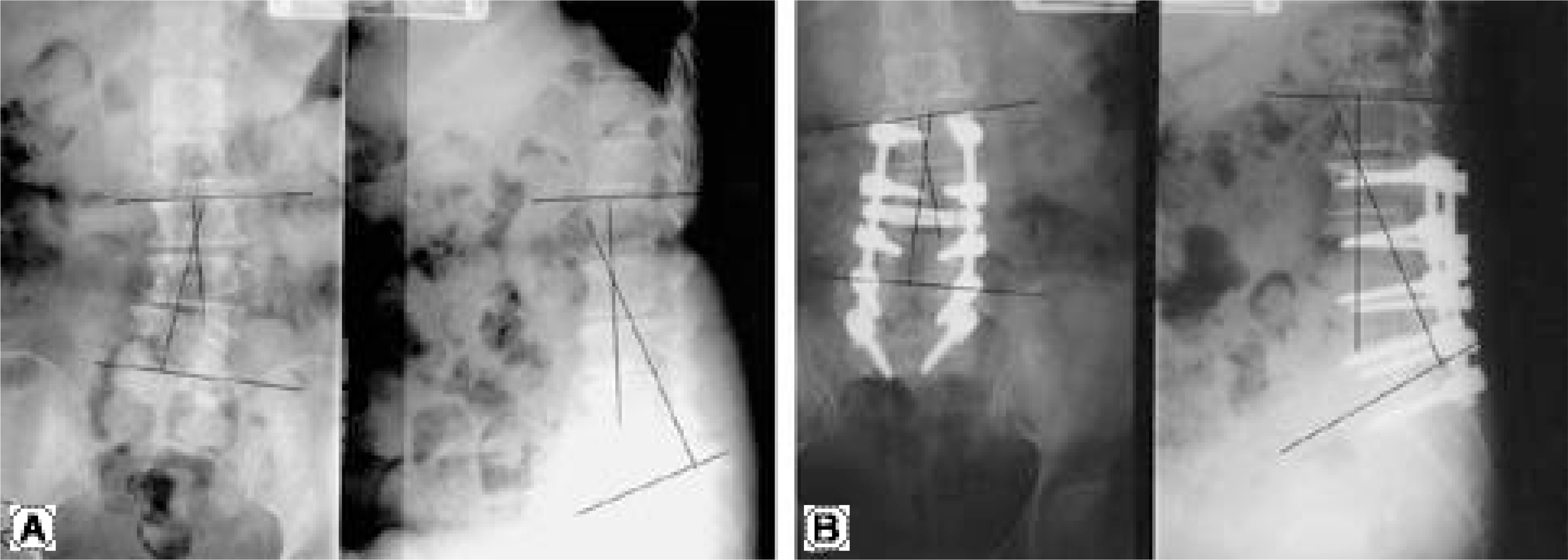
Table 1.
Deformity correction
| Preop. | Postop. | Last-F/U | Last F/U-Preop. | |
|---|---|---|---|---|
| Lordotic angle(°) | 14.2± 6.1 | 20.1± 7.3 | 19.4± 7.2 | 5.20 (37%) |
| Scoliotic angle(°) | 15.7± 4.9 | 8.9± 3.1 | 10.8± 4.7 | 4.90 (31%) |
| Pedicle rotation∗ | 0.88 | 0.62 | 0.64 | 0.24 (27%) |
Table 2.
Kirkaldy-Willis questionnaire (last follow up)
Table 3.
Correlation of total lumbar lordosis and clinical result (last follow up)
| Total lumbar lordosis | ≥ Good | ≤ Fair | Total |
|---|---|---|---|
| < 20° | 14 | 12 | 26 |
| ≥ 20° | 27 | 06 | 33 |
 XML Download