

 PDF
PDF ePub
ePub Citation
 Print
Print


Abstract
Study design
Consecutive, prospective, radiographic review of adolescent idiopathic scoliosis (A IS) patients.
Study design
Consecutive, prospective, radiographic review of adolescent idiopathic scoliosis (A IS) patients.
Summary of Background Data
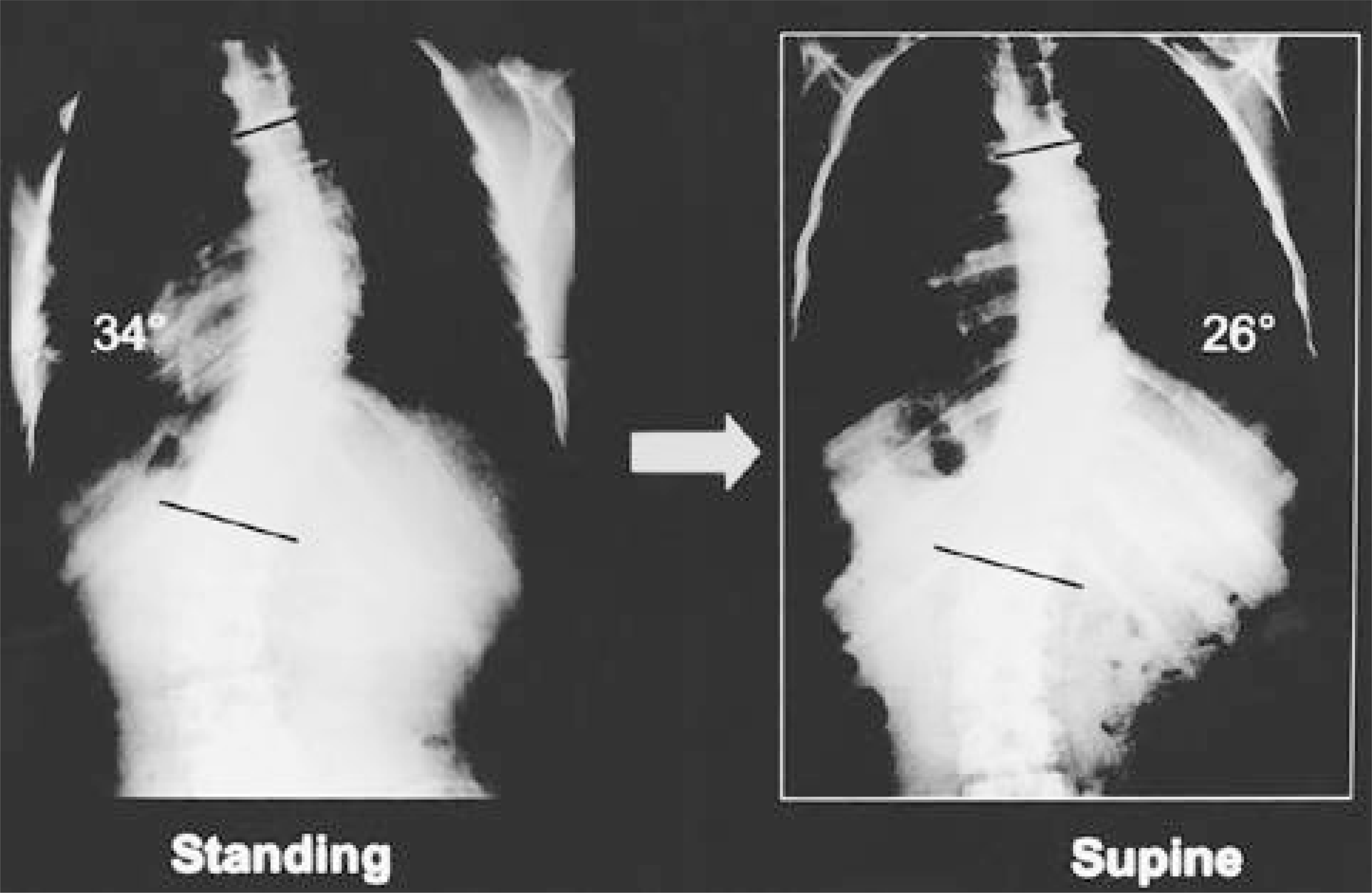
Cobb angle on standing radiographs was corrected spontaneously while the patients were in the supine position. However, there are few reports on Cobb angle in standing versus supine position in A IS.
Materials and Methods
W e checked A P plain radiographs of 101 A IS patients, 18 male and 83 female, in standing and supine position. Sixty- three cases were under Risser stage V and 38 were Risser stage V. In standing plain radiograph, 27 cases were in Cobb angle 10- 19°, 35 in 20- 29°, 15 in 30- 39°, and 24 over 40°.
A ccording to curve pattern, 31 curves were classified as King type I, 32 as type II, 8 as type III, 11 as type IV, 17 as type V, 1 thoracolumbar curve and 1 lumbar curve. Cobb angle reduction was measured on A P radiographs from each group, according to sex, maturation, Cobb angle and curve pattern.
Results
A verage reduction of Cobb angle was 8.2° (range, 1- 21°), 6.4° for male and 8.6° for female (p=0.19). The reduction value according to maturation was 8.3° for the growing group and 8.0° for the grown group (p=0.73). The average reduction value in Cobb angle 10- 19° was 5.4° (40.3%), 20- 29° was 7.1° (30.9%), 30- 39° was 8.6° (25.7%) and over 40° was 12.8°(23.6%) (p=0.001). The reduction rate decreased in proportion to Cobb angle measured in standing position. The reduction value was 8.2° in King type I curves, 8.6° in type II, 9.1° in type III, 9.1° in type IV and 6.2° in type V (p=0.238).
Concl usi on
A n average 8 ° Cobb angle reduction in supine position, compared with standing position, can influence treatment strategy in A IS patients, because a Cobb angle change more than 5- 6° is a threshold value to decide curve worsening. Thus, serial Cobb angle measurement should be performed in standing position.
Go to : 
REFERENCES
1). Duval BG. Threshold values for supine and standing Cobb angles and rib hump measurements: prognostic factors for scoliosis. Eur Spine J. 1996; 5:79–84.

2). Risser JC. The iliac apophysis; an invaluable sign in the management of scoliosis, Clin Orthop. 1958; 11:111–119.
3). King HA, Moe JH, Bradford DS, Winter RB. The selection of fusion levels in thoracic idiopathic scoliosis. J Bone Joint Surg. 1983; 65-A:1302–1313.
4). Morrissy RT, Goldsmith GS, Hall EC, Kehl D, Cowie GH. Measurment of the Cobb angle on radiographs of patients who have scoliosis. J Bone Joint Surg. 1990; 72-A:320–327.
5). Torell G, Nachemson A, Haderspeck Grib K, Schultz A. Standing and supine Cobb measures in girls with idiopathic scoliosis. Spine. 1985; 10:425–427.
6). Zetterberg C, Hansson T, Lindstrom J, Irstam L, Andersson GB. Postural and time-dependent effects on body height and scoliosis angle in adolescent idiopathic scoliosis, Acta Orthop Scand. 1983; 54:836–840.
7). Yazici M, Acaroglu ER, Alanay A, Deviren V, Cila A, Surat A. Measurement of vertebral rotation in standing versus supine position in adolescent idiopathic scoliosis. J Ped Orthop. 2001; 21:252–256.
8). Doddy MM, Lonstein JE, Stovall M, Hacker DG, Luckyanov N, Land CE. Breast cancer mortality after diagnostic radiography: Findings from the U.S. Scoliosis cohort study. Spine. 2000; 25:2052–2063.
9). Levy AR, Goldberg MS, Mayo NE, Hanley JA, Poitras B. Reducing the lifetime risk of cancer from spinal radiographs among people with adolescent idiopathic scoliosis. Spine. 1996; 21:1540–1547.
Go to :
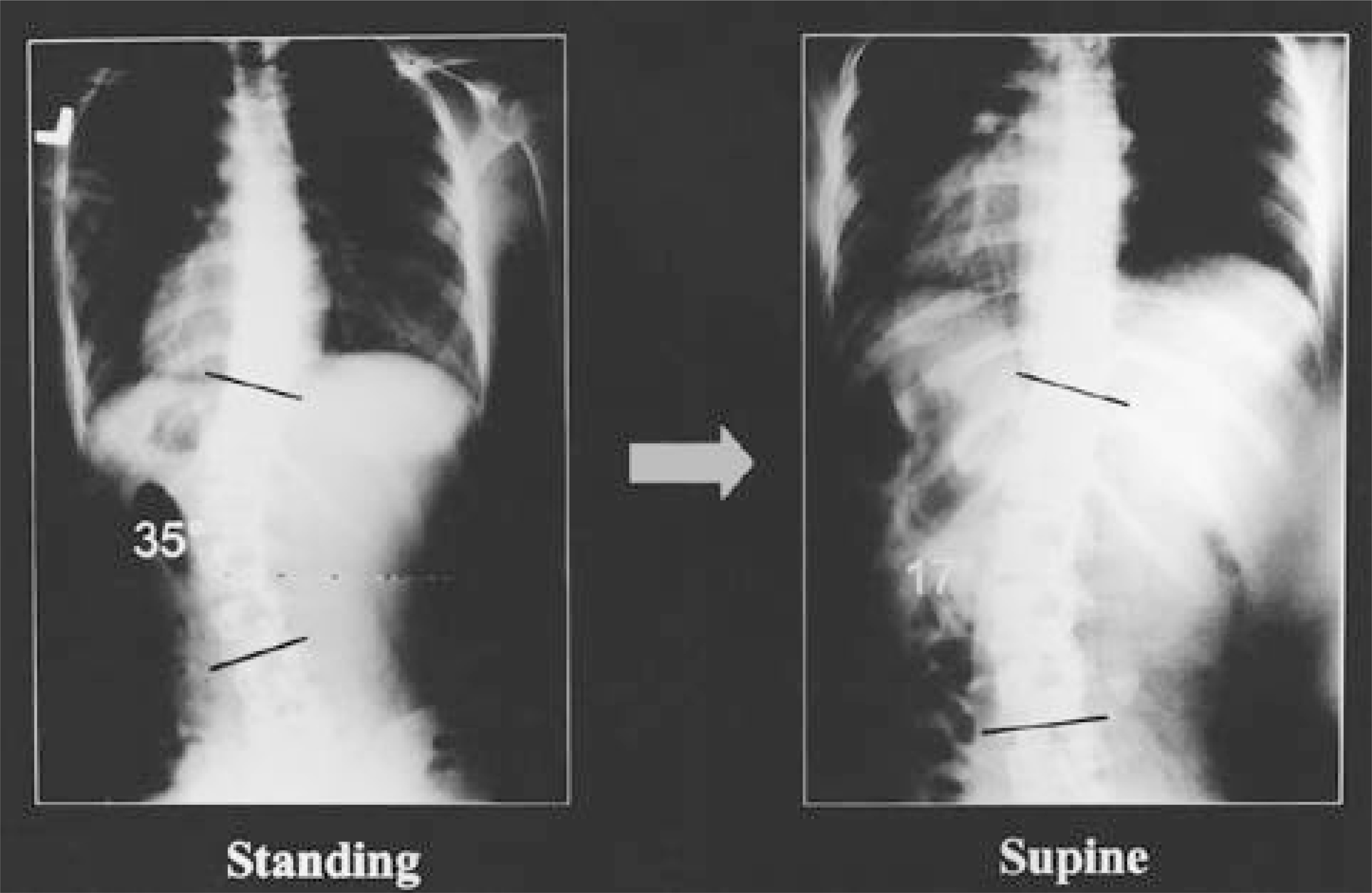
 | Fig. 2.18-year-old female with King type II curve showed 35% angle reduction in supine film |
Table 1.
Mean values for supine and standing Cobb angle and reduction rate for all samples according to sex
| Standing Cobb angle(˚) | Supine Cobb angle(˚) | Reduction angle(˚) | Reduction rate(%) | No. of patients | |
|---|---|---|---|---|---|
| Male | 25.1 | 18.7 | 6.4 | 25.5 | 18 |
| Female | 28.2 | 19.6 | 8.6 | 30.4 | 83 |
Table 2.
Mean values for supine and standing Cobb angle and reduction rate for all samples according to maturation
| Standing Cobb angle(˚) | Supine Cobb angle(˚) | Reduction angle(˚) | Reduction rate(%) | No. of patients | |
|---|---|---|---|---|---|
| Risser IV or less | 26.3 | 18.0 | 8.3 | 31.6 | 63 |
| Risser V | 29.9 | 21.9 | 8.0 | 26.8 | 38 |
Table 3.
Mean values for supine and standing Cobb angle and reduction rate for all samples according to Cobb angle of major curve in standing radiograph
Table 4.
Mean values for supine and standing Cobb angle and reduction rate for all samples according to curve pattern of deformity
 XML Download