Abstract
Objectives
A nalyzing the clinical outcome of operative treatment in lumbar degenerative kyphosis (LDK) by means of anterior and posterior operation using Wedged cage (SynCageⓇ) and pedicle screws.
Summary of Literature Review
LDK is common in old farmers who have worked in a stooping posture for decades and is a quite rigid form of kyphosis accompanied by adjacent instability, dystrophic changes of vertebral bodies and weakness of back and hip extensors. For surgical treatment, restoration and maintenance of lumbar lordosis is mandatory for global balance. A nterior release and restoration of disc space with the same morphologic cage seems to be a quite anatomic and harmonious approach.
Materials and Methods
Ten LDK patients, who underwent anterior interbody fusion using Wedged cage (SynCageⓇ) and posterior fusion with pedicle screws between 2000 to 2001, were followed up for more than 2 years. The operation was done in one or two stages. We performed anterior release, gradual widening of the intervertebral space with wedge trials of increasing size, insertion of wedged cages filled with auto-, allo- or synthetic bone and posterior pedicle fixation and fusion. We measured the lumbar lordotic angle, sacral inclination, fusion segmental angle, thoracic kyphotic angle and vertical axis line in preoperative, immediate postoperative and follow- up standing X - ray.
Results
Mean fusion segments using Wedged cage were 2.8 segments for anterior interbody fusion and 3.4 segments for posterior fusion. Mean sagittal correction angle was 40.3° with mean correction loss of 2.6°. Whole lordosis was 6.9° kyphosis pre-operatively, which was corrected to 33.4° lordosis postoperatively and 30.8° lordosis at last follow- up. Mean sacral inclination was corrected from 18.2° preoperatively to 37.8° postoperatively and 30.7° at follow- up. Vertical axis line was corrected from 11.4 cm preoperatively to 0.4 cm postoperatively and 1.3cm at follow- up. Thoracic lordosis was corrected spontaneously without any surgical extension to the thoracic spine by mean 19.9° (0.2° lordosis preoperatively to 19.7 ° kyphosis at follow- up). Loss of cardinal signs occurred in 70- 80 % of patients and satisfactory clinical results were shown in 90% of patients.
Go to : 
REFERENCES
1). Lee CS, Kim YT and Kim EG. Clinical study of lumbar degenerative Kyphosis. J Korean Spine Surg. 1997; 4(1):27–35.
2). Becker's L and Bekaert J. The role of lordosis. Acta Orthop Belg. 1991; 57(Suppl):198–202.
3). Deniel E, Geib MD, Lawrence G, Lenke MD, Keith H, Bridwell MD, Kathy Blanke RN, Levin W and McBn-ery MD. An-Analysis of Sagittal Spinal Alignment in 100 Asymptomatic Middle and Older Aged Volunteers. Spine. 1995; 20:1352–1358.
4). Yohichi Aota, Kiy oshi Kumano and Shigeru Hirab ayashi. Postfusion instability at the adjacent Segments after rigid pedicle screw fixation for degenerative lumbar spinal disorders. Journal of Spinal Disorders. 1995; 8(6):464–473.
5). Takemitsu Y, Harada Y and Iwahara T. Low back pain and aging change of spine in Japanese farmers aged more than 40 years. J Jpn Orthop Assoc. 1984; 58:551–552.
6). Takemitsu Y, Harada Y, Iwahara T, Miyamoto M and Mitatake Y. Lumbar Degenerative Kyphosis: Clinical, radiologic and epidemiological study. Spine. 1988; 13:1317–1326.
7). Andersson BJG, Ortengren R. Myoelectric back muscle activity during sitting. Scand J Rehab Med. 1974; 3(Suppl):73–90.
8). Ronald L and De Wald. Revision surgery for spinal deformity: Instructional Course Lecture. Vol XLI:235–250. 1992.
9). Kirkaldy-Willis WH, Paine KWE, Cauchoix J and Mclvor G. Lumbar Spinal Fusion: A biomechanical study, Spine. 1984; 9:574–581.
10). Doherty JH. Complication of fusion in lumbar scoliosis. J Bone Joint Surg. 1973; 55-A:438.
11). Aaro S, Ohlen G. The effect of Harrington instrumentation on the sagittal configuration and mobility of the spine in scoliosis. Spine. 1983; 8(6):570–575.

12). Winter RB. Harrington instrumentation into the lumbar spine, technique for preservation of normal lumbar lordosis. Spine. 1986; 11(9):633–635.
13). Moe JH and Denis F. The iatrogenic loss of lumbar lordosis. Orthop Trans. 1977; 1-2:131.
14). Grobler LJ, MOe JH and Winter RB, et al. Loss of lumbar lordosis following surgical collection of thoracolumbar deformity, Prthop Trans. 2(2):39. 1978.
15). Michael OLG. Loss of lumbar lordosis: A complication of spinal fusion for scoliosis. Orthop Clin. N AM. 1988; 19-2:383–393.
16). Atsuta Y. Degenerative Kyphosis in advanced age; Cause and menagement (in Japanese). J Jpn Orthop Assoc. 1994; 37-3:289–295.
17). Lee JS, Oh WH, Chung SS, Lee SG and Lee JY. Analysis of the Sagittal Alignment of Normal Spines. J Korean Orthop. 1999; 34:949–954.

18). Wambolt A, Spencer DL. A segmental analysis of the distribution of lumbar lordosis in the normal spine. Orthop Trans. 1987; 11:92–93.
19). Kim EH, Han SK and Kim HJ. A Clinical analysis of Surgical Treatment of Lumbar Degenerative Kyphosis. J Korean Spine Surg. 2001; 8(3):210–218.

20). Lee CS. Lumbar Degenerative Kyphosis. Instructional Course Lectures. 1998; 157-169.
21). Nakai O, Yamaura I, Kurosa Y, Komori H, Okawa A, Sakai H and Abe M. Posterior stabilization for Lumbar Degenerative Kyphosis:. situ fusion in maximum extension on Hall's frame. Proc 5th Conf Lumbar Fusion and Stabilization, Tokyo. Springer-Verlag;p. 135–149. 1993.
22). Fraser RD. Interbody, posterior and combined lumbar fusions. Spine. 1995; 15(20, 24Suppl):167S–177S.

23). Jahng JH, Paek DH, Chang H, Bahk WJ, Eun SP, Sohn JM and Lim GS. Anterior interbody fusion and posterior instrumentation for degenerative lumbar spondylolisthesis. J of korean Orthop. Assoc. 1996; 33(2):25–32.
24). Kuslich SD. Lumbar interbody cage fusion for back pain. Spine. 1999; 13(2):395–311.
25). Suk SI, Kim JH, Kim WJ, Lee SM, Liu Y, Chung ER and Lee CS. Treatment of fixed lumbosacral kyphosis by posterior vertebral column resection. J of Korean Spine Surg. 1998; 5:307–313.
26). Kim EH, Cho KN and Kim CH. Surgical treatment of posttraumatic kyphosis. J of korean Orthop. Assoc. 1998; 33:367–374.
27). Kim KT. Kyphosis. J of Korean Spine Surg. 1999; 6:306–315.
28). Kostuik JP, Maurais GR, Richardson WJ and Okaji-ma Y. Combined single stage anterior and posterior osteotomy for correction of iatrogenic lumbar kyphosis. Spine. 1988; 13:257–266.
Go to : 
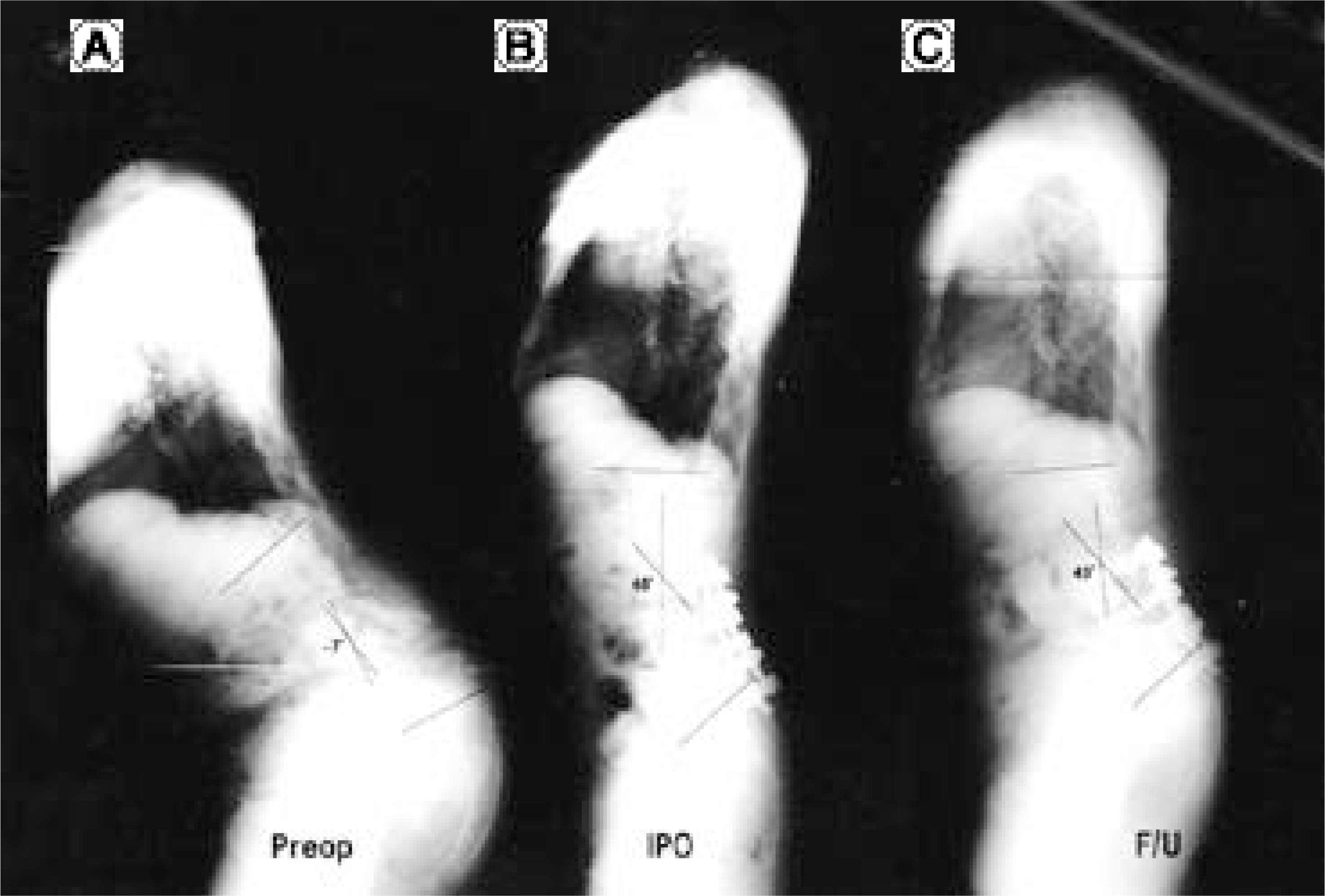
 | Fig. 1.
(A) Standing lateral radiograph of a 60 year-old female with lumbar degenerative kyphosis was showed kyphosis 7 degree of lumbar lordosis angle. (B) Immediate postoperative standing lateral radiograph was showed 48 degree lodorsis of lumbar lordosis angle. Lumbar lordosis correction was 55 degree. (C) Postoperative 24 month radiograph was showed 43 degree lordosis of lumbar lordosis angle. Correction loss was 5 degree. |
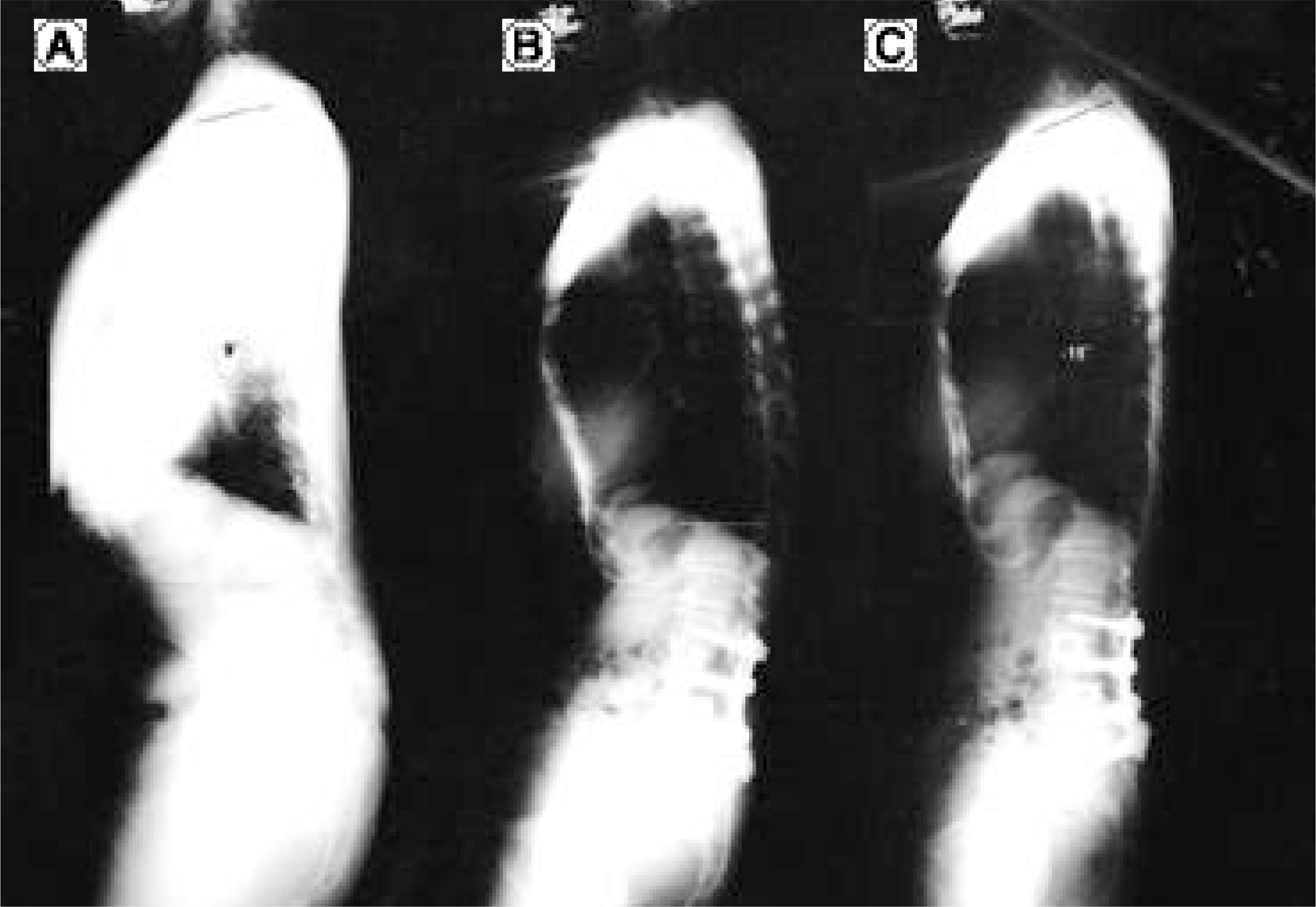
 | Fig. 2.
(A) Standing lateral radiograph of a 63 year-old female with lumbar degenerative kyphosis was showed lordosis 9 degree of thoracic angle. (B) Immediate postoperative standing lateral radiograph was showed posterior tilt of upper end vertebra. (C) Postoperative 25 month radiograph show 18 degree kyphosis of thoracic angle. Thoracic angle correction was 27 degree. |
Table 1.
Results of ten operated cases of LDK
| Case | Age | eSex | Associated disease | Method | Pre OP | IPO | F/U | ||||||||||
|---|---|---|---|---|---|---|---|---|---|---|---|---|---|---|---|---|---|
| L LA∗ | FSA† | SI∮ | C7-S1 | TKA‡ | LLA | FSA | C7-S1 | LLA | FS A | SI | C7-S1 | T KA | |||||
| 1 | 63 | F | PR§:L3-L5 Ant: L3-L5 Pos t:L3-S 1 | -2 2 | -22 | -1 0 | 8.8 | 9 | 25 | 29 23 | 0.5 | 2 6 | 30 | 22 | 3 | -18 | |
| 2 | 55 | F | Spinal stenosis | Ant:L3-L5 Post:L3-L5 | -13 | -9 | 2 5 | 15. 5 | 13 | 28 | 22 39 | 5.5 | 14 | 21 | 34 | 1 .5 | -2 |
| 3 | 71 | M | Spinal stenosis Spondylolisthesis | Ant:L3-S1 Pos t :L3-S1 | 25 | 25 | 32 | - 1.5 | -38 | 32 | 32 41 | -5.2 | 38 | 33 | 30 | -4.9 | -38 |
| 4 | 60 | F | Spondyl olisthesis | P R :L4-S1 Ant : L3-S1 Pos t:L3-S 1 | -7 | -30 | 7 | 24 | 39 | 48 | 3 4 46 | 1.3 | 43 | 24 | 36 | 9 | -8 |
| 5 | 58 | F | Instability Spo ndylolysis | Ant : L3-S1 Pos t:L3-S 1 | - 15 | -15 | 17 | 13. 4 | -9 | 29 | 2 5 43 | 6.4 | 21 | 22 | 24 | 4 | -13 |
| 6 | 57 | M | Ant:L3-S1 Post:L2-S1 | 0 | -6 | 16 | 8 | 3 | 33 | 3 4 37 | 1 | 30 | 32 | 31 | 1 | -21 | |
| 7 | 58 | F | Spinal stenosis | Ant:T12-L3 Post:T12-L3 | 25 | - 31 | 3 9 | 11 | -3 | 56 | 0 60 | -1 | 52 | - 2 | 46 | -2 .9 | -24 |
| 8 | 70 | M | Old comp. fx L3 | An t :L3-S1 Post : T12 -S1 | -34 | -32 | 14 | 19 .1 | -12 | 31 | 2 7 24 | -6.1 | 3 0 | 27 | 28 | -4.5 | -31 |
| 9 | 67 | F | Spinal stenosis | An t :L3-S1 Post : L3-S1 | 4 | -3 | 27 | 3.1 | 1 | 26 | 2 7 27 | 2.7 | 27 | 26 | 35 | 2.5 | -20 |
| 10 | 68 | F | Ant : L2-S1 Po st: L2-S1 | -32 | -25 | 15 | 13 | -1 | 26 | 3 1 38 | -1.4 | 2 7 | 28 | 21 | 4.4 | -22 | |
| Ave rage | 62.7 | Ant: 2.9 Post: 3.4 | -6.9 | -14.8 | 18.2 | 11. 4 | 0.2 | 33.4 | 26.1 37.8 | 0.4 | 30.8 | 24.1 | 3 0.7 | 1 .3 | -19.7 |


 PDF
PDF ePub
ePub Citation
 Print
Print


 XML Download