Abstract
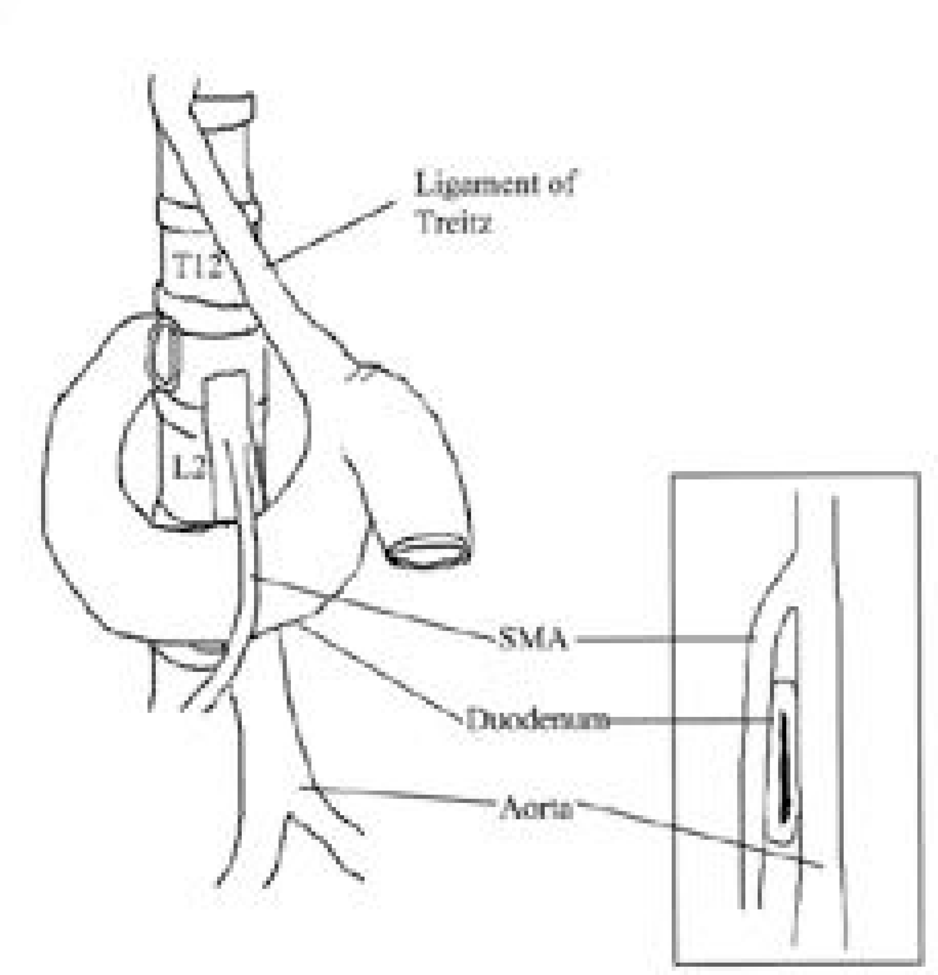
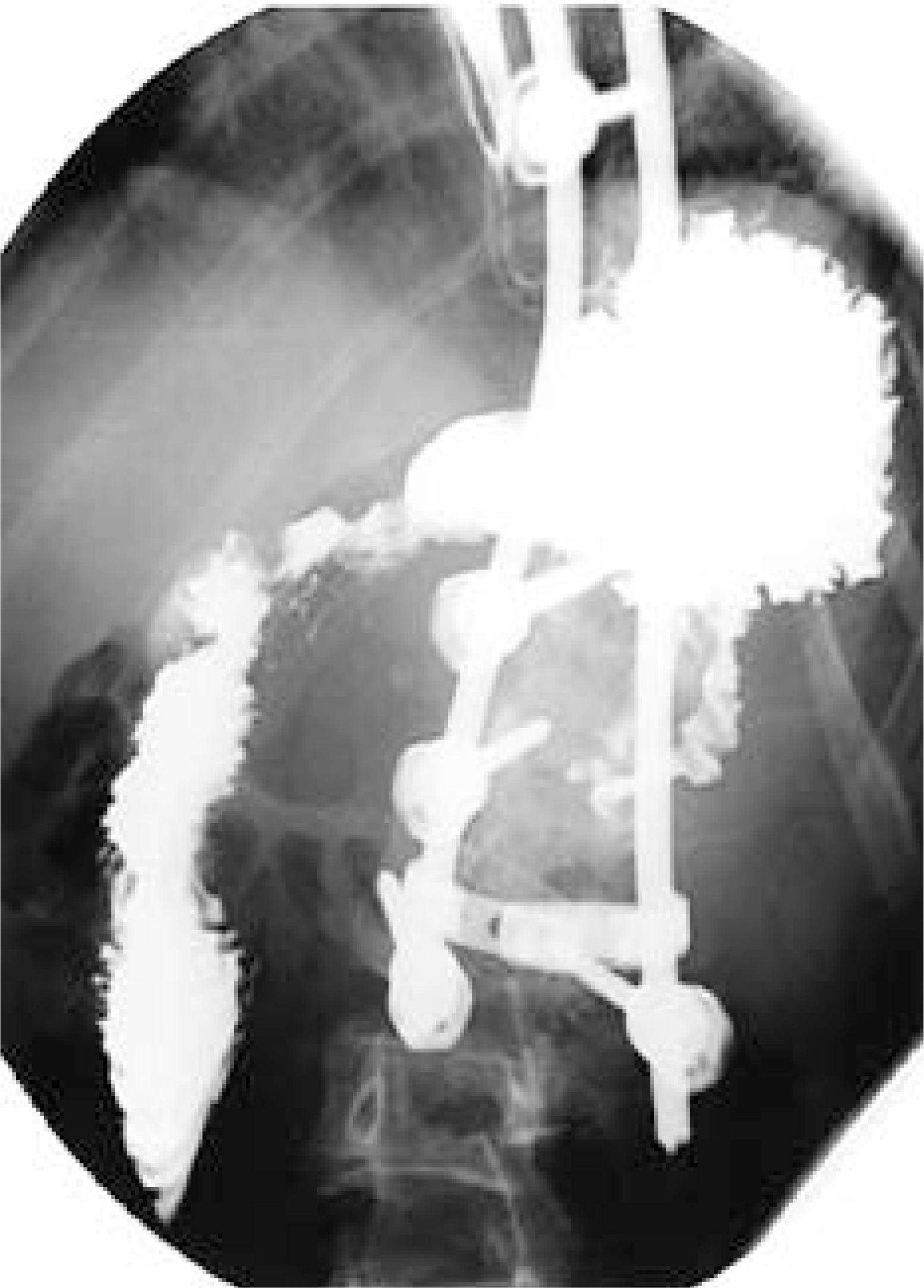
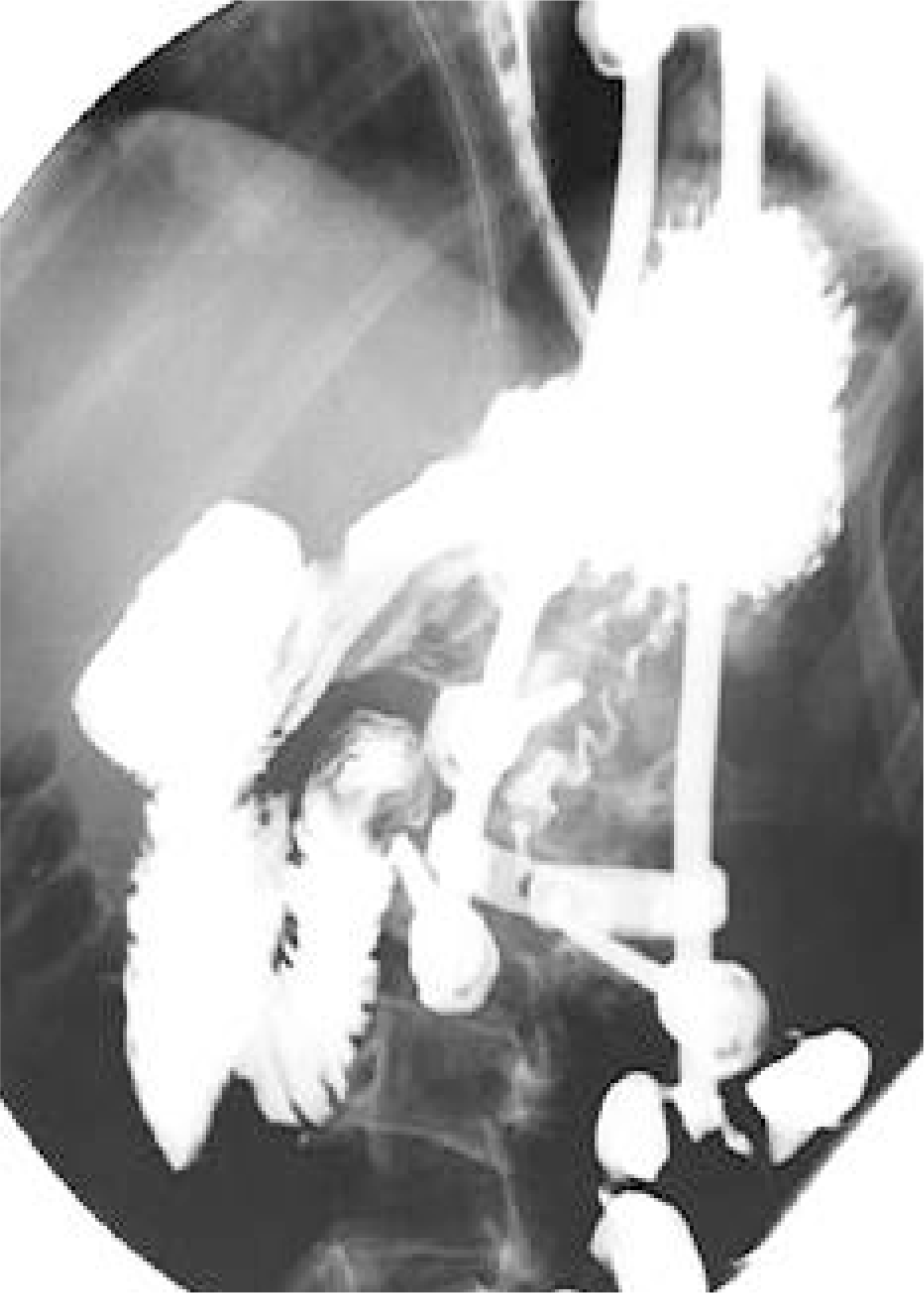
Superior mesenteric artery (SMA) syndrome is a rare condition that results from an extrinsic compression of the third portion of the duodenum, between the SMA and the aorta. The symptoms for the condition consist of abdominal pain and recurrent vomiting, caused by ileus, and can be followed by an electrolyte imbalance and nutrient deficiency. SMA syndrome can follow surgical correction of a spinal deformity, as the aorta migrates forward as the degree of the lumbar lordosis increases, and the retroperitoneal fat tissue decreases, during perioperative abstinence. A ny symptoms suggestive of SMA syndrome, after correction of a spinal deformity, should be investigated, as SMA syndrome carries a prolonged hospital stay, with the potential for mortality. A n 11year 10 month old boy, who underwent correction for thoracic kyphoscoliosis, developed postoperative abdominal distension, pain and bilious vomiting. A n upper gastrointestinal contrast study revealed SMA syndrome, which required a laparotomy.
REFERENCES
2). Bunch W., Delaney J. Scoliosis and acute vascular compression of the duodenum. Surgery,. 67:901–906. 1985.
3). Crowther MA., Webb PJ., Eyre-Brook IA. Superior mesenteric artery syndrome following surgery for scolio -sis. Spine,. 27:528–533. 2002.
5). Evarts CM., Winter RB., Hall JE. Vascular compression of the duodenum associated with the treatment of sco -liosis. J Bone Joint Surg Am,. 53:431–444. 1971.
6). Fromm S., Cash J. Superior mesenteric artery syndrome: an approach to diagnosis and management of upper gastrointestinal obstruction of unclear etiology. S D J Med,. 43:5–10. 1990.
7). Hutchinson DT., Bassett GS. Superior mesenteric artery syndrome in pediatric orthopedic patients. Clin Orthop,. 250:250–257. 1990.

9). Kellogg EL., Kellogg WA. Chronic duodenal obstruction with duodenojejunostomy as a method of treatment: Report of forty-one operations. Ann Surg,. 73:578–608. 1921.
10). Kennedy RH., Cooper MJ. An unusually severe case of the cast syndrome. Postgrad Med J,. 59:539–540. 1983.

11). Munns SW., Morrissy RT., Golladay ES., McKenzie CN. Hyperalim-entation for superior mesenteric artery (cast) syndrome follwing correction of spinal deformity. J Bone Joint Surg (Am),. 66:1175–1177. 1984.
12). Shah MA., Moreland MS. Superior mesenteric artery syndrome in scoliosis surgery: weight percentile for height as an indicator of risk. Am Acad Pediatr,. 98:567–568. 1996.

13). Shapiro G., Green DW., Fatica NS., Boachie-Adjei O. Medical complications in scoliosis surgery. Curr Opin Pediatr,. 13:36–41. 2001.

14). Strong E. Mechanics of arteriomesenteric duodenal obstruction and direct surgical attack upon etiology. Ann Surg,. 148:725–730. 1958.

15). Transfeldt EE. Moe's textbook of scoliosis and other spinal deformites. Lonstein JE, Bradford DS, Winter RB, editors. Complications of treatment. 3rd ed.Philadel -phia, W.B. Saunders, 451-482;1995.
16). Vitale MG., Higgs GB., Liebling MS., Roth N., Roye DP Jr. Superior Mesenteric artery syndrome after seg -mental instrumentation: a biomechanical analysis. Am J Orthop,. 28:461–467. 1999.
Fig. 2.
11+10-year-old male. Thoracic kyphoscoliosis. Preoperative standing anteroposterior (A) and lateral (B) radiographs.
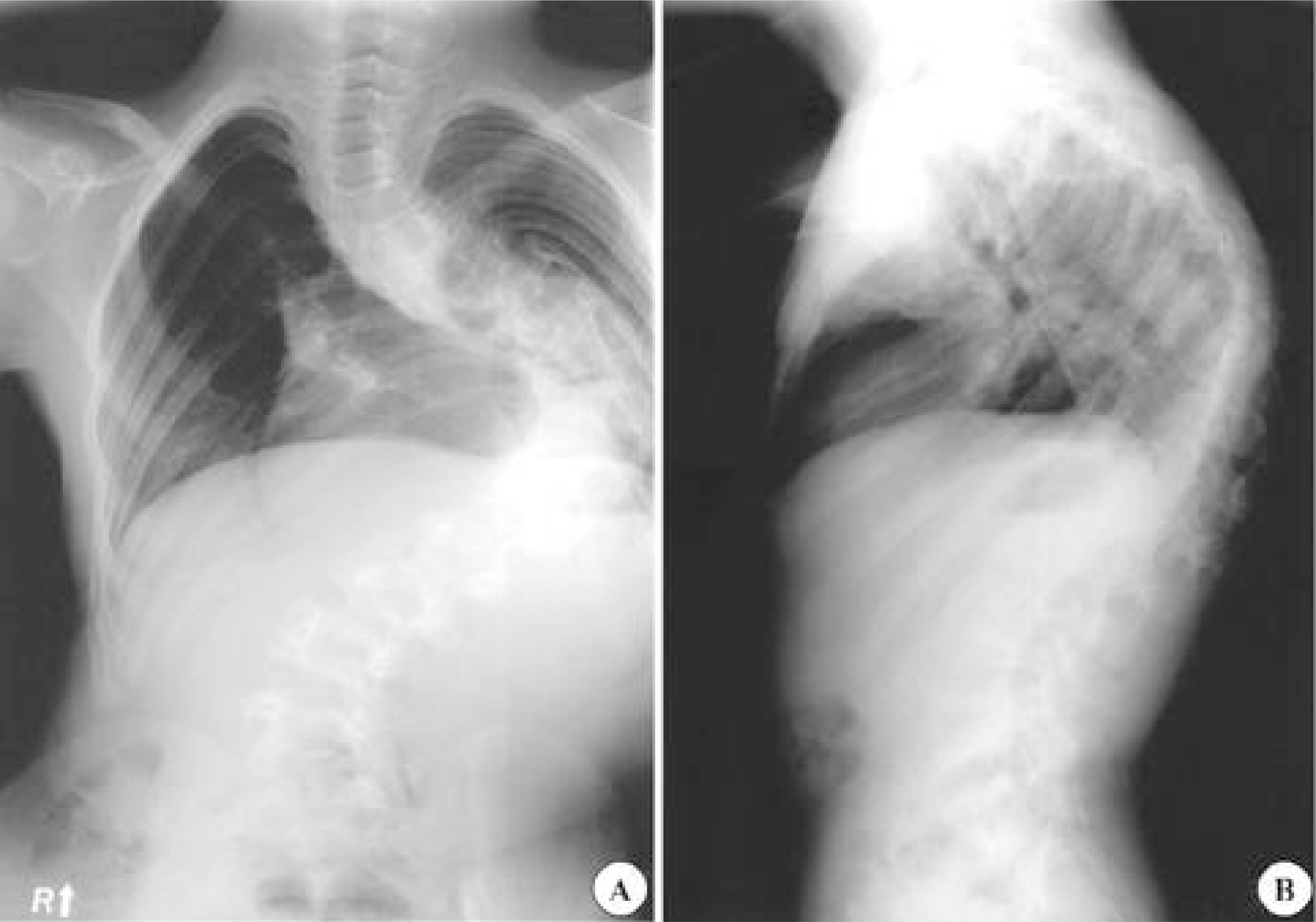
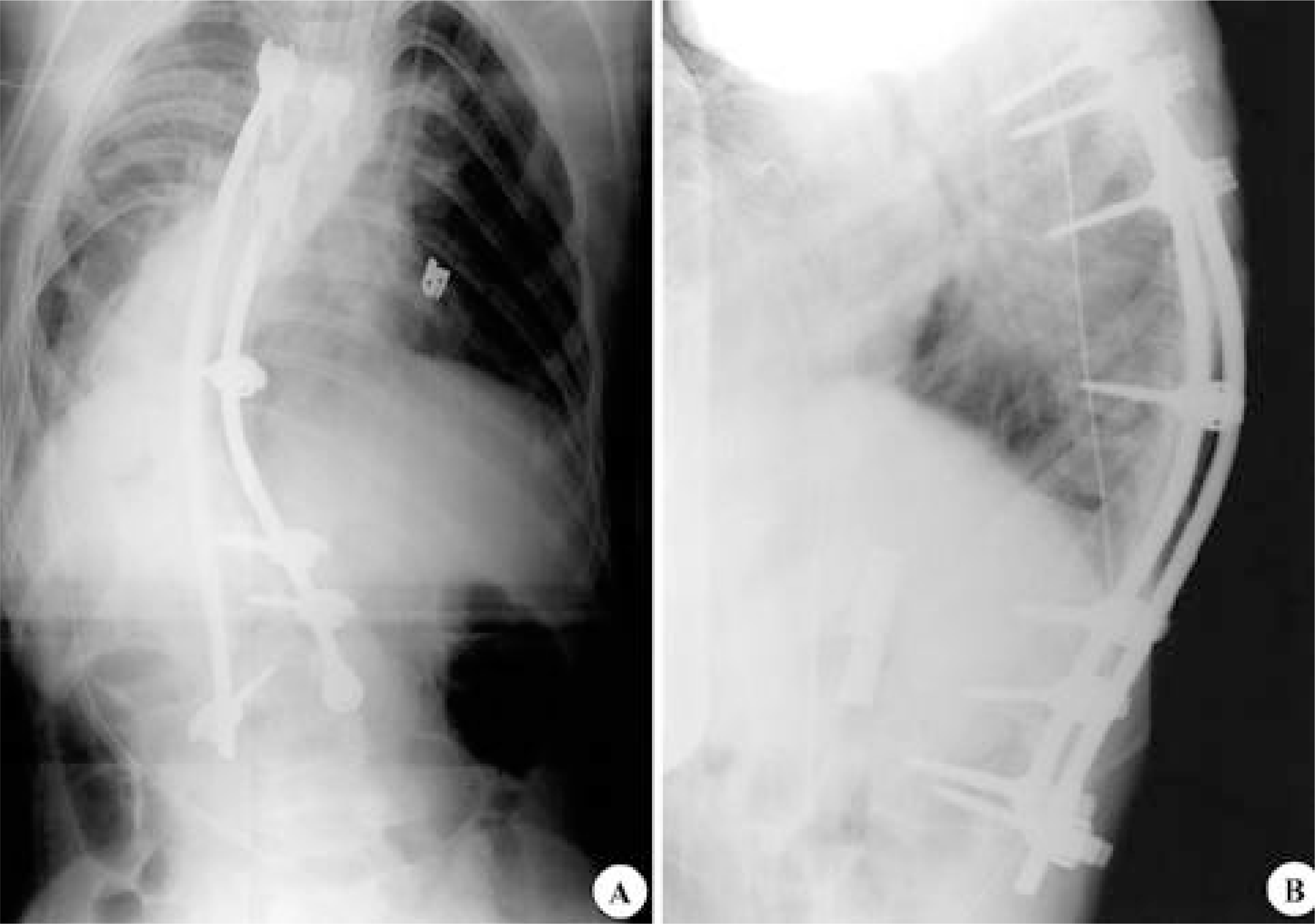
Fig. 3.
Postoperative anteroposterior (A) and lateral (B) radiographs following anteroposterior fusion from T5 to L3.


 PDF
PDF ePub
ePub Citation
 Print
Print


 XML Download