

 PDF
PDF ePub
ePub Citation
 Print
Print


Abstract
Objective
The goal of this study was to develop a novel pupil and iris segmentation algorithm. We evaluated segmentation performance based on a fractal model. Two methods were compared: Daugman's and our new proposed method.
Methods
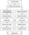
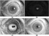
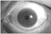
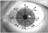
We received 200 anterior segment images with 3,872×2,592 pixels. Here we present an active contour model that accurately detects pupil boundaries in order to improve the performance of segmentation systems. We propose a method that uses iris segmentation based on a fractal model. We compared the performance of Daugman's method and the proposed new method and statistically analyzed the results.
Results
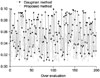
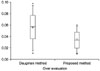
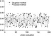
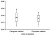
We manually compared segmentation with the Daugman's method and the new proposed method. The findings showed that the proposed segmentation accuracy was about 2.5 percent higher than Daugman's method. There was a significant difference (p<0.05) between the under and over data between the two methods.
Figures and Tables


References
1. Lim JI, LaBree L, Nichols T, Cardenas I. A comparison of digital nonmydriatic fundus imaging with standard 35-millimeter slides for diabetic retinopathy. Ophthalmology. 2000. 107(5):866–870.

2. Yen KG, Hess D, Burke B, Johnson RA, Feuer WJ, Flynn JT. The optimum time to employ telephotoscreening to detect retinopathy of prematurity. Trans Am Ophthalmol Soc. 2000. 98:145–150. discussion 150-151.
3. van Norren D, van de Kraats J. Retinal densitometer with the size of a fundus camera. Vision Res. 1989. 29(3):369–374.
4. Tuulonen A, Airaksinen PJ, Montagna A, Nieminen H. Screening for glaucoma with a non-mydriatic fundus camera. Acta Ophthalmol (Copenh). 1990. 68(4):445–449.
5. Zeimer R, Zou S, Meeder T, Quinn K, Vitale S. A fundus camera dedicated to the screening of diabetic retinopathy in the primary-care physician's office. Invest Ophthalmol Vis Sci. 2002. 43(5):1581–1587.
6. Gili Manzanaro P, Carrasco Font C, Martin Rodrigo JC, Yanguela Rodilla J, Arias Puente A. Digital analysis of the optic disc with fundus camera: a study of variability. Arch Soc Esp Oftalmol. 2004. 79(3):125–130.
7. Schmitz-Valckenberg S, Fleckenstein M, Gobel AP, Sehmi K, Fitzke FW, Holz FG, et al. Evaluation of autofluorescence imaging with the scanning laser ophthalmoscope and the fundus camera in age-related geographic atrophy. Am J Ophthalmol. 2008. 146(2):183–192.
8. Daugman J. High confidence Recognition of Persons by Rapid Analysis of Iris Texture, IEEE Conference Publication. 1995. 05. 244–251. No. 408.
9. Daugman J. New methods in iris recognition. IEEE Trans Syst Man Cybern B Cybern. 2007. 37(5):1167–1175.
10. Huang H, Hu G. Iris recognition based on adjustable scale wavelet transform. Conf Proc IEEE Eng Med Biol Soc. 2005. 7:7533–7536.
11. Monro DM, Rakshit S, Zhang D. DCT-based iris recognition. IEEE Trans Pattern Anal Mach Intell. 2007. 29(4):586–595.
12. Klemencic A, Kovacic S, Pernus F. Automated segmentation of muscle fiber images using active contour models. Cytometry. 1998. 32(4):317–326.
13. Bogdanova I, Bresson X, Thiran JP, Vandergheynst P. Scale space analysis and active contours for omnidirectional images. IEEE Trans Image Process. 2007. 16(7):1888–1901.
14. Parker JR. Algorithms for image processing and computer vision. 1997. WILEY COMPUTER PUBLISHING, 1997.
15. Gonzalez RC. Digital image processing using MatLab. 2004. Seoul: ITC..
16. Otsu N. A threshold selection method from gray_level histograms. IEEE Trans Systems Man and Cybernetics. 1979. 79:62–66.
 XML Download