

 PDF
PDF ePub
ePub Citation
 Print
Print


Abstract
Objective
The most widely used method for quantifying new blood vessel growth in tumor angiogenesis is the determination of microvessel density, which is reported to be associated with tumor progression and metastasis, and a prognostic indicator of patient outcome. In this study, we propose a method for the determination of microvessel density by image analysis, to improve the accuracy and the objectivity of determination of the microvessel density.
Methods
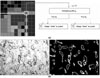
Four-micron-thick tissue sections of renal cell carcinoma samples were stained immunohistochemically for CD34. The regions with a high degree of vascularization were selected by an expert for digitization. Each image was digitized as a 24-bits/pixel image file with a resolution of 640×480 pixels. First, segmentation of the microvessels based on pixel classification using color features in hybrid color space was performed. After use of a correction process for microvessels with discontinuities and separation of touching microvessels, we counted the number of microvessels for the microvessel density measurement.
Figures and Tables
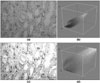 | Figure 1Color distribution of an original image and the preprocessed image. (a) Original image, (b) Preprocessed image, (c) Color distribution of (a), (d) Color distribution of (c)
|
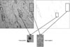
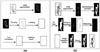 | Figure 3(a) Procedure used to connect discontinuous microvessels, (b) Procedure used to separate touching microvessels
|

References
1. Folkman J. Tumor angiogenesis: therapeutic implications. New England Journal of Medicine. 1970. 285:1182–1187.

2. Weidner N, Semple JP, Welch WR, Folkman J. Tumor angiogenesis and metastasis-correlation in invasive breast carcinoma. New England Journal of Medicine. 1991. 324:1–8.
3. Folkman J. What is the evidence that tumors are angiogenesis dependent? Journal of the National Cancer Institute. 1990. 82:4–6.
4. Zhang X, Yamashita M, Uetsuki H, Kakehi Y. Angiogenesis in renal cell carcinoma: evaluation of microvessel density, vascular endothelial growth factor and matrix metalloproteinasses. International Journal of Urology. 2002. 9:509–514.
5. Weidner N. Intratumor microvessel density as a prognostic factor in cancer. American Journal of Pathology. 1995. 147:9–19.
6. Rieder MJ, O'Drobinak DM, Greene AS. A computerized method for determination of microvessel density. Microvascular Research. 1995. 49:180–189.
7. Rodriguez RM, Alarcon TE, Navarro RW, Cuello LS. Color segmentation applied to study of the angiogenesis. Journal of Intelligent and Robotic Systems. 2002. 34:83–97.
8. Sharon AS. ANGY: a rule-based expert system for automatic segmentation of coronary vessels from digital substrated angiograms. IEEE Transaction on Pattern Analysis and Machine Intelligence. 1986. 3(2):188–199.
9. Hannen EJ, van der Laak JA, Manni JJ, Freihofer HP, Slootweg PJ, Koole R, et al. Computer assisted analysis of the microvasculature in metastasized and non-metastasized squamous cell carcinomas of the tongue. Head and Neck. 2002. 24:643–650.
10. Wild R, Ramakrishnan S, Sedggewick J, Griffioen AW. Quantitative assessment of angiogenesis and tumor vessel architecture by computer-assisted digital image analysis: effects of VEGF-toxin conjugate on tumor microvessel density. Microvascular Research. 2000. 59:368–376.
11. Duda RA, Hart PE, Stork DG. Pattern classification. 2001. 2nd ed. New York: John Wiley & Sons Inc.;215–245.
12. Cheng HD, Jiang XH, Sun Y, Wang J. Color image segmentation: advances and prospects. Pattern Recognition. 2001. 34:2259–2281.
13. Ohta YI, Kanade T, Sakai T. Color information for region segmentation. Computer Graphics and Image Processing. 1980. 13:222–241.
14. Berns RS. Principles of color technology. 2000. 3rd ed. New York: John Wiley & Sons Inc.;44–62.
15. Johnson RA, Wichern DW. Applied multivariate statistical analysis. 1998. 4th ed. Singapore: Prentice-Hall;314–343.
16. Vincent L, Soille P. Watersheds in digital spaces: an efficient algorithm based on immersion simulations. IEEE Transaction on Pattern Analysis and Machine Intelligence. 1991. 13:583–397.
17. Jeong HS, Sung RH. Microvessel quantitation and assessment of its utility by CD34 staining in invasive breast carcinoma. Korean Journal of Pathology. 1997. 31:298–307.
18. Kido H, Kaneko K, Makinouchi A. Semiautomatic segmentation of lung region from three dimensional color images of visible human. Journal of Korean Society of Medical Informatics. 2007. 13(2):171–176.
19. Gao H, Xue P, Lin W. A new marker based watershed algorithm. Proceedings of 2004 IEEE international symposium on circuits and systems. May. Vancouver, Canada: 81–84.
 XML Download