

 PDF
PDF ePub
ePub Citation
 Print
Print


INTRODUCTION
Scrub typhus is a mite-borne human bacterial infection caused by Orientia tsutsugamushi. The disease has become a public health problem in Asia, where about 1 million new cases are identified annually and 1 billion people may be at risk [1]. A recent epidemiologic study conducted in Korea reported an almost 4-fold increase in the number of patients observed in 2013 (10,485 cases) compared to in 2001 (2,637 cases) [2]. The study also showed that the incidence of scrub typhus has been increasing in all age groups, especially in children aged less than 10 years. Interestingly, 389 cases (0.5%) were reported between January and February in 2013.
Scrub typhus occurs in individuals who engage in occupational or recreational behavior that brings them into contact with mite-infested habitats such as bush and grass. The onset of disease is characterized by fever, headache, myalgia, cough, and gastrointestinal symptoms. The symptoms are usually mild and self-limited. However, some cases show more severe clinical manifestations and the disease may be fatal. Reported complications of scrub typhus include interstitial pneumonia, acute renal failure, meningoencephalitis, gastrointestinal bleeding, and multiple organ failure [3456]. Herein, we present a patient with scrub typhus complicated with a splenic infarction.
CASE REPORT
A 40-year-old man visited the emergency medical center of Inha University Hospital in late October of 2014 complaining of fever. His fever had started 1 week ago, and was accompanied by headache and cough. Gastrointestinal symptoms such as nausea, vomiting, abdominal discomfort, and diarrhea followed. He was prescribed amoxicillin-clavulanate by the doctor at a local clinic, but his symptoms persisted. He was a 20 pack-year smoker with an unremarkable medical history. He was a tour guide with a history of contact with bush and grass when he travelled to Anmyeondo in Taean, Chungcheongnam-do 3 weeks ago. He had observed a bite mark on his abdomen 2 weeks ago.
His initial blood pressure was 125/63 mmHg, pulse was 93 beats per minute, respiratory rate was 20 breaths per minute, and temperature was 38.9°C. Physical examination revealed a non-pruritic maculopapular rash on his trunk and a 1 cm x 1 cm eschar on his epigastrium. Tenderness was noted on the left upper quadrant of his abdomen. Laboratory examinations yielded the following values: leukocyte count, 6,780 cells/mL (64% neutrophils); hemoglobin, 14.9 g/dL; platelet count, 117,000 cells/mL; erythrocyte sedimentation rate, 31 mm/hr; C-reactive protein, 8.01 mg/dL; aspartate aminotransferase, 75 IU/L; alanine aminotransferase, 79 IU/L; alkaline phosphatase, 407 IU/L, lactate dehydrogenase, 484 IU/L, total bilirubin, 0.8 mg/dL; prothrombin time (PT), 13.6 sec, activated partial thromboplastin (aPTT), 43.4 sec; and D-dimer, 1.90 µg/mL (reference range: 0.00-0.50 µg/mL). To evaluate the cause of his left upper abdominal pain, computerized tomography (CT) of the abdomen was performed, which revealed splenomegaly and a wedge-shaped low attenuated lesion in the upper pole of the spleen, suggestive of splenic infarction (Fig. 1). CT of the thorax showed enlarged lymph nodes in both the axilla and small lymph nodes at the supraclavicular areas and mediastinum.
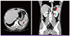 | Figure 1Contrast-enhanced computerized tomography (CT) of the abdomen revealed splenomegaly and a wedge-shaped low attenuated lesion in the upper pole of the spleen (red arrow), suggestive of splenic infarction.
|
With a clinical diagnosis of scrub typhus, we initiated treatment with 100 mg of oral doxycycline twice daily upon admission. The patient became afebrile in 48 hours and showed marked improvement in tenderness in the left upper quadrant of his abdomen. An indirect immunofluorescence (IF) test for Orientia tsutsugamushi indicated immunoglobulin G 1:160, and a nested polymerase chain reaction of the blood buffy coat was positive for an O. tsutsugamushi 56-kDa protein-encoding gene. A comparative analysis of the O. tsutsugamushi DNA sequence from the patient with those in the GenBank repository confirmed that he was infected with the Boryong genotype [7]. He was discharged on the fourth day of hospitalization and oral doxycycline was maintained for a total duration of 2 weeks. He was free of symptoms and showed no abdominal tenderness 1 week after discharge.
DISCUSSION
The main pathologic findings of scrub typhus are systemic vasculitis and perivasculitis. Cecilia and colleagues reported the target cells of O. tsutsugamushi to be endothelial cells in all major organs [8]. Endothelial dysfunction and disseminated intravascular coagulation (DIC) may occur in response to endothelial cell injury and severe immune reaction against O. tsutsugamushi has been reported in a patient with scrub typhus [9]. Corresponding to this pathologic finding, hemorrhage and infarction in various organs such as atraumatic hemoperitoneum [10], massive gastrointestinal bleeding [4], cerebral hemorrhage [11], acute myocardial infarction [12] and cerebral infarction [1113] have also been reported. In our presented case, the patient showed thrombocytopenia and increased level of d-dimer, yet coagulation tests including PT and aPTT were within normal range. Given that infarction was localized to the spleen and there were no clinical signs of disseminated thrombosis or bleeding, we concluded that thrombocytopenia was secondary to splenomegaly and increased level of d-dimer was due to endothelial injury and subsequent systemic vasculitis by O. tsutsugamushi rather than the manifestation of DIC.
To the best of our knowledge, only 4 clinical cases of splenic infarction due to scrub typhus have been reported [141516]. In all clinical cases reported, including our own, pain or tenderness in the left hypochondrial area showed improvement with doxycycline treatment. Moreover, follow-up ultrasound performed in 2 cases a few months after treatment revealed that the splenic infarct had disappeared or decreased, which suggests that the vascular complications of scrub typhus may be reversible with effective antibiotic therapy [14]. Follow-up CT scan was not performed in our patient since he was free of symptoms and showed no abdominal tenderness 1 week after discharge. Meanwhile, we planned follow-up IF test after he completed a total duration of 2 weeks of oral doxycycline, yet he was lost to follow-up on the next visit.
It is highly likely that splenic infarction has been clinically underdiagnosed in Korea considering 2 recent radiologic studies. They retrospectively reviewed CT images of patients with scrub typhus which were taken within 1 week after the manifestation of symptoms. Splenic infarcts were revealed in 3 of total 19 patients (16%) [17] and 6 of 94 patients (6.4%) in abdominal CT images [18]. Considering increasing incidence of scrub typhus and possibility of under-diagnosis of splenic infarction in Korea, it is important for clinicians to have a high degree of suspicion. Accordingly, in endemic areas, early radiologic evaluation should be considered in a patient of scrub typhus with abdominal symptoms or signs, and inversely, scrub typhus should be considered as one of the causes of splenic infarction. Moreover, public education through the media on the risk factors and the symptoms of scrub typhus is necessary because early diagnosis may not only prevent development of complications of scrub typhus, but also reverse the course of vascular complications with early treatment.
 XML Download