

 PDF
PDF ePub
ePub Citation
 Print
Print


Positron emission tomography/computerized tomography (PET/CT) is a functional image modality using its ability to metabolize glucose and concentrate specific molecules that have been labeled with a positron-emitting radionuculide. 18F-2-deoxy-2fluoro-D-glucose (18F-FDG), a glucose analog tagged with a positron-emitting isotope of fluorine, is taken up by metabolically active cells such as malignant cells. This metabolic activity is measured using standardized uptake value (SUV) and is expressed in images. This unique characteristic of 18F-FDG PET/CT may be used for differential diagnosis of fever of unknown origin (FUO). 18F-FDG PET/CT was helpful in approximately 50% of patients with FUO [123]. In a prospective study performed in Germany, 18F-FDG PET/CT was non-diagnostic for 43.3% (104/240) of patients with FUO or inflammation of unknown origin. These non-diagnostic findings included 30% (72/240) of false positive results that were non-specific uptakes [4]. However, prognosis, diagnostic and therapeutic strategies have not been known in patients with FUO whose 18F-FDG PET/CT finding is non-diagnostic. Hence, we described the outcome of patients with FUO that underwent 18F-FDG PET/CT.
This study was conducted in a tertiary hospital with 900 beds located in Bucheon, Korea. Institutional Review Board of the hospital approved this study. Patients with FUO that underwent 18F-FDG PET/CT were retrospectively identified from January 2016 - June 2017. Based on patient records, all adults were included that fulfilled a modified definition of FUO including (1) fever higher than 38.3ºC on several occasions (2) duration of fever for at least three weeks (3) uncertain diagnosis after three days of study in the hospital [5]. Complete blood cell count (including differential count), peripheral blood morphology, and routine blood work (lactic dehydgrogenase, bilirubin, and liver enzyme), urinalysis with microscopy, blood and urine culture, antinuclear antibodies, rheumatoid factor, human immunodeficiency virus antibody, chest radiography, and hepatitis serology were done as first-level work-up [6]. Enhanced computerized tomography and transthoracic echocardiography were done as second-level work-up. We recommended 18F-FDG PET/CT as a second-level work-up for patients with FUO when diagnosis was not confirmed after these tests were done. All patients fasted for at least six hours whose blood glucose level was less than 140 mg/dL before administration of 18F-FDG PET/CT. After intravenous injection of body weight-adapted 18F-FDG (4.4 MBq/kg), patients rested for 60 minutes. PET/CT images were acquired from mid-thigh to vertex of the skull. PET data were acquired by using of two-three minutes per bed position and field of view of 700 mm. CT was conducted by low dose non-enhanced and dose reducing protocol (100 KeV) and reconstructed with SAFIRE (Sinogram Affirmed Iterative Reconstruction) with slice thickness of 3 mm. Attenuation corrected axial PET images (4.07 × 4.07 × 3 mm) were reconstructed using 3D OSEM (two iterations, 21 subsets) with Siemens UltraHD PET. Scans were conducted using an integrated PET/CT system (Biograph mCT 128; Siemens Healthcare, Knoxville, TN, USA). A nuclear medicine physician analyzed PET/CT images under the knowledge of patient’s clinical history and other imaging studies. Data on demographics (age, sex), abnormal findings (laboratory, radiologic, and pathologic findings), treatment, final diagnosis, and outcome (resolution of symptoms) were searched. Results of 18F-FDG PET/CT were divided into four groups: (1) True negatives, 18F-FDG-PET/CT was normal without any underlying disease detected by other investigations, (2) True positives, 18F-FDG-PET/CT detected a specific disease process causing FUO which was then confirmed by additional investigations, (3) False negatives, 18F-FDG-PET/CT was normal but a specific disease process could be detected with another diagnostic test or response to specific treatment, (4) False positives, 18F-FDG-PET/CT showed tracer uptake that could not be identified as the cause of FUO by additional tests [4].
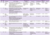
A total of eight patients with FUO who had complaints of non-specific symptoms such as fever, chills, myalgia, and headache underwent 18F-FDG PET/CT during the study period. Clinical characteristics, 18F-FDG PET/CT findings, and outcomes are summarized in Table 1. Except one patient (case No. 7) who was transferred to other hospital, all patients had high erythrocyte sedimentation rate (median, 120 sec; range, 3-120 sec) and C-reactive protein (median, 13.57 mg/dL; range, 0.13 – 21.16 mg/dL). The media duration from the first visit date to the day of 18F-FDG PET/CT test date was 12 days (range, 2 -29 days). 18F-FDG PET/CT was done in three (37.5%) patients one week after the first visit date. The following tests were conducted as second-level work-up: ultrasonography in four patients, spinal magnetic resonance in four patients, gastroscopy in three patients, colonoscopy in three patients, electromyography/nerve conduction velocity in two patients, bone scan in one patient, and bone marrow biopsy in one patient. Sural nerve biopsy, lymph node biopsy, and temporal artery biopsy were done according to 18F-FDG PET/CT findings. 18F-FDG PET/CT findings were categorized as true positives in two (25%) patients. These two patients were diagnosed with microscopic polyangitis and Kikuchi’s disease, respectively. Results of other six patients were categorized as false positives. After excluding one patient who was transferred to another hospital, for five patients whose diagnoses were not confirmed, their symptoms resolved during the follow-up.
Our descriptive study reveals that patients with FUO whose 18F-FDG PET/CT finding is non-diagnostic seems to be favorable. Previous studies have been limited to usefulness of 18F-FDG PET/CT as a diagnostic tool of FUO. There was no information on how to manage patients with non-diagnostic 18F-FDG PET/CT result in FUO. Even though 18F-FDG PET/CT is an expensive test, diagnosis is not confirmed in approximately half of patients [123]. Clinicians face major issues with these patients. These experiences cause clinicians to be reluctant to recommend 18F-FDG PET/CT to patients with FUO. 18F-FDG PET/CT can be a more useful test in FUO if prognosis of patients with non-diagnostic 18F-FDG PET/CT are anticipated. This first study revealing prognosis of patients with non-diagnostic 18F-FDG PET/CT findings might be valuable for establishing diagnostic and therapeutic strategies in FUO.
There are three possible paths for patients in whom FUO remains undiagnosed after extensive evaluation: early or late spontaneous resolution, delayed diagnosis, and mortality with sustained fever. In a previous report by Knockaert DC, et al. [7], about half (31/61) of undiagnosed FUO patients’ symptoms resolved spontaneous within 2 months after discharge. Ten (16.4%) patients became without symptoms during corticosteroid or non-steroidal anti-inflammatory drug usage. Delayed diagnosis was conducted with 12 (19.7%) patients. Of eight patients that died, deaths were related with FUO in two patients. Finally, in their report, mortality rate in patients with undiagnosed FUO followed up for five years or more was 3.2%. This favorable outcome of undiagnosed FUO after extensive evaluation was in accordance with results of our study.
Choosing 18F-FDG PET/CT as a second stage work-up might be useful for ruling out possible diagnosis in FUO due to its high sensitivity. A recent systemic review has reported 95.5% (107/112) of diagnostic yield from 18F-FDG PET/CT for diagnosing malignancy in FUO [1]. A recent study has reported that the sensitivity (79% vs. 45%) and clinical contribution (72% vs. 55%) of 18F-FDG PET/CT in diagnosing FUO are significantly higher than those of 67Ga SPECT/CT [8]. High sensitivity of 18F-FDG PET/CT has resulted in high false positive rate [48]. Our study also showed the presence of non-specific findings not leading to diagnosis in six (75%) patients. Clinical implication of false positive results of 18F-FDG PET/CT is currently unknown. Nonetheless, high sensitivity might be useful as a tool for decision making of FUO based on good prognosis in undiagnosed patients with FUO. Revest et al. have also insisted that symptoms of patients experiencing FUO with negative first-line investigations and negative 18F-FDG PET/CT almost always disappear spontaneously despite the lack of evidence [9].
In our study, only two (25%) patients had true positive finding in 18F-FDG PET/CT. Compared to previous studies on diagnostic usefulness of 18F-FDG PET/CT in FUO [123410], its contribution to diagnosis for FUO was low. A possible explanation is that different populations are used between the present study and previous studies. 18F-FDG PET/CT has high contribution rate in patients finally diagnosed as malignancy [111]. Based on these previous results, it can be assumed that contribution rate of 18F-FDG PET/CT in FUO might be higher in studies that included more patients with malignancy. In our study, no one had malignancy. This might have resulted in a low contribution rate of 18F-FDG PET/CT. Another possible explanation is the relatively short follow-up duration in our study. This might have led to the inclusion of more patients with spontaneous resolution but less patients with delayed diagnosis in the study. When more patients with spontaneous resolution are included, the diagnostic contribution of 18F-FDG PET/CT is lower. Although 18F-FDG PET/CT had a low contribution to diagnosis for FUO, it would be still valuable to evaluate its usefulness as “fast-tract” work-up for diagnosis of FUO. Analyzing the safety and cost-benefit of conducting 18F-FDG PET/CT as a second-level work-up that may replace other tests is necessary in the future.
This study has limitations. It is a small-size retrospective study. The number of enrolled patients are insufficient for conclusive results. In addition, owing to its retrospective nature, there is possibility of miss-categorization of FUO. Short-term follow-up period was also a significant limitation not to ensure outcome of enrolled patients. Finally, it was impossible to compare with patients who were not tested by 18F-FDG PET/CT, because our study was single-arm descriptive study.
In conclusion, outcome of patients with FUO whose 18F-FDG PET/CT finding is non-diagnostic seems to be favorable. Well-designed prospective trials should be conducted to establish 18F-FDG PET/CT-guided diagnostic/treatment strategies for FUO.
 XML Download