

 PDF
PDF ePub
ePub Citation
 Print
Print


Introduction
Non-tuberculous mycobacteria (NTM) are mycobacteria other than Mycobacterium tuberculosis complex and Mycobacterium leprae. NTM are ubiquitous organisms, which are frequently isolated from environmental sources, including surface water, tap water, and soil [12]. NTM disease is closely related to a host's immune status. The most common manifestation of human NTM infection is lung disease, especially in patients with bronchiectasis or chronic obstructive pulmonary disease. Disseminated infections can also develop in immunocompromised patients such as human immunodeficiency virus-infected patients [3]. Mycobacterium kansasii is one of the slow growing NTM species that constitutes only 2–4% of all NTM organisms isolated from clinical specimens; however, M. kansasii is the second most common species that cause NTM lung disease in the United States and Japan [34]. In Korea, M. kansasii is relatively uncommon, which accounts for only 2% of the NTM lung disease [56]. While M. kansasii usually involves the lungs, concomitant lymphadenopathy or pleural effusion have been rarely documented [7].
Resistant organisms, nonbacterial infections (i.e., fungal or viral), unresolved infection foci (i.e., abscess, catheter), and non-infectious fevers (i.e., drug fever or metabolic fever) are documented causes of persistent neutropenic fever [8]. Since NTM disease is a rare cause of neutropenic fever, the diagnosis can be often delayed in hematologic patients. The recommended treatment duration for pulmonary M. kansasii disease is at least 12 months if sputum cultures are negative [3]. However, the treatment duration should be individualized according to each patient's immune status and clinical course of NTM disease [3]. In patients with acute leukemia, the optimal duration of NTM treatment prior to stem cell transplantation (SCT) has not been documented.
Here, we describe the successful treatment of a case of M. kansasii pneumonia with necrotizing lymphadenitis mimicking invasive fungal pneumonia that developed in an acute myeloid leukemia (AML) patient. This study was approved by the Institutional Review Board of Seoul St. Mary's Hospital at the Catholic University of Korea with a waiver of informed consent (Subject number: KC15RISE0399).
Case Report
A 46-year-old man diagnosed with AML underwent induction chemotherapy with cytarabine 100 mg/m2 for 7 days and idarubicin 12 mg/m2 for 3 days. On day 10 after induction chemotherapy, neutropenic fever developed (axillary temperature of up to 38.3°C). No other symptoms were reported. Laboratory data showed a white blood cell (WBC) count of 150/mm3 (absolute neutrophil count [ANC] 0/mm3) and C-reactive protein (CRP) level of 0.33 mg/dL. Liver enzymes were within their normal ranges. Chest X-ray was normal and no definite infection focus was noted during the physical examination. Cefepime (2 g twice/day) and isepamicin (400 mg once/day) was started empirically and posaconazole prophylaxis (200 mg 3 times/day) was continued. On day 14, cefepime was changed to imipenem/cilastatin (500 mg 4 times/day) due to persistent neutropenic fever. On day 20, ANC was still 0/mm3, and fever persisted despite broad-spectrum antibiotics. Posaconazole prophylaxis was stopped and amphotericin B deoxycholate (1 mg/kg/day) was started.
On day 25, the patient had recovered from neutropenia (WBC 11,930/mm3; ANC 7,160/mm3), but the chest X-ray showed new infiltrates in the left hilar area. Low dose chest computed tomography (CT) was performed, which showed a 3.7 cm diameter, ill-defined, round consolidation with adjacent ground-glass opacity (Fig. 1A). Repeated serum galactomannan assays performed twice per week were consistently negative during the hospital stay. Although the pathogen had not yet been identified, it met the criteria for possible category of invasive fungal pneumonia as defined by the European Organization for the Research and Treatment of Cancer/Mycoses Study Group [9]. On day 33, the fever subsided, and amphotericin B deoxycholate was changed to oral itraconazole capsules (200 mg twice/day). The patient was discharged with itraconazole, which was maintained until next admission episode for consolidation chemotherapy. The patient's clinical course is presented in Figure 2.
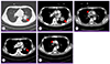 | Figure 1Radiologic findings of this case.
(A) Low dose chest computed tomography (CT), performed on day 24 after induction chemotherapy, shows ill-defined, round consolidation (3.7 cm diameter, arrow) in left hilar area, with adjacent linear opacities and ground glass opacities. (B, C) Follow-up enhanced chest CT, performed before consolidation chemotherapy, shows increased size of consolidation (3.7 cm to 4.1 cm diameter, arrow) and newly noted necrotizing right paratracheal lymphadenopathy (4.5 cm diameter, arrowhead). (D, E) Enhanced chest CT after 6 months of treatment for nontuberculous mycobacteria shows decreased size of left hilar lesion (arrow) and right paratracheal lymphadenopathy (arrowhead).
|
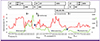 | Figure 2Schematic presentation of the patient's clinical course.
CIP, ciprofloxacin; FEP, cefepime; ISP, isepamicin; IPM, imipenem/cilastatin; CFP, cefoperazone; MEM, meropenem; PSC, posaconazole; AMB, amphotericin B, ITC, itraconazole; MIF, micafungin; INH, isoniazid; RIF, rifampicin; ETB, ethambutol; CT, computed tomography; CTx, chemotherapy; SCT, stem cell transplantation; Adm, admission; ANC, absolute neutrophil count.
|
Before starting consolidation chemotherapy, follow-up enhanced chest CT showed increased size of consolidation (3.7 cm → 4.1 cm diameter) with newly noted right paratracheal necrotizing lymphadenitis (4.5 cm diameter) (Fig. 1 B, C). Itraconazole was changed to amphotericin B deoxycholate (1 mg/kg/day), and a bronchoscopy was performed to identify the pathogen. Necrotic mucosal change was noted at a superior segment of left lower lobe (LLL) bronchus. Bronchial washing from LLL bronchus and a biopsy at the necrotic lesion was performed. The bacterial culture, fungus culture, concentration of acid-fast bacilli, and tuberculosis polymerase chain reaction (PCR) from the bronchial washing fluid were all negative. In the pathology specimen, chronic granulomatous inflammation without caseous necrosis was observed. The Ziehl-Neelsen stain and tissue PCR for M. tuberculosis were negative. The patient began receiving consolidation chemotherapy (cytarabine 2 g/m2 for 5 days and idarubicin 12 mg/m2 for 3 days) while continuing amphotericin B deoxycholate. Fifteen days after consolidation chemotherapy, the growth of NTM from the bronchial washing fluid was reported. The growth was confirmed as M. kansasii via PCR method (GenoType Mycobacterium CM assay, Hain Lifescience, Nehren, Germany). Therefore, oral isoniazid (300 mg/day), rifampicin (600 mg/day), and ethambutol (800 mg/day) were started. Approximately 2 months later, follow-up chest CT showed a decrease in the size of left hilar lesion (4.1 cm to 2.2 cm) and right paratracheal lymphadenopathy (4.5 cm to 3.5 cm).
After 70 days of anti-NTM therapy, allogeneic SCT was successively completed without acute complications. Antimicrobial susceptibility testing for M. kansasii revealed susceptibility to rifampicin. Anti-NTM medications were continued in addition to cyclosporine for immunosuppression after SCT. At post-SCT day 56, a blood chemistry test revealed an aspartate aminotransferase (AST) level of 180 IU/L, an alanine aminotransferase (ALT) of 207 IU/L, and a total bilirubin level of 5.49 mg/dL. The cyclosporine drug level was below 50 ng/mL. After changing rifampicin to rifabutin (450 mg/day), AST/ALT, and total bilirubin were normalized and cyclosporine drug levels were in therapeutic range.
During anti-NTM therapy, fever and respiratory symptoms were not noted. Chest CT performed 6 months post anti-NTM therapy showed interval regression (Fig. 1D, E). Furthermore, M. kansasii was not isolated from the respiratory specimen after starting NTM treatment. After 12 months of anti-NTM treatment, the patient achieved complete resolution of NTM diseases with clinical, radiologic, and microbiological improvement. He did not experience reactivation of the M. kansasii infection nor related complications. In addition, complications related to SCT, such as graft-versus-host disease or cytomegalovirus reactivation, were not reported.
Discussion
In this case report, M. kansasii disease was diagnosed during chemotherapy for AML. Initial CT findings indicated invasive fungal pneumonia, which may have resulted in a delayed diagnosis of NTM lung disease. After 70 days of treatment with isoniazid, rifampicin, and ethambutol, the patient successfully received SCT and recovered without any complications.
NTM is a very rare cause of fever in leukemic patients during chemotherapy [10]. M. kansasii is known to cause pneumonia, skin infection, or disseminated infection while it has not been commonly documented to cause lymphadenitis [71112]. Hilar or mediastinal lymphadenopathies have been found in only 8% of patients with M. kansasii lung disease, and the most common pulmonary radiographic findings are nodules and consolidation [6]. There have been several case reports of M. kansasii infection in hematologic patients; 6 cases in hairy cell leukemia patients, 1 in a chronic myelogenous leukemia patient, 1 in chronic lymphocytic leukemia patient, 1 in a follicular lymphoma patient, and 1 in a hemophagocytic syndrome patient [13141516]. Of these 10 cases, 6 cases were disseminated infection with lung involvement, 2 cases were pneumonia, and 2 cases were skin infection [13141516].
The American Thoracic Society guidelines for the treatment of NTM disease caused by M. kansasii recommend a 3-drug regimen of rifampicin, isoniazid, and ethambutol [3]. However, few studies have examined the optimal management for M. kansasii in hematologic patients who need repetitive chemotherapy or SCT. In addition, the optimal duration of anti-NTM medication, both prior to SCT and post-SCT, has not been documented for patients with leukemia. One study has reported that SCT administered after 100 days of anti-TB medication may be feasible and safe for treating tuberculosis in patients with various hematologic diseases [17].
In this case, the patient had left hilar consolidation and right mediastinal lymphadenitis. However, bronchoscopic biopsy was performed at the necrotic lesion of left bronchus alone. Our patient's radiologic finding was not the typical finding of M. kansasii lung disease described in the ATS/IDSA guidelines, but there are reports that M. kansasii lung disease presented as consolidation as well as nodules, cavitation, or bronchiectasis [36]. Based on the positive culture for NTM from bronchial washing fluid, and granuloma from the pathology specimen of left bronchus, the patient could be diagnosed as NTM lung disease [3]. However. transbronchial aspiration of right mediastinal necrotic LN should have been considered to differentiate other concomitant causes, such as fungus, M. tuberculosis, or other rare pathogens. The patient suffered from neutropenic fever before anti-NTM diagnosis. However, the clinical symptoms and radiological findings dramatically improved after 2 months of treatment with a 3-drug NTM regimen. In addition, interferon-γ release assays (IGRA), which cannot be used for diagnosis of tuberculosis, was negative in our patients. Negative predictive value for pulmonary tuberculosis of IGRA is known to be 91% ~ 99% [1819]. Repetitive aspergillic antigen tests were also negative in this case. Our patient initially received rifampicin; however, this was changed to rifabutin due to the metabolic interaction in the cytochrome P-450 system [20].
To our knowledge, this is the first report of an M. kansasii infection that developed before SCT in an AML patient. Clinicians should be aware that the active diagnostic efforts for pathological or microbiological confirmation could be essential during neutropenia, especially in cases with persistent neutropenic fever. In conclusion, we present here a case of pneumonia with necrotizing lymphadenitis caused by M. kansasii that developed during the period of febrile neutropenia after chemotherapy for AML. Although the initial lesion looked like invasive fungal pneumonia, the clinical presentation differed. The patient recovered from NTM without reactivation and experienced no complications related to SCT. This case demonstrates the guidance of scheduling SCT in addition to simultaneous NTM treatment. However, additional clinical experience and prospective studies are necessary to determine the optimal treatment of M. kansasii-associated NTM disease during chemotherapy or SCT.
 XML Download