Journal List > Infect Chemother > v.49(4) > 1035482

Sim, Lee, Park, Kim, Choi, Choi, Kim, Sohn, Ahn, and Yoon: Infective Endocarditis Presenting as Endogenous Endophthalmitis Secondary to Streptococcus agalactiae in a Healthy Adult: Case Report and Literature Review

Abstract

Endogenous endophthalmitis secondary to group B Streptococcus (GBS) is extremely rare, particularly in healthy adults. However, the visual prognosis is poor. We report the first South Korean case of GBS infective endocarditis presenting as endogenous endophthalmitis and skin and soft tissue infection. Cultures of blood, vitreous humor, and pus from skin aspirates yielded a penicillin-susceptible serotype V strain of Streptococcus agalactiae. After 6 weeks, the patient completely recovered from GBS infective endocarditis. However, despite early antibiotic treatment and early surgical intervention, the patient's right eye developed phthisis bulbi and was a candidate for evisceration.

INTRODUCTION

Streptococcus agalactiae, a group B Streptococcus (GBS), is part of the normal flora of the skin, throat, lower gastrointestinal tract, and female genital tract. It is recognized as a major human pathogen in neonatal sepsis and postpartum infection. However, the incidence of invasive infection by S. agalactiae has increased in recent years in non-pregnant adult patients, elderly patients, and patients with chronic immunosuppressive diseases [1]. In particular, GBS endogenous endophthalmitis is an extremely rare but visually devastating disease [234]. The clinical profile of GBS endophthalmitis is not well characterized because little literature is available. Here, we report the first South Korean case without evidence of immunosuppression, presenting as endophthalmitis complicating GBS infective endocarditis. Furthermore, we describe the clinical, prognostic, and therapeutic characteristics of 22 patients with this infection in the literature.

CASE REPORT

A previously healthy 43-year-old woman visited our emergency room complaining of a 10-hour history of decreased visual acuity with ocular pain in the right eye. She was taking oral antibiotics (cefaclor) for a skin abscess of the right buttock area, which developed 2 weeks earlier. She also underwent dental scaling 3 months earlier. She underwent a bilateral laser in-situ keratomileusis (LASIK) procedure 10 years earlier, and her final corrected visual acuity was 20/20 in both eyes.
On admission, she had a blood pressure of 120/60 mmHg, heart rate of 88 beats/min, body temperature of 37.9°C, and respiratory rate of 20/min. Physical examination did not reveal a cardiac murmur. No Osler's nodes, Janeway lesions, or splinter hemorrhages were observed. There was a 3-cm skin swelling with purulent discharge on the right buttock area that was tender and erythematous. The results of the initial laboratory investigations were normal, except for a leukocyte count of 13.9 × 10³/mm3, with 74% neutrophils and a C-reactive protein level of 26.7 mg/dL. Ophthalmic examination revealed an initial visual acuity of 20/100 and 20/20 in the right and left eye, respectively. There was full ocular motility. Slit-lamp examination showed severe conjunctival injection and chemosis, total corneal epithelial defect, corneal edema, and keratin precipitate on the corneal endothelium. The anterior chamber was poorly visible because of keratin precipitate and intense fibrinous reaction. Intraocular pressure was 25 and 10 mmHg using Goldman applanation tonometry. The right fundus was poorly visible, and ultrasonography revealed the presence of vitritis. The results of external, slit-lamp, and fundus examinations of the left eye were normal. Clinical samples of blood, the aqueous humor of the right eye, and discharge from the buttock lesion were obtained on admission. Gram-positive cocci in chains was cultured from all of these samples, and the patient was diagnosed with endogenous endophthalmitis of the right eye as well as a skin and soft tissue infection on the right buttock secondary to streptococcal sepsis. Initial transthoracic echocardiography was normal. For the first 12 days of hospitalization, before infective endocarditis was confirmed, intravenous ceftriaxone (2 g/day) was prescribed as an empirical and definite antibiotic therapy with a regimen of intravitreal vancomycin (1 mg/0.1 mL) and ceftazidime (2.25 mg/0.1 mL).
Despite intravitreal vancomycin and ceftazidime injection, her visual acuity rapidly deteriorated to light perception within a day. On the third day of hospitalization, she underwent right core vitrectomy, which revealed a pus-filled vitreous cavity. Culture of the vitreous humor was performed. Group B β-hemolytic streptococci (S. agalactiae) was identified as the causative organism in the vitreous specimen, blood, and skin pus of the right buttock, using a VITEK II system (bioMérieux, Hazelwood, MO, USA), with a probability of 99.0%. The GBS was highly susceptible to penicillin, vancomycin, and cephalosporins according to the Clinical Laboratory Standards Institute (CLSI) guidelines. Ten days after initial transthoracic echocardiography, transesophageal echocardiography revealed a 5-mm vegetation to the anterior mitral leaflet. The patient was finally diagnosed with an invasive GBS infection with infective endocarditis presenting with endogenous endophthalmitis and a skin and soft tissue infection. A MicroScan MICroSTREP plus panel was used to determine antimicrobial susceptibility and measure minimum inhibitory concentrations. The results showed susceptibility to penicillin (0.06 μg/mL), ampicillin (0.12 μg/mL), ceftriaxone (0.25 μg/mL), cefotaxime (0.25 μg/mL), cefepime (0.25 μg/mL), meropenem (0.12 μg/mL), azithromycin (0.25 μg/mL), clindamycin (0.06 μg/mL), vancomycin (0.5 μg/mL), and levofloxacin (0.5 μg/mL). To further confirm the identity of this isolate, sequencing of a 438-base pair 16S rRNA gene fragment according to CLSI recommendations revealed a 97% identity match to S. agalactiae strain sequence type 1 (SS1) associated with serotype V (accession number: CP010867.1). According to the clinical guidelines under the clinical diagnosis of infective endocarditis, intravenous ampicillin (3 g q6 hrs) and gentamicin (5.1 mg/kg q24 hrs) were administered for 28 and 14 days, respectively, with intravitreal antibiotic therapy [5]. The patient was discharged after completing the 28-day intravenous ampicillin treatment. At the 6-month follow-up visit after the completion of antibiotic therapy, the patient remained free of complications from infective endocarditis or skin and soft tissue infection. She had no infectious signs on her right eye but had lost perception to light, and phthisis bulbi had developed.

DISCUSSION

Endophthalmitis is defined as an internal infection of the eye involving the vitreous or aqueous humor that can be classified as either endogenous or exogenous depending on the route of infection. Exogenous bacterial endophthalmitis occurs most commonly, generally after penetrating ocular trauma, intraocular surgery, or a corneal ulcer. However, endogenous endophthalmitis, also known as metastatic endophthalmitis, can arise from hematogenous spread of bacteremia or other remote septic foci. Endogenous endophthalmitis accounts for only 5–7% of endophthalmitis cases and has a high mortality rate of up to 29% with poor visual acuity outcomes [6]; it is associated with underlying medical conditions such as diabetes or cardiac diseases and malignancy in up to 90% of patients [7]. However, the present endogenous endophthalmitis patient was a middle-aged immunocompetent woman with no evidence of underlying disease.
Systemic infections such as liver abscesses, meningitis, and infective endocarditis may subsequently develop endogenous endophthalmitis. The present patient was diagnosed with infective endocarditis. However, this was likely due to bacteremia rather than the source of sepsis. Considering her oral antibiotic treatment for a skin abscess on the right buttock without systemic symptoms for 2 weeks, the skin and soft tissue infection may be regarded as the primary infection focus that caused GBS bacteremia complicated with infective endocarditis. Although the patient underwent dental scaling prior to infection, there was no evidence that dental scaling increased her risk of infective endocarditis.
Infective endocarditis caused by S. agalactiae is an uncommon but aggressive disease with a high mortality rate of ~40% [8]. It is generally characterized by an acute onset, the presence of large vegetations, rapid valvular destruction, and frequent development of complications. Therefore, it is considered very different from infective endocarditis caused by other Streptococci. However, there is limited information regarding metastatic endophthalmitis.
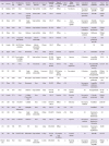
Endogenous endophthalmitis caused by GBS is very rare in non-pregnant adults, accounting for only 5.2% of bacterial endogenous endophthalmitis cases, only 21 of which have been reported in the literature worldwide [2 910111213141516171819]. In contrast to the present case, 18 (85.7%) of these 21 cases had predisposing factors such as old age, diabetes mellitus, cardiac diseases, malignant diseases, and immunosuppression (Table 1). Including the present case, the most common septic focus was skin and soft tissue infection (n = 5, 22.7%) followed by septic arthritis (n = 3; 13.6%), urinary tract infection (n = 3; 13.6%), central nervous system infection (n = 2; 9.0%), pharyngitis (n = 2; 9.0%), pneumonia (n = 2; 9.0%), or no evident foci of infection (n = 5; 22.7%). Among them, 5 (18.1%) cases exhibited infective endocarditis, but none exhibited vegetation in the initial transthoracic echocardiography. The present case exhibited cardiac vegetation in the transesophageal echocardiography 7 days after the initial transthoracic echocardiography. Therefore, transesophageal echocardiography should be recommended for patients with endogenous endophthalmitis caused by GBS. Septic arthritis (n = 10, 45.4%) was one of the most common manifestations in previously reported cases of endophthalmitis secondary to GBS; this may be because of the role of the synovial space and vitreous humor as culture media for facilitating bacterial seeding, entrapment, and proliferation [11].
Table 1

Clinical characteristics and treatment outcomes of 22 cases of endogenous endophthalmitis due to Streptococcus agalactiae

ic-49-286-i001
Ref Country Sex/age Predisposing illness Infection source Other sites of infection Time to eye signs Laterality & visual acuity Type of endophthalmitis Culture (B/V) Other culturepositive sites Vitrectomy Intravitreal antibiotics Systemic antibiotics Visual outcome Serotype
[2] Malaysia M/55 None Foot abscess Septic arthritis 2 weeks OS-LP Diffuse +/− Foot abscess + Vancomycin, gentamicin Vancomycin, penicillin Phthisis bulbi
[2] China M/67 None Cervical epidural abscess Osteomyelitis 1 week OS-CF Posterior +/+ + Vancomycin, gentamicin Penicillin Phthisis bulbi
[2] China M/53 DM Septic arthritis Septic arthritis 5 days OD-LP Diffuse +/+ Knee synovial aspirate Vancomycin, ceftazidime Penicillin, gentamicin Evisceration
[2] China M/82 DM Unknown Septic arthritis Concurrent OD-NLP Diffuse +/+ Vancomycin, ceftazidime Penicillin, ciprofloxacin Phthisis bulbi
[2] China F/63 None Unknown Septic arthritis 2 days OD-LP Diffuse +/+ Vancomycin, amikacin Ceftriaxone Phthisis bulbi
[3] USA M/42 Congenital heart disease Pharyngitis Infective endocarditis 10 days OD-LP Posterior +/NA NA Cefazolin, gentamicin, penicillin Died
[4] Canada F/80 AF, HTN, Knee prosthesis Unknown Infective endocarditis, meningitis 12 hours OS-LP Diffuse +/+ CSF NA NA Died
[9] Japan M/83 None Pneumonia None 4 days OD-LP, OS-NLP Posterior −/+ Aqueous NA Ceftazidime Enucleated
[10] UK M/55 Renal canaliculi, multiple myeloma UTI Septic arthritis Concurrent OD-HM, OS-6/36 Posterior, diffuse +/+ Aqueous Cefuroxime Amoxicillin, gentamicin LP, 6/6 Ib
[10] UK F/76 None UTI Pneumonia 2 days OD-LP, OS-NLP OS-Pan +/NA Urine, conjunctiva NA Benzylpenicillin, gentamicin, acyclovir LP, NLP Ib/c
[10] UK M/65 DM Septic arthritis Infective endocarditis Concurrent OD-LP, OS-6/5 OD-Posterior diffuse, OS-Focal +/+ Kneeaspirate Cefuroxime Amoxicillin, gentamicin, Benzylpenicillin Phthisis bulbi, 6/5 Ib
[10] UK M/60 None Pharyngitis Septic arthritis Concurrent OS-6/60 Pan +/NA NA Benzylpenicillin, gentamicin Evisceration Ia/c
[11] China F/95 None Pneumonia None 3 days OS-NLP Diffuse +/NA + Vancomycin, ceftazidime Ampicillin/sulbactam, vancomycin OU-NLP
[12] UK F/70 None Unknown Septic arthritis 4 days OD-6/18, OS-6/36 NA +/NA Synovial aspirate NA Benzylpenicillin OU-Recovery V
[13] Canada F/74 None Cellulitis, Chronic foot ulcer None 2 days OS-LP Posterior NA/+ Foot ulcer pus + Vancomycin, amikacin Cefazolin, gentamicin Phthisis bulbi
[14] USA F/62 DM, HTN Cellulitis None 1 weeks OD-20/70, OS-LP Diffuse +/+ Foot tissue Vancomycin, amikacin Penicillin G LP, 20/100
[14] USA M/48 HIV, Splenectomy Meningitis None 2 day OD-CF Pan +/NA CSF Penicillin G NLP
[15] USA F/75 None Septic arthritis Infective endocarditis 20 days OD-20/70 Posterior +/NA Urine NA Vancomycin, ceftriaxone, Clindamycin NA
[16] USA M/56 Leukemia Unknown None 3 days OD-LP Diffuse +/+ Aqueous + Vancomycin, amikacin Vancomycin, clindamycin. Cefotaxime LP
[17] USA F/81 HTN UTI None 3 days OD-20/400 Diffuse +/+ + Vancomycin Vancomycin, amikacin, amphotericin 20/200
[18] USA F/61 DM, HTN, MI Endarteritis Septic arthritis 12 days OU-Not sited. OD-Anterior, focal OS-Diffuse +/+ Synovial aspirate + NA Penicillin OS-NLP
Present case Korea F/43 None Cellulitis Infective endocarditis 2 weeks OD-20/100 Pan +/+ Pus of hip abscess + Vancomycin, ceftazidime Ceftriaxone, gentamycin, ampicillin Phthisis bulbi V
Ref, reference; B, blood culture; V, vitreous culture; M, male; OS, left eye; LP, perception of light; CF, counting fingers; DM, diabetes mellitus; OD, right eye; NLP, no perception of light; F; female; NA, net applicable; AF, atrial fibrillation; HTN, hypertension; CSF, cerebrospinal fluid; UTI, urinary tract infection; HM, hand motion; OU, both eye; HIV, human immunodeficiency virus; MI, myocardial infarction.
Previous cases of endophthalmitis secondary to GBS exhibited dramatic loss of vision within a few hours to a few days; this rapid progression is associated with the virulence of GBS. Hemolytin and cytolysin produced by GBS may result in the development of the necrotic vitreous cavity, total retinal detachment, and microabscesses on the choroid [2]; this eventually leads to poor visual prognosis despite prompt antibiotic therapy and surgical intervention such as in the present case.
S. agalactiae is classified as serotype Ia/c, Ia/b, II, III, IV, V, VI, VII, or VIII. GBS serotypes Ia, III, and V account for more than two-thirds of cases [20]. The present patient had serotype V, which accounts for >25% of cases of invasive GBS infection [20].
The most important component in the treatment of endogenous endophthalmitis caused by GBS is prompt and appropriate systemic antibiotic therapy. Unlike exogenous endophthalmitis with a relatively intact blood–ocular barrier, endogenous endophthalmitis has a disrupted barrier due to the transmural passage of the hematogenously spreading organism [19]. Consequently, intravenous antibiotics can easily reach the ocular tissue with endogenous endophthalmitis and the primary infection site. On the contrary, intravitreal antibiotic injection has not been shown to be an effective treatment for endogenous endophthalmitis. However, it may be reasonable to select intravitreal antibiotics on a case-by-case basis, because the existing negative perception could be a consequence of selection bias [19]. Therapeutic vitrectomy can be 3 times more beneficial in retaining useful vision and decreasing the requirement of evisceration or enucleation [19]. However, the advantages of vitrectomy can be limited, because GBS causes rapid and extensive destruction of ocular structure in the early stage of infection, such as in the present case. In the literature, of the 9 eyes that underwent vitrectomy, 7 developed phthisis bulbi and loss of light perception. Although most patients recovered from GBS sepsis (except 2 cases of death; fatality rate: 9.0%), the visual outcome is poor in most patients. Among 30 cases, 22 involved the eyes, in which only 7 eyes recovered useful vision while the others had either only light perception or no light perception and phthisis bulbi.
In conclusion, the present case suggests GBS infections should be included in the differential diagnosis of endogenous endophthalmitis in healthy adults. The present case also highlights the requirement of a high index of suspicion for infective endocarditis in patients despite normal results from initial transthoracic echocardiography. Aggressive antibiotics and surgical therapy may be needed in these cases because of rapid and extensive ocular destruction in the early stage of infection.

Notes

Conflict of Interest No conflicts of interest.

References

1. Skoff TH, Farley MM, Petit S, Craig AS, Schaffner W, Gershman K, Harrison LH, Lynfield R, Mohle-Boetani J, Zansky S, Albanese BA, Stefonek K, Zell ER, Jackson D, Thompson T, Schrag SJ. Increasing burden of invasive group B streptococcal disease in nonpregnant adults, 1990-2007. Clin Infect Dis. 2009; 49:85–92.
crossref
2. Lee SY, Chee SP, Group B. Streptococcus endogenous endophthalmitis: case reports and review of the literature. Ophthalmology. 2002; 109:1879–1886.
3. Farber BP, Weinbaum DL, Dummer JS. Metastatic bacterial endophthalmitis. Arch Intern Med. 1985; 145:62–64.
crossref
4. Buglass TD, Romanchuk KG. Fatal case of group B streptococcal endogenous endophthalmitis. Can J Ophthalmol. 1995; 30:149–150.
5. The Korean Society of Infectious Diseases, Korean Society for Chemotherapy, The Korean Society of Clinical Microbiology, The Korean Society of Cardiology, The Korean Society for Thoracic and Cardiovascular Surgery. Clinical guideline for the diagnosis and treatment of cardiovascular infections. Infect Chemother. 2011; 43:129–177.
6. Schiedler V, Scott IU, Flynn HW Jr, Davis JL, Benz MS, Miller D. Culture-proven endogenous endophthalmitis: clinical features and visual acuity outcomes. Am J Ophthalmol. 2004; 137:725–731.
crossref
7. Okada AA, Johnson RP, Liles WC, D'Amico DJ, Baker AS. Endogenous bacterial endophthalmitis. Report of a ten-year retrospective study. Ophthalmology. 1994; 101:832–838.
8. Sambola A, Miro JM, Tornos MP, Almirante B, Moreno-Torrico A, Gurgui M, Martinez E, Del Rio A, Azqueta M, Marco F, Gatell JM. Streptococcus agalactiae infective endocarditis: analysis of 30 cases and review of the literature, 1962-1998. Clin Infect Dis. 2002; 34:1576–1584.
crossref
9. Matsuo K, Nakatuka K, Yano Y, Fujishima W, Kashima K. Group B streptococcal metastatic endophthalmitis in an elderly man without predisposing illness. Jpn J Ophthalmol. 1998; 42:304–307.
crossref
10. O'Brart DP, Eykyn SJ. Septicaemic infection with group B streptococci presenting with endophthalmitis in adults. Eye (Lond). 1992; 6:396–399.
11. Wu Z, Huang J, Huynh S, Sadda S. Bilateral endogenous endophthalmitis secondary to group B streptococcal sepsis. Chin Med J (Engl). 2014; 127:1999.
12. Galloway A, Deighton CM, Deady J, Marticorena IF, Efstratiou A. Type V group B streptococcal septicaemia with bilateral endophthalmitis and septic arthritis. Lancet. 1993; 341:960–961.
crossref
13. Ing EB, Erasmus MJ, Chisholm LD. Metastatic group B streptococcal endophthalmitis from a cutaneous foot ulcer. Can J Ophthalmol. 1993; 28:238–240.
14. Nagelberg HP, Petashnick DE, To KW, Woodcome HA Jr. Group B streptococcal metastatic endophthalmitis. Am J Ophthalmol. 1994; 117:498–500.
crossref
15. Pokharel D, Doan AP, Lee AG. Group B streptococcus endogenous endophthalmitis presenting as septic arthritis and a homonymous hemianopsia due to embolic stroke. Am J Ophthalmol. 2004; 138:300–302.
crossref
16. Gupta SR, Agnani S, Tehrani S, Yeh S, Lauer AK, Suhler EB. Endogenous Streptococcus agalactiae (Group B Streptococcus) endophthalmitis as a presenting sign of precursor T-cell lymphoblastic leukemia. Arch Ophthalmol. 2010; 128:384–385.
crossref
17. Saffra N, Rakhamimov A, Husney R, Ghitan M. Streptococcus agalactiae endogenous endophthalmitis. BMJ Case Rep. 2013; 2013:pii:bcr2013008981.
18. Siddiqui MA, Lester RM. Septic arthritis and bilateral endogenousendophthalmitis associated with percutaneous transluminal coronary angioplasty. J Am Geriatr Soc. 1996; 44:476–477.
crossref
19. Jackson TL, Eykyn SJ, Graham EM, Stanford MR. Endogenous bacterial endophthalmitis: a 17-year prospective series and review of 267 reported cases. Surv Ophthalmol. 2003; 48:403–423.
crossref
20. Edwards MS, Baker CJ. Group B streptococcal infections in elderly adults. Clin Infect Dis. 2005; 41:839–847.
crossref
TOOLS
ORCID iDs

Yu Ra Sim
https://orcid.org/0000-0003-3330-5807

Young Kyung Yoon
https://orcid.org/0000-0001-8435-935X

Similar articles