Abstract
Primary sternal osteomyelitis (PSO) is a rare condition that may develop without any contiguous focus of infection. Due to the rarity of the disease, early diagnosis and appropriate treatment are often delayed. Herein, we describe a patient with PSO caused by Staphylococcus aureus that presented with chest pain and fever. The patient had no predisposing factors for sternal osteomyelitis. The chest pain was thought to be non-cardiogenic, as electrocardiography and cardiac enzyme did not reveal ischemic changes when he visited the emergency room. After blood culture revealed the presence of S. aureus, every effort was made to identify the primary focus of infection. Bone scan and magnetic resonance imaging revealed osteomyelitis with soft tissue inflammation around the sternum. After 8 weeks of antibiotics treatment, the patient recovered without any complications.
Sternal osteomyelitis usually occurs after cardiac surgery or chest trauma and is called secondary sternal osteomyelitis [1]. In contrast, primary sternal osteomyelitis (PSO) has no contiguous focus of infection, which is uncommon; it occurs in intravenous drug abusers and patients with diabetes or those infected with human immunodeficiency virus (HIV), which predisposes individuals to infection [2]. Without any predisposing factors, it is difficult to suspect sternal osteomyelitis with merely symptoms, such as fever or chest pain [3]. This can lead to a delay in the exact diagnosis of sternal osteomyelitis, finally resulting in sequelae after treatment with inappropriate antibiotics [4].
In this study, we report a patient with chest pain and fever who was finally diagnosed as a PSO with Staphylococcus aureus bacteremia, an extremely rare condition among immunocompetent adults without any predisposing factors.
A 69-year-old man visited the emergency room (ER) due to a 3-day history of chest pain. Three days prior, the pain was dull on the substernal area and developed during resting; it was aggravated by cough or positional change. On the presenting day, it turned into a stabbing pain for 5 hours and fever developed. He had been taking amlodipine and enalapril for hypertension for 2 years. He had no specific familial or recent travel history. He denied recent chest trauma and any invasive procedure, including acupuncture. He was acutely ill-looking in appearance. On physical examination at the ER, blood pressure was 170/88 mmHg, heart rate was 95 beats/min, and body temperature was 38.5℃. Laboratory findings were as follows: white blood cells (WBC) count of 10.38×10⁹/mm3 (neutrophils, 65.6%), hemoglobin of 14.5 g/dL, and platelet count of 229×109/ mm3. Erythrocyte sedimentation rate (ESR) was 35 mm/h (normal range, 0-10 mm/h), high sensitive C-reactive protein (hsCRP) was 21.51 mg/L (normal range, 0.1-5.0 mg/L) and procalcitonin was 0.046 ng/mL (normal range, 0-0.046 ng/mL). Blood chemistry showed that aspartate aminotransferase/alanine aminotransferase was 23/20 IU/L, total protein/albumin was 8.0/4.52 g/dL, creatine phosphokinase/lactate dehydrogenase was 160/430 IU/L, and blood urea nitrogen/creatinine was 11.5/0.92 mg/dL. Troponin-I was 0.1 ng/mL (normal range, 0.0-0.3 ng/mL) and CK-MB 1.02 ng/mL (normal range, 0-5 ng/mL). Chest X-ray was normal. Electrocardiography (EKG) showed normal sinus rhythm with no evidence of ischemic change. After a blood culture was performed, the patient was discharged by an emergency physician with cefditoren (100 mg three times per day) as an empirical antibiotic treatment for fever and acetaminophen (300 mg three times per day).
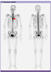
Three days later, he returned to an outpatient clinic for follow-up. He was still complaining of chest pain, but fever had subsided. Physical examination revealed that blood pressure was 150/80 mmHg, heart rate was 80 beats/min, and body temperature was 36.3℃. Grade 3 systolic murmur was auscultated on the second intercostal space that was accentuated in sitting position. Although direct tenderness at the sternomanubrial angle area was observed, there was no redness and swelling on the anterior chest wall. Additionally, there was no skin rash, such as petechia, splinter hemorrhage, and anything suggestive of Osler’s node. He denied any intravenous drug use. The blood cultures performed at the ER revealed methicillin-susceptible S. aureus (MSSA, 4 out of 4 bottles) by using the VITEK II (bioMérieux, Marcy l’Étoile , France) system. The patient was hospitalized promptly and was treated with intravenous nafcillin (2 g every 4 hours) and gentamicin (1 mg/kg every 8 hours, for initial 5 days) after blood culture. On admission, laboratory findings were as follows: WBC count was 5.68×10⁹/L (neutrophils, 63.5%), hemoglobin was 13.5 g/dL, and platelet count was 193×109/L. ESR increased to 50 mm/h and hsCRP increased to 84.55 mg/L; procalcitonin was 0.229 ng/mL. Antibody testing for HIV and hepatitis B and C returned negative results. EKG showed no interval change. Transthoracic echocardiography (TTE) showed a high density round structure in the non-coronary cusp of the aortic valve (AV). For further evaluation of the AV lesion, transesophageal echocardiography (TEE) was performed on the second hospital day (HD) which revealed that thickening of the AV seemed to be AV sclerosis rather than AV vegetation. We planned to perform follow up echocardiography to investigate the change in the AV lesion after antibiotic therapy. On the third HD, Tc99m bone scan was performed to detect any metastatic septic lesion. Increased uptake at the sternomanubrial junction was observed, which is compatible with inflammatory change (Figure 1). Sternum series showed subtle sclerosis along the manubriosternal junction, probably representing degenerative change and/or inflammation (Figure 2A). Chest magnetic resonance imaging (MRI) on the 7th HD showed an ill-defined high signal intensity in the bone marrow of sternomanubrial joint with extension to surrounding soft tissue (Figure 2B). Blood culture on the day of admission revealed MSSA (3 out of 4 bottles), and the blood culture result was negative after 3 days of nafcillin treatment. The TTE performed on the 14th HD did not show any interval change in the AV thickness. However, chest pain and tenderness on the sternum gradually subsided with the use of intravenous antibiotics, and then disappeared on the 8th HD. We considered that this patient had PSO caused by MSSA without distal seeding rather than MSSA bacteremia with infective endocarditis. After excluding the possibility of infective endocarditis, we changed nafcillin to oral levofloxacin (500 mg once daily) on the 17th HD, and he was discharged. In total, he completed 8 weeks of susceptible antibiotic therapy without any complications or relapse.
PSO is a rare condition that accounts for 0.3% of all osteomyelitis cases reported in the literature [256]. PSO typically occurs secondary to predisposing factors, such as immunodeficiency, intravenous drug abuse, subclavian vein catheterization, or liver cirrhosis [2]. The most common bacterial cause of PSO is S. aureus, although Pseudomonas aeruginosa is predominant among intravenous drug abusers [78]. Salmonella typhi, S. epidermidis, Nocardia nova, Mycobacterium tuberculosis, and even Aspergillus spp. have also been reported as the causes of PSO [6910111213]. In Korea, three cases of PSO have been reported; these were caused by S. aureus, Actinomyces israelii, and an unidentified organism [91415].
PSO usually presents with chest pain, fever, chills, a painful chest mass, redness, tenderness, and swelling [4]. However, these symptoms are non-specific and may not always be present. Owing to the rarity of the disease and its non-specific symptoms, early diagnosis and appropriate treatment are often delayed [1]. Since our patient did not have any mass-like lesion, redness, or swelling in the sternal area, we had to differentiate between cardiogenic and non-cardiogenic causes of chest pain. We were not sure whether our patient had a systolic murmur when he visited the ER. Clinicians should consider infectious causes of chest pain when symptoms, such as fever, cardiac murmur, and elevated WBC count are combined. The differential diagnosis should include skin and soft tissue infections, including cellulitis and shingles; musculoskeletal sources of chest pain, such as costochondritis and fibromyalgia; pericarditis, myocardial infarction, valvular abnormalities and cardiomyopathy; benign soft tissue tumors; and bone sarcomas [35].
In addition, the diagnosis of PSO encompasses a combination of clinical information, radiologic imaging and microbiological test results, and other laboratory findings. Leukocytosis and elevated C-reactive protein (CRP) may be helpful in making a diagnosis. Conventional radiography is a basic, inexpensive, and readily available modality, although it is not sensitive and the results may be limited. Clinicians should be aware that radiographic abnormalities are usually observed 10 to 21 days after the onset of infection [16]. Although bone scans are more expensive than plain radiographs and are sometimes nonspecific, they are sensitive and can help detect disseminated lesions. Technetium Tc99 methylene disphyrophosphate (DPD) bone scans and gallium citrate Ga67 and indium In111 labeled white blood cells are commonly used. Computed tomography (CT) and magnetic resonance imaging (MRI) are now considered a standard for the diagnosis of osteomyelitis [16]. These techniques are very sensitive but lack specificity. A bone biopsy can provide the definitive diagnosis and is recommended when feasible [7]. We did not perform a bone biopsy because our patient did not have any mass-like lesions in the sternal area and showed clinical improvement after targeted antibiotic therapy to the isolated organism.
The treatment of sternal osteomyelitis requires identification of the causative microorganism based on blood culture or direct bone biopsy [917]. Although antibiotics are the mainstay of PSO treatment, early surgical intervention can provide definitive treatment resulting in cure and decreased morbidity [56]. In particular, surgical intervention may be necessary for P. aeruginosa infection, in cases where antibiotic treatment has failed, or when abscesses, foreign material, or extensive bone necrosis are present.
In conclusion, PSO is a rare condition that is difficult to diagnose early in immunocompetent patients without apparent predisposing factors. To the best of our knowledge, this is the fourth case of PSO reported in Korea. The patient was immunocompetent without any risk factors for infection, and mild fever and chest pain were the only symptoms; thus, PSO was not suspected at the time of admission. This case provides an important lesson. In patients presenting with chest pain, a thorough history should be obtained and a careful physical examination should be carried out to differentiate cardiogenic and non-cardiogenic origins. It is necessary to consider infectious causes of chest pains presenting with fever.
References
1. Platt MA, Ziegler K. Primary sternal osteomyelitis with bacteremia and distal seeding. J Emerg Med. 2012; 43:e93–e95.
2. Mofredj A, Guerin JM, Leibinger F, Masmoudi R. Primary sternal osteomyelitis and septicaemia due to Staphylococcus aureus. Scand J Infect Dis. 1999; 31:98–100.
3. Vacek TP, Rehman S, Yu S, Moza A, Assaly R. Another cause of chest pain: Staphylococcus aureus sternal osteomyelitis in an otherwise healthy adult. Int Med Case Rep J. 2014; 7:133–137.
4. Gill EA Jr, Stevens DL. Primary sternal osteomyelitis. West J Med. 1989; 151:199–203.
5. Lin JC, Miller SR, Gazzaniga AB. Primary sternal osteomyelitis. Ann Thorac Surg. 1996; 61:225–227.
6. Kara A, Tezer H, Devrim I, Caglar M, Cengiz AB, Gür D, Secmeer G. Primary sternal osteomyelitis in a healthy child due to community-acquired methicillin-resistant Staphylococcus aureus and literature review. Scand J Infect Dis. 2007; 39:469–472.
7. Lo WK, Whimbey EE, Walsh GL. Primary sternal osteomyelitis presenting as a pleural-based mass. Chest. 1993; 103:1912–1913.
9. Lee JH, Jeon SC, Jang HJ, Kim H, Kim YH, Chung WS. Primary sternal osteomyelitis caused by Actinomyces israelii. Korean J Thorac Cardiovasc Surg. 2015; 48:86–89.
10. Chen YL, Tsai SH, Hsu KC, Chen CS, Hsu CW. Primary sternal osteomyelitis due to Peptostreptococcus anaerobius. Infection. 2012; 40:195–197.
11. Baraboutis IG, Argyropoulou A, Papastamopoulos V, Psaroudaki Z, Paniara O, Skoutelis AT. Primary sternal osteomyelitis caused by Nocardia nova: case report and literature review. Braz J Infect Dis. 2008; 12:257–259.
12. de Carli DM, Severo MD, Haygert CJ, Guollo M, Omairi A, Pedro VD, Silva EP, Rodrigues AT. Sternal osteomyelitis caused by infection with Mycobacterium tuberculosis
. J Bras Pneumol. 2009; 35:709–712.
13. Cherif E, Ben Hassine L, Boukhris I, Khalfallah N. Sternal tuberculosis in an immunocompetent adult. BMJ Case Rep. 2013; 2013:pii:bcr2013008810.
14. Yi IH, Youn HC, Kim DH, Kim SC, Kim BS, Cho KS, Park JC, Kwak YT. Primary sternal osteomyelitis: a case report. Korean J Thorac Cardiovasc Surg. 2006; 39:340–342.
15. Im HJ, Kim YK, Lee SM, Lee WW, Kim SE. Chronic osteomyelitis in sternum mimicking bone metastasis of lung cancer patient. Nucl Med Mol Imaging. 2009; 43:245–249.
16. Pineda C, Vargas A, Rodriguez AV. Imaging of osteomyelitis: current concepts. Infect Dis Clin North Am. 2006; 20:789–825.
17. Korean Society for Chemotherapy, Korean Society of Infectious Diseases, Korean Orthopaedic Association. Clinical guidelines for the antimicrobial treatment of bone and joint infections in Korea. Infect Chemother. 2014; 46:125–138.