

 PDF
PDF ePub
ePub Citation
 Print
Print


INTRODUCTION
The genus Massilia is a fastidious, aerobic, non-spore forming, and motile Gram-negative rod, belonging to the family Oxalobacteraceae [1]. Members of the genus have been mainly isolated from environmental sources [2]; however, some species are known to cause infections in immunocompromised patients [3]. Due to insufficient identification methods, infections due to Massilia are still challenging to clinical microbiologists and clinicians. Here, we report the first case of Massilia varians isolated from a deep finger wound that was successfully identified using 16S rDNA sequencing.
CASE REPORT
A 40-year-old woman was admitted to the orthopedic surgery department of a tertiary-care hospital in Seoul, Korea. This patient had injured her left thumb with a kitchen knife right before visiting the hospital. She had a history of hypertension, but did not have any other diseases. There was no redness, swelling, pus or serous discharge from her wound upon physical examination. Radiological evaluation showed no evidence of left thumb fracture. The size of the wound was 1 cm in width and 1 cm in depth. Her vital signs were as follows: body temperature, 36.5°C; blood pressure, 120/82 mmHg; pulse, 96/min; and respiratory rate, 20/min. The laboratory results were as follows: white blood cell count, 9.69 ⨯ 103/mm3 (reference value, 4.0 ⨯ 103 to 10.8 ⨯ 103/mm3) with 74.4% neutrophils (reference value, 40 to 73% of total leukocytes); and erythrocyte sedimentation rate, 16 mm/h (reference value, 0 to 20 mm/h). Routine blood chemistry and electrolytes showed values within reference ranges. She was diagnosed with a rupture of the extensor pollicis longus tendon of the left thumb, and she underwent tenorrhaphy on the second hospital day. A deep wound culture was taken before the operation. She was discharged home the day after surgery without any complications.
The deep wound sample was inoculated onto 5% sheep blood agar (Asan Pharmaceutical, Seoul, Korea) and MacConkey agar (Asan Pharmaceutical) plates. The cultures were incubated in 6% CO2 at 35°C for 2 days. Grayish mucoid colonies were grown on the 5% sheep blood agar, and colorless colonies were grown on the MacConkey agar after 48 h incubation. Both cultures were positive for oxidase and catalase reactions. Gram-negative short rods were seen in stained preparations, which were confirmed using Scanning Electron Microscopy (SEM) (FE SEM S-800, Hitachi, Tokyo, Japan) at the acceleration voltage of 20 Kv. The organism was 1.5-2.0 μm in length and 0.5-1.0 μm in width with a single flagella (Figure 1). Motility was confirmed using motility-indole-ornithine (MIO) semi-solid agar (Asan Pharmaceutical).
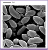 | Figure 1Electron micrograph of the Massilia varians isolate from the deep finger wound showing a single flagellum. Magnification, × 20,000.
|
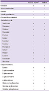
Biochemical features were investigated using VITEK® 2 Gram-negative (GN) Identification cards with the Vitek 2 system (bioMérieux, Marcy l'Etoile, France) and API 20NE cards (bioMérieux). The GN card identified the isolate as Sphingomonas paucimobilis, but with low probability (91%). The API 20NE card identified the isolate as within the Shewanella putrefaciens group, with low probability (90%). Data on the assimilation of carbon-sources, hydrolysis of chromogenic substrates, and enzyme activities of the isolate are described in Table 1. Matrix-assisted laser desorption/ionization time of flight mass spectrometry (MALDI-TOF MS) (MicroflexTM MALDI Biotyper, Version 3.1, Bruker Daltonics, Bremen, Germany) identified the organism as Massilia sp. with a score of 1.621. The analysis of 16S rDNA sequence was also performed with the universal bacterial primers 27F (5’-AGA GTT TGA TCC TGG CTC AG -3’) and 1541R (5’-AAG GAG GTG ATC CAG CCG CA-3’), amplifying a 1,362-bp segment. Sequences of the partial 16S rDNA fragment were 100% identical to those of strain CCUG 35299T (GenBank accession number NR_042652.1), the type strain of M. varians, when compared using BLAST (http://blast.ncbi.nlm.nih.gov/).
Table 1
Biochemical features of the type strain CCUG 35299T and the Massilia varians deep finger wound isolate.
![]()
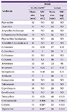
Antimicrobial susceptibility tests were performed by disk diffusion method, according to Clinical and Laboratory Standards Institute (CLSI) guideline [4]. And minimal inhibitory concentrations (MICs) were determined by Etest® (bioMérieux) (Table 2). The antimicrobial susceptibility patterns of the isolate were similar with those of the CCUG 35299T type strain.
Table 2
Antimicrobial susceptibilities and E test MICs of the type strain CCUG 35299T and Massilia varians isolated from deep wound
![]()
DISCUSSION
The genus Massilia was first proposed by La Scola et al. in 1998 [5]. Phylogenetically, it is placed in the vicinity of Naxibacter, Telluria, Duganella, and Janthinobacterium based on 16S rDNA gene sequences. However, Massilia and Naxibacter show different phenotypic characteristics from Telluria, Duganella, and Janthinobacterium [156]. The genus Naxibacter was first described in 2005 [6], and at the time was comprised of four species; Naxibacter alkalitolerans [6], Naxibacter haematophilus [7], Naxibacter suwonensis [8], and Naxibacter varians [7]. However, the Massilia and Naxibacter genera are now grouped together based on their phenotypic and biochemical features as well as 16S rDNA gene sequence comparisons. Therefore, a proposal was made to transfer all species of the genus Naxibacter to the genus Massilia by Kämpfer et al. in 2011 [1]. The genus Massilia has mainly been isolated from soil, air, or water, and is comprised by at least 23 species [125678910111213141516171819] (Supplementary Table 1). However, several species belonging to the genus Massilia have been recovered from clinical specimens [13571420] (Supplementary Table 2). N. varians was first identified from the eye of 90-year-man in Tromsø, Norway in 2008 [7]. After this date, N. varians was referred to as M. varians [1]; however, there have been no further reports of M. varians isolations since this first report.
The deep wound isolate was misidentified as S. paucimobilis (91%) and S. putrefaciens group (90%) by the GN and API 20NE cards respectively, most likely due to lack of database information for the genus Massilia. MALDI-TOF MS identified the isolates at the genus level, but with a low score. Therefore, when a certain unknown bacterial isolate is identified as the genus Massilia, 16S rDNA sequencing is strongly recommended.
To the best of our knowledge, this is the first case of M. varians isolated from clinical specimen since the first report in 2008.
 XML Download