

 PDF
PDF ePub
ePub Citation
 Print
Print


Introduction
Varicella-zoster virus (VZV) is a human herpesvirus that causes chickenpox and herpes zoster [12]. The most common neurologic complication of VZV reactivation is herpetic neuralgia, which is usually self-limited. However, VZV reactivation in immunocompromised patients can cause disseminated infections and severe neurologic dysfunctions, including meningitis, neuropathy, myelitis, stroke, and encephalitis [3]. Transverse myelitis is an unusual complication caused by VZV reactivation in immunocompetent patients [4]. To date, few cases of transverse myelitis were reported in Korea and most of them were not confirmed microbiologically [56789]. Herein we report a case of transverse myelitis caused by VZV in an immunocompetent older patient, and this case was confirmed microbiologically by detection of VZV DNA in the cerebrospinal fluid (CSF) by polymerase chain reaction (PCR).
Case Report
A 79-year-old woman was admitted to our Institution with weakness in the right lower leg and numbness in the lower limbs for three days. Ten days before admission, the patient felt a throbbing pain in the right flank, and the pain was not relieved by analgesics. Three days before admission, the patient’s right leg was paralyzed, and she felt numbness in both legs, lost the control of micturition, and presented with multiple skin eruptions, which appeared on the right flank and gradually spread to the trunk, face, and extremities.
Upon admission, blood pressure was 140/110 mmHg, the heart rate was 66 beats per minute, and body temperature was 36.3°C. Multiple vesicles and pustules were observed on the whole body (Fig. 1). The patient had never had contact with chickenpox or herpes zoster patients. In addition, she had no history of herpes zoster infection and vaccination. Neurologic examination indicated that the strength of the proximal half (grade 3/5) and the distal half (grade 4/5) of the right lower limb decreased. Furthermore, the sense of pain and temperature below level T9 on the right side of the body and below T6 on the left side of the body decreased. Magnetic resonance imaging (MRI) indicated diffuse hyperintensity of the spinal cord at level T4–T11 on T2-weighting (Fig. 2A).
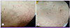 | Figure 1Development of multiple vesicles and pustules in the right flank (A) and their spread to the trunk (B).
|
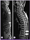 | Figure 2Whole spine MRI (T2-weighted sagittal image) on admission (A) shows high-signal intensity from level T4 (upper arrow) to T11 (lower arrow). On admission day 36 (B), the extent of diffuse hyperintensity decreased, with faint enhancement of the spinal cord from level T8–T9 (upper arrow) to T9 (lower arrow).
|
The results of blood tests were as follows: white blood cell count, 7.09 ⨯ 103/mL; hemoglobin, 13.3 g/dL; platelet count, 187 ⨯ 103/mL; C-reactive protein, 1.06 mg/dL; erythrocyte sedimentation rate, 16 mm/hour; and positive serum IgG and IgM antibodies against VZV.
CSF analysis indicated abnormal values for white blood cell count (1076 cells/μL; 0% of neutrophils, 96% of lymphocytes, and 4% of monocytes), and protein (442.5 mg/dL), and a negative result for bacterial culture. VZV DNA was detected by PCR amplification in CSF. The PCR results for IgG and IgM of herpes simplex virus-1 (HSV-1), herpes simplex virus-2 (HSV-2), and cytomegalovirus (CMV) were negative in the CSF.
Intravenous acyclovir was initiated at 500 mg every 8 hours. The dosage of acyclovir was adjusted to 250 mg every 8 hours for three days after initiation of therapy because of deterioration of renal function. On admission day 3, the patient presented deterioration of consciousness and convulsions; therefore, brain MRI was performed. MRI indicated increased signal intensity at the right temporo-occipital lobe and left frontal lobe on T2 weighting, suggesting the occurrence of encephalitis. The patient was maintained on antiviral and antiepileptic therapy for three weeks. After this period, her mental status was recovered, and the function of both legs improved with a rehabilitation program involving strength training. Repeat MRI showed partial improvement of myelopathy (Fig. 2B). However, sensory impairment below level T10 persisted for four months after initiation of therapy.
Discussion
VZV is a human neurotropic alphaherpesvirus that causes chickenpox (varicella) in children [12]. After primary infection, the virus becomes latent in cranial nerve and sensory root ganglia [23]. However, VZV reactivation may occur with advanced age or immunosuppression, particularly in cases of cell-mediated immunosuppression [123].
VZV reactivation may cause neurological complications such as chronic pain (postherpetic neuralgia), cranial nerve palsy, zoster paresis, meningoencephalitis, cerebellitis, myelopathy, multiple ocular disorders, and stroke [234]. The most common manifestation of VZV reactivation is herpes zoster. Unvaccinated individuals aged 85 years or older have a 50% risk of developing herpes zoster [10]. However, transverse myelitis is one of the rarest complications, particularly in immunocompetent patients [123]. To date, five cases of VZV myelitis have been reported in Korea; however, most of them were clinically suspicious cases with consistent image findings [56789]. Only one microbiologically confirmed case of transverse myelitis caused by VZV was reported approximately 20 years ago [6]. Four other reported cases of VZV in Korea were diagnosed by classical imaging findings and clinical examination.
This report describes a microbiologically confirmed case of transverse myelitis caused by VZV in an immunocompetent older patient. Myelitis and encephalitis due to VZV reactivation are more common in immunocompromised patients [1]. In these patients, VZV myelitis may occur without typical skin lesions and can occur far different level of skin lesion [411]. By contrast, in immunocompetent patients, VZV myelitis has a typical presentation (dermatomal rashes followed by myelitis at the corresponding level) and good outcomes [1411]. However, our patient showed an atypical presentation, characterized by generalized and disseminated eruptions on the body.
The diagnosis of VZV myelitis can be challenging [12]. Older patients may show a variety of neurologic symptoms from local paralysis to severe neurologic dysfunction due to multiple causes; therefore, thinking of several possibilities is critical and various examinations are needed to differentiate the causes. To date, no predictable markers of disease progression are available to patients with VZV myelitis [4]. Therefore, clinical suspicion and aggressive evaluation are crucial for the early diagnosis of VZV myelitis [12].
The detection of VZV antibodies and VZV DNA in CSF are confirmatory diagnostic tests [111213]. However, Rosenfeld et al. reported that patients showed clinical signs of severe VZV myelitis, although the VZV antibody tests and PCR results for VZV DNA were all negative [12]. Imaging studies are useful for the diagnosis of VZV myelitis. MRI of VZV myelitis is likely to show T2-hyperintensity in the spinal cord [1213]. Although the standard treatment regimen for VZV myelitis is not yet established, there is anecdotal evidence for treatment of VZV myelitis with acyclovir [412131415]. Moreover, there is little evidence that early antiviral treatment reduces the risk of VZV myelitis. Therefore, the early diagnosis and antiviral treatment of VZV is essential to recovery from myelitis and minimize its complications, and this treatment is crucial to prevent the development of postherpetic neuralgia [14]. Our case and some other cases reported previously also support the advantages of early antiviral treatment for VZV myelitis. We did not use corticosteroids because the additional benefit of the steroid was not clear although the combination of high-dose acyclovir and corticosteroids have shown a good prognosis in previous case reports [11].
In conclusion, this is the second confirmed case of VZV myelitis in immunocompetent patients in Korea. Even in immunocompetent older patients, VZV myelitis may be severe and involve atypical skin lesions. Therefore, early diagnosis and aggressive antiviral treatment may be necessary.
 XML Download