

 PDF
PDF ePub
ePub Citation
 Print
Print


Introduction
Hemophagocytic syndrome (HPS), also called hemophagocytic lymphohistiocytosis (HLH), is an uncommon but severe disease associated with various infectious, genetic, neoplastic, and autoimmune diseases [1]. It is characterized clinically by fever, cytopenia, splenomegaly, and hemophagocytosis in the bone marrow (BM), liver, or lymph nodes [2].
Scrub typhus is a disease caused by Orientia tsutsugamushi, and is treated mostly with antibiotics. However, it can be fatal for some patients because of acute renal failure, acute respiratory distress, and septic shock [34]. HPS is a rare complication of scrub typhus, although it can be treated using adequate antibiotics. Here, we report a case of Epstein-Barr virus (EBV)-associated HPS after scrub typhus infection that was not improved using adequate anti-rickettsial treatment.
Case report
A 73-year-old male, who was diagnosed with scrub typhus according to an eschar on the axilla and a positive result of indirect immunofluorescence assay for O. tsutsugamushi (antibody titer of 1:1,280), was transferred to our institution from a local hospital because of a persistent fever despite 7-day doxycycline therapy. The patient had no previous history of illness, blood transfusion, and did not take any medications before he visited the hospital. His occupation was a farmer, and he resided in a rural area of Goseong-gun, Gyeongsangnam-do, Korea. On admission, he had a body temperature of 36.9°C, a pulse of 72 beats/min, a respiration rate of 21 breaths/min, and a blood pressure of 97/54 mmHg. Hepatosplenomegaly, a maculopapular rash on the abdomen, and a 1-cm sized eschar on the left axillary area were observed. Laboratory results were as follows: Complete blood counts revealed a white blood cell (WBC) count of 2,610/mm3 (41.7% segmented neutrophils and 55.8% lymphocytes), a hemoglobin (Hb) level of 11.9 g/dL, and a platelet count of 237,000/mm3. The blood biochemical profile included 1,500 U/L lactate dehydrogenase, 675 U/L alkaline phosphatase, 245 U/L aspartate aminotransferase (AST), 106 U/L alanine aminotransferase (ALT), 6.6 g/L total protein, 2.3 g/L albumin, 444 U/L γ-glutamyl transpeptidase, 0.65 mg/dL total bilirubin, 61 mg/dL total cholesterol, 2,000 ng/mL ferritin, 70 mg/dL fibrinogen, 138 mg/dL triglyceride, 85.2 mg/L C-reactive protein, 18.4 mg/dL blood urea nitrogen, 0.67 mg/dL creatinine, and 3.4 mg/dL uric acid. Serological tests for human immunodeficiency virus, cytomegalovirus, parvovirus, Hantaan virus, leptospira, hepatitis A virus, hepatitis B virus, and hepatitis C virus were all negative. Anti-nuclear and rheumatoid factor antibodies were also negative. Blood and urine cultures were negative. However, indirect immunofluorescence antibody IgG for O. tsutsugamushi titer was positive at 1:5,120. An abdomen computerized tomography revealed hepatosplenomegaly. Azithromycin (500 mg daily) was provided for 5 days in consideration of an adverse drug reaction or doxycycline-resistant scrub typhus.
Four days after admission, his fever remained and his general condition had deteriorated. Laboratory data revealed a WBC of 1,800/mm3, 9.8 g/dL hemoglobin, a platelet count of 105,000/mm3, 591 U/L AST, and 365 U/L ALT. BM aspiration and biopsy were performed, which revealed hypercellular marrow with hemophagocytosis and histiocyte infiltration (Fig. 1). EBV was detected in BM aspirates using polymerase chain reaction (PCR), with a titer of 2,094 copies/μg DNA in peripheral blood mononuclear cells. A subsequent DNA test for O. tsutsugamushi in the blood was reported as negative by the Korea Center for Disease Control and Prevention.
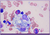 | Figure 1Microscopic finding of bone marrow aspiration shows hemophagocytosis of neutrophils, normoblasts, and platelets (Wright-Giemsa stain, ×1,000)
|
After a diagnosis of HPS was made, 60 mg methylprednisolone was prescribed for 1-week, and the dose was then tapered by 10 mg every 3 days. His fever subsided on hospital day 8, and subsequent tests revealed that his complete blood count profile recovered and his elevated liver enzymes declined to near normal levels within 3-weeks of starting steroid treatment.
Discussion
HPS is a fatal hyperinflammatory syndrome that is characterized by the activation and proliferation of histiocytes or lymphocytes with uncontrolled hemophagocytosis and cytokine overproduction [5]. The diagnosis of HPS is established by fulfilling five of the following eight criteria: 1) fever, 2) hepatosplenomegaly, 3) cytopenia (affecting two cell lineages), 4) hypertriglyceridemia and/or hypofibrinogenemia, 5) hemophagocytosis in the BM, spleen, or lymph nodes, 6) low or absent natural killer (NK) cell cytotoxicity, 7) hyperferritinemia, and 8) elevated soluble CD25 (sCD25) levels [6]. Because scrub typhus and HPS have similar clinical characteristics, such as cytopenia, hepatosplenomegaly, and abnormal liver function tests, scrub typhus infection can make diagnosing HPS challenging.
EBV is a causative organism of HPS, and is particularly notorious for its severe morbidity and mortality [7]. Therefore, the early diagnosis and treatment of EBV-associated HPS (EBV-HPS) is essential for a good outcome [8]. The pathogenesis of EBV-HPS is that EBV-infected B cells stimulate cytotoxic T lymphocytes, which leads to hypercytokinemia and the stimulation of histolytic cells [9]. EBV causes the stimulation, generation, and uncontrolled secretion of T and NK cells, as well as the generation of IL-2, INF-α, and IL-6, which are responsible for HPS [10]. Of these immunological profiles, an increased plasma concentration of the soluble IL-2 receptor (sCD25) and impaired NK cell activity are two important diagnostic parameters in HPS [7]. The elevation of sCD25 suggests the activation of T lymphocytes, and is closely related with prognosis of HPS [11]. However, NK cell activity and CD25 level were not performed in this case, because those tests were not supported at our laboratory facility.
According to the HLH-2004 guidelines, the treatment for EBV-HPS is usually composed of etoposide-based chemotherapy and high-dose steroids to relieve the hyperinflammatory state, which is based on the clinical data of familial HPS [6]. Therefore, the effectiveness of the HLH-2004 guidelines for secondary HPS is not established definitely. A previous case of EBV-HPS after scrub typhus infection was reported in a pediatric patient, which was improved after chemotherapy including dexamethaxone, etoposide, and clarithromycin [7]. The current case was improved without intensive chemotherapy, which might be harmful due to its adverse effects. The relatively low copy numbers of EBV-DNA suggest that the HPS might have been less severe in the current case. The EBV viral loads in EBV-HPS cases reported previously range from >1,000-1,000,000 copies/μg; therefore, measuring the level of viral DNA using quantitative PCR might be useful to demonstrate the treatment response and predict mortality [12].
Because EBV can be reactivated in critically ill patients [13], some might argue that EBV served as a bystander in the current case, and that HPS arises from scrub typhus itself. While most cases of HPS associated with scrub typhus infection are improved by adequate antibiotic treatment, as shown in Table 1, the current patient did not improve with azithromycin until steroids were initiated.
Table 1
Cases of scrub typhus associated hemophagocytic syndrome
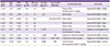
| Case [Ref] | Age/Sex | WBC (/mm3) | Hb (g/dL) | Platelet (/mm3) | Ferritin (ng/mL) | Associated Infection | Treatment drug | Outcome |
|---|---|---|---|---|---|---|---|---|
| 1 [14] | 75/F | 5,600 | 12.8 | 14 | 183 | - |
Doxycycline Prednisolone 1 mg/kg |
Improved within 10 days |
| 2 [14] | 69/F | 1,300 | 11.3 | 75 | 282 | - | Minocyline | Improved within 10 days |
| 3 [15] | 74/F | 6,700 | 12.3 | 165 | - | Parvovirus B19 | Minocyline | Improved within 1 days |
| 4 [16] | 58/F | 2,300 | 6.5 | 56 | - | - | Doxycycline | Improved within 3 days |
| 5 [16] | 37/F | 3,300 | 9.6 | 106 | - | - | Doxycycline | Improved within 2 days |
| 6 [17] | 81/F | 9,780 | 6.9 | 72 | 1,530 | - |
Doxycycline Prednisolone 1 mg/kg |
Expired at 39 hospital day |
| 7 [18] | 7/M | 2,520 | 7.9 | 17 | >1,650 | EBV | Etoposide, Dexamethasone | Improved within 4 weeks |
| 8 [19] | 34/F | 3,500 | 8.4 | 16 | 3,212 | EBV, CMV | Ceftriaxone, Minocycline | Expired at 13 hospital day |
| Case | 73/M | 2,610 | 11.9 | 237 | 2,000 | EBV |
Azithromycin Methylprednisolone 1 mg/kg |
Improved within 3 weeks |
![]()
In addition, previous case reports showed that viruses such as EBV, CMV and parvovirus can trigger HPS after scrub typhus infection (Table 1) [141516171819]. Considering the O. tsutsugamushi PCR results in blood was negative in the current case and there have been no reports of doxycycline-resistant O. tsutsugamushi in South Korea, it is unlikely that scrub typhus is the only reason for HPS. Therefore, we regarded that EBV triggered HPS after a scrub typhus infection in the current case. We could not determine whether it was primary infection or a reactivation because an EBV serological test was not performed. However, it is possible that the current case was caused by the reactivation of EBV because more than 87.2% of adults in South Korea have EBV-viral-capsid antigen (VCA) IgG antibodies [20].
The current case suggests that secondary HPS should be considered as a differential diagnosis when there is no improvement after treating scrub typhus using adequate antirickettsial therapy.
 XML Download