

 PDF
PDF ePub
ePub Citation
 Print
Print


Introduction
Abiotrophia defectiva is part of the normal human microbiota, colonizing the oral, genitourinary, and intestinal tracts [1]. It is a rare, yet important, cause of infective endocarditis, and is estimated to cause approximately 5-6% of all cases of infective endocarditis, including being a major cause of blood culture-negative infective endocarditis [2]. It affects diseased valves in 90% of cases and it is implicated in embolic complications and valvular destruction, despite being sensitive to antibiotics [3]. Previous studies have shown mortality and relapse rates as high as 17% despite antibiotic treatment, and this makes accurate and quick identification important [456]. Herein we report the first case of infective endocarditis caused by A. defectiva in Korea.
Case report
A 62-year-old female was admitted to the emergency department (ED) after two consecutive episodes of syncope. The patient was diagnosed with severe rheumatic mitral stenosis, and underwent a mitral valve replacement (MVR). Two months previously, the patient had undergone a simple extraction of her #16 tooth due to secondary dental caries and had taken prophylactic antibiotics (amoxicillin 2,000 mg).
Upon admission to the ED the patient was alert and had a body temperature of 38.6°C, a pulse rate of 66 beats/min, and blood pressure of 149/51 mmHg. Upon physical examination, no cardiac murmur was auscultated and no other evidence, such as clubbed fingers, Janeway lesions or petechiae were found. Laboratory studies showed a white blood cell count of 14,260/mm3(neutrophil 79.6%), erythrocyte sediment rate of 92 mm/h and C-reactive protein of 111.1 mg/L. Chest X-ray revealed mild cardiomegaly and electrocardiography showed a newly developed complete atrioventricular block. Chest CT and abdominal-pelvic CT was performed to evaluate fever focus, which showed no significant finding.
A transthoracic echocardiogram (TTE) showed that the mechanical prosthetic mitral valve functioned well, and there was no visible vegetation, however, infective endocarditis could not be completely ruled out because of the patient’s previous MVR. Thus, empirical antibiotics (vancomycin 1 g intravenous q12hr, gentamicin 60 mg intravenous q8hr and rifampin 600 mg per oral q24hr) were administered, and a transesophageal echocardiogram (TEE) performed the day after admission showed mitral valve vegetation and mild transvalvular mitral regurgitation (Fig. 1), leading to a conclusive diagnosis of infective endocarditis according to Duke criteria (one major and three minor criteria).
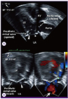 | Figure 1Transesophageal echocardiography performed the day after admission.
(A) Long-axis view (135°) of a transesophageal echocardiography showing two examples of vegetation attached to the prosthetic mitral valve. The larger one measures 0.7 ☓ 0.5 cm (large arrow head) and the smaller one measures 0.5 ☓ 0.3 cm (small arrow head). (B) Two-chamber view (59°) of a transesophageal echocardiography (zoomed view) showing vegetation attached to the prosthetic mitral valve (arrowhead). A color Doppler shows the jet flow with mild transvalvular mitral regurgitation in systole (arrow).
LA, left atrium; LV, left ventricle; RV, right ventricle.
|
Three sets of blood cultures taken on admission showed tiny, non-hemolytic colonies on a blood agar plate (ASAN Pharmaceutical, Hwaseong, Korea), and pleomorphic Gram-positive cocci from smear preparation of the blood agar plate (Fig. 2). On the third day after admission, A. defectiva was identified by MALDI-TOF-MS (Matrix-assisted laser desorption/ionization time-of-flight mass spectrometry, Bruker Daltonics Inc., MA, USA), not identified in VITEK 2 (bioMérieux, Marcy l'Etoile, France) systems. However, because the amount of bacteria was insufficient, cultures were sub-cultured on a medium containing vitamin B6 to assess antibiotic susceptibility.
 | Figure 2Colony and microscopic characteristics of Abiotrophia defective. Tiny, non-hemolytic colonies on a blood agar plate (ASAN Pharmaceutical, Hwaseong, Korea) after 48 hour of incubation at 35°C with 5% CO2 (left), and pleomorphic Gram-positive cocci from smear preparation of the blood agar plate (Gram stain, ×1,000, right).
|
Susceptibility to cefotaxime, penicillin G and vancomycin was tested by the E-test method; the isolate tested cefotaxime-sensitive (MIC 0.75 μg/ml), penicillin G-intermediate (MIC 1.0 μg/ml) and vancomycin-sensitive (MIC 0.38 μg/ml). An adjusted antibiotic regimen of vancomycin 1 g q12hr and gentamicin 60 mg q8hr was initiated, after which the patient’s fever subsided. Vancomycin Therapeutic drug monitoring was performed, and vancomycin dosage was adjusted to 400 mg q12hr.
On day 12 of admission, the patient became feverish once more and in the subsequent TEE the mitral valve vegetation on the medial side seemed to have resolved, but there was an increase in the size of the vegetation on the lateral annulus (0.5 ☓ 0.3 cm − 0.8 ☓ 0.3 cm) which indicated a perivalvular infection (Fig. 1). Based on these findings, a second MVR was performed on day 19 of admission.
Operation finding showed small vegetation in prosthetic MV and annulus, which was removed, and MVR was conducted using 23 mm ATS medical open pivot heart valve (ATS Medical, Inc., Minneapolis, MN, USA). Pathology finding showed myxoid degeneration, acute and chronic inflammation, fibrosis and calcification, and tissue culture showed no growth of organism (It was on the 19th day on antibiotics, and blood culture was negatively converted at this time).
On day 23 of admission, a follow-up TTE showed a well-functioning prosthetic mitral valve. The patient was treated with antibiotics for 5 weeks and was discharged on day 36. The patient was followed-up at an out-patient clinic for 3 months after discharge with no significant complications.
Discussion
Organisms of the genus Abiotrophia were first classified as nutritionally variant streptococci (NVS) in 1961 [6]. Bouvet et al. identified two species of NVS; Streptococcus defectivus and Streptococcus adjacens [7]. In 1995, Kawamura et al. identified Abiotrophia as a separate genus, based on 16S rRNA analyses [8]. NVS are part of the normal flora of the mouth and urogenital and intestinal tracts [1]. Recently, an increasing number of cases have been described of A. defectiva isolates recovered from invasive and non-invasive infections following dental procedures [9].
Abiotrophia spp., has fastidious culturing and the unspecific colony morphology that they presents on primary detection, such strains have caused major diagnostic difficulties. Thus, it has been supposed that many culture-negative endocarditis could have been caused by these species, which could have lead to an underestimation as pathogens of infective endocarditis [10]. In this case report, we identified A. defectiva by MALDI-TOF-MS. MALDI-TOF-MS is a new technology for routine identification of bacteria in clinical microbiology laboratories. Much of the work using MALDI-TOF-MS for microbial identification has focused on demonstrating that reproducible mass spectra can be obtained using intact cells and developing algorithms for interpretation and comparison of these spectra. By testing colonies, it takes only a few minutes to have a correct identification which makes not only possible to identify the microorganisms at the species levels but sometimes at the sub-species and strains levels, allowing the detection of epidemic lineages [11].
A. defectiva can cause serious infections such as bacteremia, osteomyelitis, brain abscess, pancreatic abscess, septic arthritis, crystalline keratopathy and in rare cases, infective endocarditis [312]. Endocarditis caused by NVS is implicated in 5−6% of all streptococcal endocarditis cases, and <1% of all endocarditis cases are caused by A. defective [3]. However, A. defectiva has a higher affinity for the endocardium because of its ability secrete exopolysaccharide, enabling it to adhere to fibronectin in the extracellular matrix [2]. Of all the NVS-induced endocarditis patients, 90% suffered from heart disease, and in most cases, bacteremia associated with the pre-existing valvular heart disease led to the development of endocarditis with subacute prognoses [13].
Infective endocarditis caused by A. defectiva and other NVS had higher mortality, morbidity and complication rates than those caused by other viridans streptococci [14]. Most deaths were due to refractory congestive cardiac failure or major systemic emboli [15]. A mortality rate of up to 17% has been reported, which is higher compared to endocarditis caused by viridians streptococci (0−12%) [4]. The relapse rate can also be up to 17% in some cases. Previous studies have shown a treatment failure rate as high as 41%, despite the use of appropriate antibiotics [5].
The antibiotic regimen to combat A. defectiva endocarditis includes penicillin or ampicillin, plus an aminoglycoside or vancomycin for cases of antimicrobial resistance, taken for 4−6 weeks [16]. Infective endocarditis due to A. defectiva progresses slowly, but despite its sensitivity to antimicrobials ~50% of cases need surgical management. Surgical treatment combined with concurrent antimicrobial therapy result in better prognoses, and the main indications for surgery are persistent sepsis and vegetation, severe congestive heart failure and recurrent embolism [151718].
In Korea, 2 cases of infective endocarditis caused by S. adjacens were first reported in 1996. Since then, a case of infective endocarditis by Grnulicatella adjacens was reported in 2010. All cases were successfully treated with antibiotics and surgical treatment [1920]. After classified as A. defectiva, this is the first report as the pathogen of infective endocarditis in Korea.
The case presented here is an example of the successful treatment of infective endocarditis caused by A. defectiva following a tooth extraction in a post-MVR patient, and represents the first reported case of infective endocarditis caused by A. defectiva in Korea. This case shows that A. defectiva could be considered as a causative organism of infective endocarditis in Korea.
 XML Download