

 PDF
PDF ePub
ePub Citation
 Print
Print


Introduction
Tropical diseases from overseas have been imported every year recently [1]. But most clinicians were not familiar with these tropical diseases. So, the exact diagnosis and appropriate treatment often could be delayed.
Chikungunya has been reported from Africa, the Indian Ocean islands, the Pacific region and South and South-East Asia, with intervals of 7 to 8-year to 20-year between consecutive outbreaks in these regions [2, 3]. In North-East countries including Korea, China, and Japan, the first imported case from Japan was reported in 2007 [4], and in China, multiple sporadic non-indigenous cases have already been reported and an outbreak developed in Guandong region in 2010 [5].
In recent times, with an increase in international travel and global warming, the risk for spreading Chikungunya to non-endemic regions has enhanced [6]. Recently, the positive results for Chikungunya virus came out from retest of the samples of patients diagnosed or suspected with dengue fever in Korea [7]. Since Chikungunya classified as communicable disease group IV at December 30, 2010, until now there has been no official report for symptomatic patients. We report the first imported case infected with Chikungunya virus in Korea.
Case Report
A 23-year-old man was admitted to our emergency room with fever and back pain, which was occurred abruptly in June 30th 2013. Before our emergency room, he had been Manila to teach Korean language in the Republic of the Philippines as volunteer worker from June 18th to June 25th, and returned to Korea in June 25th. On admission, he also reported severe headache, myalgia, and chills. In history taking, he said that he had disc herniation in his spine before 4 years. During his stay in Manila, he said that he was bitten by mosquitos. His body temperature was 38.5℃, heart rate was 78 beats per minute, respiratory rate was 18 breaths per minute, and blood pressure was 130/80 mmHg. On physical examination, his face, especially around his eyelids, was swollen. But tenderness, swelling, and heating sense on any joint were not observed. On laboratory, a complete blood cell count showed a white blood cell of 5,390/mm3 (neutrophil 73.1%), hemoglobin of 13.8 g/dL, platelet count of 224,000/mm3. On chemistry, aspartate aminotransferase was slightly increased to 41 IU/L, alanine aminotransferase 14 IU/L, total bilirubin 0.33 mg/dL, total protein 6.9 g/dL, albumin 4.5 g/dL, blood urea nitrogen 5 mg/dL, Cr 0.75 mg/dL, glucose 98 mg/dL, and lactate dehydrogenase 361 IU/L. Erythrocyte sedimentation rate and C-reactive protein were 3 mm/hr and 6.74 mg/L respectively. There is no evidence of malaria in peripheral blood smear. The peripheral blood culture showed the negative result for salmonellosis. The serologic test of dengue and hepatitis A virus were not conducted.
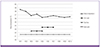
On the third day, he reported three times of diarrheas, and complained that myalgia on whole body and back pain persisted (Fig. 1). We performed the stool culture and parasite study, there was no positive results. On repeat physical examination, there is no evidence of tenderness, swelling, and heating sense on any joint. The direct and indirect tenderness were not observed, and bowel sound was normal. We performed magnetic resonance imaging (MRI) for the region of lumbar spine to differenciate the causes of back pain other than his disc herniation.
On the fifth day, diarrhea stopped spontaneously, but a maculopapular rash developed with itching in his abdomen, chest, back, both upper and lower extremity (Fig. 2). Facial edema especially around both eyelids sustained. Because any drugs were not administered except intravenous tramadol, we thought that drug rash was less likely possible. Considering chickungunya virus infection, we performed serology and reverse transcription polymerase chain reaction (RT-PCR). On the eighth day, skin rash disappeared, and back pain persisted. The result of MRI on lumbar spine revealed the focal protrusion of disc in L4-5 level. On next day, the patient was discharged.
The result of specific IgM for chickungunya virus in IgM capture enzyme immunoassays (MAC-ELISA) was reported by Korea Centers for Disease Control and Prevention (KCDC) as positive, and RT-PCR negative. MAC-ELISA kit (NovaTec, Dietzenbach, Germany) was used to detect chickungunya specific IgM antibody [7]. The viral RNA was extracted by Viral Gene-spin DNA/RNA extraction kit (iNtRON Biotechnology, Suwon, Korea) and amplified by the one-step duplex RT-PCR assay [7]. This case met the laboratory criteria that was presented in World Health Organization guideline [2], which was defined at least one of the following tests in the acute phase; 1) virus isolation, 2) presence of viral RNA by RT-PCR, 3) presence of virus-specific IgM antibodies in single serum sample collected in acute or convalescent stage, 4) four-fold increase in IgG values in samples collected at least three weeks apart. Finally, this case was diagnosed with confirmed case infected with chikungunya virus. After two weeks, we identified that back pain remained without other arthralgia.
Discussion
Chikungunya virus is an RNA virus that belongs to the Alphavirus genus of the Togaviridae family, and is primarily transmitted by bites of mosquitoes of the genus Aedes such as Aedes aegypti and Aedes albopictus [8]. The clinical characteristics are presented with fever, joint pain, headache, skin rash, myalgia. Clinical symptoms, therefore, are similar with dengue, the initial differentiation among both diseases is difficult [7]. The virus was first isolated in 1953 in the southern province of Tanzania [9]. Before 2000, large outbreaks of chikungunya were not common, but since 2000 epidemics have become more frequent, and chikungunya virus presents with global distribution including Africa, Asia, and Europe [2]. Chikungunya has also been confirmed in travelers returning to Europe, Australia, the UK, and the USA from the Indian Ocean Islands and Asia [10]. Despite the increasing trend of traveling abroad, any cases were not reported after designation as communicable disease group IV in Korea [11]. We experienced a 23-year-old man, the imported first case confirmed, infected with chikungunya virus returning from the Republic of the Philippines.
But, in the past the four hundred eighty eight dengue-suspected serum samples during 2009-2010 were already retested for identification of chikungunya virus through ELISA and RT-PCR and twenty positive results in ELISA, two positive results in RT-PCR, and one virus isolation came as a result [7]. Total 21 patients were diagnosed for confirmed cases on the base of laboratory critereia [2]. This result means that some patients infected with chikungunya virus after returning a foreign travel already existed in the past and there was the difficulty of differenciation from dengue in the early stage because of similarity of clinical symptoms with each other.
The diagnosis of chikungunya is on the basis of clinical, epidemiological and laboratory criteria [10]. Among 3 diagnostic methods, laboratory examination needs for confirmation of chikungunya [2, 12]. The RT-PCR, a confirmatory test with rapid and sensitive technique, is valuable during early stage of illness, namely initial viremic phase [13]. Therefore, serologic test such as ELISA plays a important role four to seven days after the onset of fever [12, 14]. Blood sample was extracted from our patient at six days after the onset of fever. Although RT-PCR from KCDC showed the negative result, MAC-ELISA present positive finding: positive in chikungunya-specific IgM. According to a recent study, sensitivity five to six days after the development of fever and specificity were 100% and 95.6%, respectively [15]. Crossreactivity with other flavivirus may occur in the MAC-ELISA, but which is known to be very rare [2, 13]. Therefore, we think this patient is the confirmed case on the basis of diagnostic criteria of World Health Organization and Centers for Disease Control and Prevention [12, 13].
The Republic of the Philippines is one of countries with reported current or previous local transmission of chikungunya virus [16]. Outbreaks of 561 cases in 2012 and 1609 in 2013 arised in multiple areas [17]. Especially in metro manila where the patient had resided mainly, 275 cases sustained chikungunya, the epidemiologic factor was more likely to be suitable to him [18]. On admission to our hospital, thrombocytopenia was not shown in the patient, and skin rash unlike that of dengue developed with fever subsiding on the fifth day. The skin rash in chikungunya generally emerges between the second and fifth day of onset on fever [2], and was reported to last for 2-3 days [19]. The frequency of skin rash was reported to 40.1% and 50% [19, 20]. Given fever usually lasts for 3 to 4 days in chikungunya, skin rash seems to develop at the end of the fever, which is similar in comparison with dengue [21]. But, dengue presents with maculopapular rash with the sparing of small islands of normal skin, the development of skin rash in dengue is less frequent than that of chikungunya [2, 21]. The intravenous tramdol was administered due to his back pain from the second day to the tenth day of hospitalization. Considering the duration of his skin rash, tramadol was less likely to be the cause of skin rash because the skin rash was only transient for 3 days. In our case, we suspected the possibility of chikungunya on the basis of the different pattern of skin rash and no thrombocytopenia compared with dengue.
There was no clear evidence of joint pain, swelling, heating sense, and tenderness on ankle, knee, and wrist. Instead, he complaint back pain and headache mainly. Considering the disc herniation in L4-5 level, we presumed that back pain may have been chiefly due to chikungunya because back pain had not been observed before several months the visit of the Republic of the Philippines although there was some contribution of back pain by disc herniation.
With the increase of the distribution of mosquitos by global warming and more frequent foreign travel, the number of the domestic patients infected with chikungunya virus would be increased although this patient is the first imported case in Korea [6]. Chickungunya has been classified as communicable disease group IV since December 30, 2010 in Korea [11]. But, since then the requests for examination of chikungunya virus were few until March 2013; the number of request was only twenty five [7]. This suggests that the recognition for chikungunya among clinicians may be not sufficient. The reported number of the patients diagnosed for dengue are on the rise from 2006 to 2012 according to KCDC [1]. Given that dengue virus and chikungunya virus share the same vector, Aedes aegypti and Aedes albopictus and co-infections of humans with two viruses occurred, the number of imported cases infected with chikungunya virus in Korea could not be overlooked [22]. Therefore, clinicians should consider serological and molecular test for chikungunya virus as well as dengue virus for febrile patients returning from foreign travel because of the difficulty of differenciation from chikungunya and dengue fever initially through clinical symptoms.
In conclusion, we report the first imported case infected with chikungunya virus. In case of febrile patients after the travel of tropical and subtropical region, we suggest the possibility of chikungunya viral infection and request the laboratory test for chikungunya.

 XML Download