Abstract
Recently, serotype K1 Klebsiella pneumoniae has been a major agent of an invasive syndrome characterized by liver abscess and its metastatic infection. Extrahepatic infection and its characteristics in patients with renal abscess caused by K. pneumoniae are poorly understood, and few cases of central nervous system infection have been reported. This is a report of 80-year-old woman with uncontrolled type 2 diabetes mellitus with renal abscess caused by serotype K1 K. pneumoniae, complicated with ventriculitis despite of appropriate use of antibiotics. Physicians need to be aware of possibility of metastatic infection in patients with serotype K1 K. pneumoniae infection, if they develop neurologic symptom and focus of infection is still present.
Klebsiella pneumoniae, a member of the Enterobacteriaceae family, has been implicated as a major cause of pneumonia since the 19th century and is also an established cause of urinary tract infection, hepatobiliary infections, and osteomyelitis, mainly in alcoholics and diabetics [1]. Since the 1980s, a new distinctive invasive syndrome has been reported in Taiwan, which is characterized by community-acquired liver abscess and metastatic spread to distant sites in patients without underlying hepatobiliary disease [1]. Lung abscess, prostate abscess, soft tissue abscess, endophthalmitis/uveitis, and central nervous system (CNS) infection could occur as metastatic complications [2, 3].
Compared to those on liver abscess, there are only a few reports on metastatic infection and virulence factor in perinephric or renal abscess caused by K. pneumoniae, in the medical literature. In particular, there are no reports on CNS infections such as ventriculitis in patients with K. pneumoniae renal abscess. Here, we report, to our knowledge, the first case of renal abscess caused by serotype K1 K. pneumoniae that spread to the CNS and presented as ventriculitis.
An 80-year-old woman presented with decreased mentality, a 3-day history of polyuria, general weakness, and poor oral intake. She had been diagnosed with type 2 diabetes mellitus more than 30 years before and was treated with an oral hypoglycemic agent.
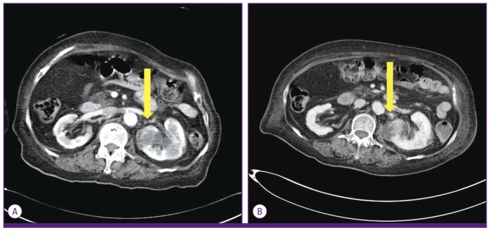
At the current hospital admission, the patient's temperature was 38.0℃. Her blood pressure was 143/92 mmHg, heart rate was 123/min, respiratory rate was 28/min, and oxygen saturation was 99% in room air. She was dehydrated, and the rest of the physical examination findings were unremarkable. No definite abnormality was detected through neurologic examination. Her laboratory values were notable as follows: white blood cell (WBC) count of 16,540 cells/mm3, segmented neutrophil concentration of 88.4%, bicarbonate level of 17.3 mmol/L, C-reactive protein (CRP) level of over 34.1 mg/dL, glucose level of 648 mg/dL, hemoglobin A1c concentration of 8.4%, sodium level of 151 mEq/L, osmolality of 357 mOsm/Kg, and creatinine level of 1.19 mg/dL. Routine urinalysis showed WBC 30-49/HPF and some bacteria. Computer tomography (CT) scan of the abdomen showed multiple ill-defined nodular hypodense lesions in the left kidney that suggest acute pyelonephritis of the left kidney with multiple abscess formation (Fig. 1). No definite abnormality was observed on brain CT.
The patient was admitted to the hospital and treated with intravenous hydration and insulin and started treatment empirically on piperacillin/tazobactam (4.5 g q8hr, IV). We attempted percutaneous drainage of renal abscess, but did not succeed because of inadequate liquefaction. Cultures from peripheral blood and urine revealed K. pneumoniae, which was susceptible to most antibiotics including amikacin, cephalosporins, and ciprofloxacin. The string test was positive, showing a hypermucoviscosity phenotype. The patient was afebrile and in a stable condition on day 3 of hospitalization.
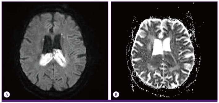
On day 6 of hospitalization, the patient developed a fever, her mental state deteriorated, and she developed stupor. At that time, her WBC count and CRP level decreased to 13,860 cells/mm3 and 5.99 mg/dL. The patient showed seizure-like activity, and neurologic examination revealed objective changes with positive pyramidal and Babinski signs. Magnetic resonance imaging (MRI) of the brain showed elevated signals in the lateral ventricles, Sylvian fissure, suprasellar, interpeduncular cistern, and posterior fossa, suggestive of ventriculitis (Fig. 2). Cerebrospinal fluid (CSF) showed a very turbid and milky appearance, and the WBC count was 7,200/mm3 segmented neutrophil concentration was 44%, protein level was 359.3 mg/dL, glucose level <10 mg/dL. The antibiotic regimen was changed from piperacillin/tazobactam to ceftriaxone (2 g q12hr, IV) and vancomycin (1 g q12hr, IV), and emergency extraventricular drainage (EVD) was performed. Although the blood culture at that time showed negative result, the CSF culture revealed K. pneumonia. We continued ceftraixone alone. K. pneumoniae isolated from the CSF was found to be of hypermucoviscosity phenotype and was shown same antibiotic susceptibility as those drawn from first blood culture. Abdomen CT scan was performed, and abscess of left kidney increased in size and protruded (Fig. 1), and other disseminated infection was not found. Percutaneous drainage for renal abscess was performed. Two weeks after admission, the EVD catheter and pig tail catheter of the left renal abscess were removed. The patient's general condition and laboratory data remained favorable. After 28 days of intravenous antibiotics, the patient was discharged to the long-term care facility with oral cefditoren (600 mg/day) of two weeks, showing improvement of neurologic state slowly, and after 1 month, could express in words.
We evaluated the serotyping and virulence genes of K. pneumoniae from blood and CSF including serotype K1, rmpA. For serotyping, we use PCR detection of serotype K1 specific alleles at the wzx loci, with the following primers, as previously described [4]. Consequently, serotype K1-specific alleles at the wzx were positive in the strains of the blood and CSF, indicating that the serotype of this K. pneumoniae was K1. The presence of genes encoding virulence factors was determined by polymerase chain reaction using primers documented previously [5] and rmpA gene was confirmed in the microorganism from CSF and blood. We didn't analyze the presence of magA gene which was known as one of factor associated virulence.
Perinephric and renal abscesses are rare among the various existing intra-abdominal abscesses. More than three-quarters of the cases arise from a urinary tract infection (UTI) and known risk factors are urinary flow obstruction, renal stones, other structural abnormalities of the urinary tract, prior surgery, trauma, and diabetes mellitus [6]. Our patient had diabetes mellitus controlled by administration of an oral hypoglycemic agent for 30 years, and she presented with a hyperosmolar hyperglycemic state at admission. Patients with diabetes have greater frequency, severity, and complication of infection. For reasons that are not fully understood, there are abnormalities of cell-mediated immunity and phagocytosis in the presence of hyperglycemia, with diminished vascularization [6]. Neutrophils are an essential part of the innate immunity against bacterial infection. Impaired neutrophil activity in type 2 diabetes mellitus is partly responsible for the increased susceptibility to infection [7].
K. pneumoniae is one of the major causes of community-acquired liver abscess in Southeast Asia, and capable of causing UTI in both healthy people and in-hospital patient [8]. Since the 1980s, a few case reports of secondary CNS infection due to liver abscess have been published. In Korea, there have been a few reports of disseminated infection due to K. pneumoniae. Jung et al. [9] reported a case of endogenous endophthalmitis and septic pulmonary embolism in K. pneumoniae renal abscess, and this report also have summarized cases of K. pneumoniae infection with multiple foci, involving endoophthlmitis, renal, liver or prostate infection and CNS infection with concomitant liver abscess or spondylitis. However, no cases of metastatic CNS infection with K. pneumoniae renal abscess have been reported. K. pneumoniae CNS infection is a life-threatening disease and the case fatality rate of K. pneumoniae meningitis remains high at 30-40%, with severe neurological sequelae, especially serotype K1 isolates is found in this patients [10]. Ventriculitis is inflammation in the ventricular system of the CNS and occurs as a complication of meningitis, which is a result of severe meningitis affecting the basal cisterns or rupture of a cerebral abscess into a ventricle. Although antimicrobial treatment with surgical intervention is known to the mainstay of treatment [11], prognosis is poor in most of cases. Generally, the presence of inflammation in the setting of bacterial meningitis enhances the ability of a given antimicrobial to penetrate the blood-brain barrier. However, this phenomenon may be less observed in case of ventriculitis, in which the ventricles can do as a persistent source of infection, as a potential blockade of CSF outflow tracts [12]. We used proper antibiotics continuously, and performed prompt external ventricular drainage. Early invasive treatment in our patient resulted to favorable outcome. In cases with K. pneummoniae CNS infection, concomitant septic metastatic infection involving liver, eyes is commonly found [13]. However in our patient, other metastatic septic site was not shown.
Serotype K1 K. pneumoniae is known as one of the subtypes associated with manifestations of distinct invasive syndrome in the presence of liver abscess. Capsular serotype K1 has been generally considered to be the predominant virulent strain of K. pneumoniae capable of producing a capsular polysaccharide-presenting hypermucoviscosity, which is more resistant to phagocytosis [14,15,16]. A recent report has documented that serotype K1 K. pneumoniae is also capable of causing ocular or CNS complications from pyogenic liver abscess [17]. Other factors associated with the invasive syndrome are hypermucoviscous phenotype, presence of virulence-associated genes such as rmpA and magA; magA is a part of the gene cluster for serotype K1 capsular formation [14,15,16], and rmpA is a regulatory gene responsible for the hypermucoviscous phenotype [1]. Yu et al. [18] had reported previously that the presence of the rmpA gene may contribute to expression of the hypermucoviscosity phenotype, in addition, the metastatic infection. In our case, we identified the serotype K1, hypermucoviscosity and rmpA, which would contribute to invasive syndrome.
Treatment of renal abscess includes drainage of pus and antibiotic therapy [6]. In this case, we immediately started proper antibiotic therapy, but we could not perform drainage of the abscess due to inadequate liquefaction of the abscess at the time of admission. Our patient developed metastatic infection presenting as ventriculitis despite the use of proper antibiotics with in vitro susceptibility. In our case, undrained abscess could cause metastatic CNS infection as well as virulence of serotype K1 and rmpA gene.
This is a report of a patient with renal abscess caused by serotype K1 K. pneumoniae, complicated with ventriculitis. Physicians need to be aware of possibility of metastatic infection in patients with serotype K1 K. pneumoniae if the patients develop neurologic symptom and focus of infection is still present.
References
1. Shon AS, Bajwa RP, Russo TA. Hypervirulent (hypermucoviscous) klebsiella pneumoniae: a new and dangerous breed. Virulence. 2013; 4:107–118. PMID: 23302790.
2. Saccente M. Klebsiella pneumoniae liver abscess, endophthalmitis, and meningitis in a man with newly recognized diabetes mellitus. Clin Infect Dis. 1999; 29:1570–1571. PMID: 10585817.
3. Cheng DL, Liu YC, Yen MY, Liu CY, Wang RS. Septic metastatic lesions of pyogenic liver abscess: association with Klebsiella pneumoniae bacteremia in diabetic patients. Arch Intern Med. 1991; 151:1557–1559. PMID: 1872659.
4. Lin WH, Wang MC, Tseng CC, Ko WC, Wu AB, Zheng PX, Wu JJ. Clinical and microbiologic characteristics of Klebsiella pneumoniae isolates causing community-acquired urinary tract infections. Infection. 2010; 38:459–464. PMID: 20734217.
5. Brisse S, Fevre C, Passet V, Issenhuth-Jeanjean S, Tournebize R, Diancourt L, Grimont P. Virulent clones of Klebsiella pneumoniae: identification and evolutionary scenario based on genomic and phenotypic characterization. PLoS One. 2009; 4:e4982. PMID: 19319196.
6. Kasper DL, Baron MJ. Intraabdominal infections and abscesses. In : Kasper DL, Braunwald E, Hauser S, Longo D, Jameson JL, Fauci AS, editors. Harrison's principles of internal medicine. 18th ed. New York: McGraw Hill;2011. p. 1077–1083.
7. Lin JC, Siu LK, Fung CP, Tsou HH, Wang JJ, Chen CT, Wamg SC, Chang FY. Impaired phagocytosis of capsular serotypes K1 or K2 Klebsiella pneumoniae in type 2 diabetes mellitus patients with poor glycemic control. J Clin Endocrinol Metab. 2006; 91:3084–3087. PMID: 16720670.
8. Podschun R, Ullmann U. Klebsiella spp. as nosocomial pathogens: epidemiology, taxonomy, typing methods, and pathogenicity factors. Clin Microbiol Rev. 1998; 11:589–603. PMID: 9767057.
9. Jung J, Jang Y, Choi G, Min K, Han K, Park J. A Case of renal abscess associated with endogeneous endophthalmitis and septic pulmonary embolism by Klebsiella pneumoniae. Infect Chemother. 2011; 43:485–489.
10. Liliang PC, Lin YC, Su TM, Rau CS, Lu CH, Chang WN, Lee TC, Chen HJ. Klebsiella brain abscess in adults. Infection. 2001; 29:81–86. PMID: 11339480.
11. Lee TH, Chang WN, Su TM, Chang HW, Lui CC, Ho JT, Wang HC, Lu CH. Clinical features and predictive factors of intraventricular rupture in patients who have bacterial brain abscesses. J Neurol Neurosurg Psychiatry. 2007; 78:303–309. PMID: 17012340.
12. Ziai WC, Lewin JJ 3rd. Update in the diagnosis and management of central nervous system infections. Neurol Clin. 2008; 26:427–468. PMID: 18514821.
13. Chang WN, Huang CR, Lu CH, Chien CC. Adult Klebsiella pneumoniae meningitis in Taiwan: an overview. Acta Neurol Taiwan. 2012; 21:87–96. PMID: 22879119.
14. Fang CT, Chuang YP, Shun CT, Chang SC, Wang JT. A novel virulence gene in Klebsiella pneumoniae strains causing primary liver abscess and septic metastatic complications. J Exp Med. 2004; 199:697–705. PMID: 14993253.
15. Lin JC, Chang FY, Fung CP, Xu JZ, Cheng HP, Wang JJ, Huang LY, Siu LK. High prevalence of phagocytic resistant capsular serotypes of Klebsiella pneumoniae in liver abscess. Microbes Infect. 2004; 6:1191–1198. PMID: 15488738.
16. Maruno T, Ooiwa Y, Takahashi K, Kodama Y, Takakura S, Ichiyama S, Chiba T. A liver abscess deprived a healthy adult of eyesight: endogenous endophthalmitis associated with a pyogenic liver abscess caused by serotype K1 Klebsiella pneumoniae. Intern Med. 2013; 52:919–922. PMID: 23583997.
17. Fang CT, Lai SY, Yi WC, Hsueh PR, Liu KL, Chang SC. Klebsiella pneumonia genotype K1: an emerging pathogen that causes septic ocular or central nervous system complications from pyogenic liver abscess. Clin Infect Dis. 2007; 45:284–293. PMID: 17599305.
18. Yu WL, Ko WC, Cheng KC, Lee HC, Ke DS, Lee CC, Fung CP, Chuang YC. Association between rmpA and magA genes and clinical syndromes caused by Klebsiella pneumoniae in Taiwan. Clin Infect Dis. 2006; 42:1351–1358. PMID: 16619144.