

 PDF
PDF ePub
ePub Citation
 Print
Print


Introduction
Tuberculosis (TB) continues to be a major cause of morbidity and mortality throughout the world [1,2]. Rapid diagnosis and timely treatment of TB are important but not easy to accomplish, especially in cases of extrapulmonary TB (E-TB), due to technical difficulties in collecting specimens; these clinical samples are sometimes paucibacillary, decreasing the sensitivity of diagnostic tests [3]. Recently, two commercial interferon (IFN)-γ release assays (IGRAs), QuantiFERON-TB Gold or In-Tube (QFT-TB) and T-SPOT.TB, which use Mycobacterium tuberculosis (TB)-specific stimulants in an enzyme-linked immunosorbent assay (ELISA) and an enzyme-linked immunospot assay (ELISPOT), respectively, have been shown to be useful diagnostic tools for latent TB infection [2,4]. Furthermore, the diagnostic sensitivity of IGRAs for certain forms of E-TB, such as lymph node TB or skeletal TB, has been reported as greater than 90%; thus, IGRAs appear to be promising as a diagnostic adjuvant to rule out active TB [3, 5-17].
However, IGRAs sometimes produce indeterminate test results (ITRs), which limit their clinical utility. ITRs have been reported with a frequency of 0% to 5.4% for the T-SPOT.TB test [18-20] and 7.2% to 21.4% for the QFT-TB test [21-24]. While several studies have identified risk factors for ITRs in the QFT-TB test, such as extreme age, immunosuppressive treatment, lymphopenia, hypoalbuminemia, underlying immunocompromised condition, and prolonged processing time [22,24-26], few studies have evaluated the risk factors for ITRs in the T-SPOT.TB test. Therefore, we evaluated the risk factors associated with ITRs in the T-SPOT.TB test in routine clinical practice.
Methods and Materials
1. Study population
All adult patients with suspected E-TB admitted between April 2008 and August 2010 to the Asan Medical Center, a 2,700-bed tertiary hospital in Seoul, South Korea, were prospectively enrolled. Patients were included if they had any clinical symptoms, signs, or radiographic evidence of E-TB; there were no exclusion criteria.
Microbiological and pathological specimens for diagnosing E-TB were processed by standard techniques and procedures, as previously described [3,6,7,11,14]. Decisions regarding antituberculous therapy were the responsibility of the attending physicians based on each patient's initial clinical features, blood tests, image findings, and extracellular fluid profiles. The results of the T-SPOT.TB tests were concealed from the attending physicians because these results could have affected decisions regarding empirical antituberculous therapy. The study protocol was approved by the Institutional Review Board of our hospital.
2. Clinical categories of E-TB and definition of terms
All patients were independently classified by two independent study investigators (O-H Cho and S-H Kim), without knowledge of the T-SPOT.TB test results, on the basis of clinical, histopathological, radiological, and microbiological information collected over at least 3 months of follow-up. The clinical categories of patients with suspected E-TB have been previously described [3,6,7,11,14]. Briefly, patients classified as having confirmed TB were those with clinical specimens positive for M. tuberculosis by culture or by PCR. Patients were classified as having probable TB if they responded to anti-TB therapy and met one of the following criteria: (1) histologic examination of biopsy samples showing caseating granulomas; (2) a clinical picture of pleural, peritoneal, or pericardial disease associated with extrasanguinous fluid changes consistent with pleural, peritoneal, or pericardial TB; (3) a clinical picture of meningitis associated with cerebrospinal fluid changes consistent with tuberculosis meningitis; or (4) a clinical picture of unexplained urinary symptoms or persistent pyuria, along with radiologic findings consistent with genitourinary TB. Patients were classified as having "not active TB" if another diagnosis was made or if there was clinical improvement without any anti-TB therapy. Patients were classified as having possible TB if they did not fulfill any of the above criteria but active TB could not be excluded. For patients with suspected TB meningitis, we followed the proposed consensus case definition of TB meningitis with some modifications [27]. Briefly, patients with suspected TB meningitis were classified as confirmed (M. tuberculosis based on culture or PCR assays of cerebrospinal fluid and/or extraneural specimens), probable (diagnostic score of 12, including cerebral image criteria), possible (diagnostic score of 6 ± 11, including cerebral image criteria), or not active TB (alternative cause identified).
Patients with confirmed or probable TB were regarded as positive for E-TB, and those with inactive TB were regarded as negative for E-TB. Patients were classified as having indeterminate forms of E-TB if they did not fulfill the above criteria. Immunocompromised patients were defined as those with an underlying disease, such as HIV infection, malignancy, liver cirrhosis, or chronic renal failure, or those receiving immunosuppressive treatment [11].
3. T-SPOT.TB
The ELISPOT assays (T-SPOT.TB; Oxford Immunotec, Abingdon, UK) were performed as previously described [3,6,7,11,14]. Briefly, a peripheral venous blood sample was collected from each patient for T-SPOT.TB to measure the IFN-γ-producing T-cell response. Peripheral blood mononuclear cells (PBMCs) were separated from peripheral venous blood, and 2.5 × 10⁵ PBMCs were plated per well. These wells were precoated with anti-human IFN-γ antibody. The PBMCs were cultured at 37℃ for 18 hours, and the spots were counted with an automated microscope (ELiSpot 04 HR; Autoimmune Diagnostika GmbH, Strassberg, Germany). The criteria for positive, negative, and indeterminate outcomes were those recommended by the manufacturer. ITR was defined as < 20 spots (low mitogen response) in the positive control well or > 10 spots (high nil response) in the negative control well.
4. Statistical analyses
Statistical analyses were performed with SPSS for Windows (version 18.0K; SPSS Inc., Chicago, IL). Categorical variables were compared using the Fisher's exact test or Pearson Chi-square test, as appropriate. Continuous variables were compared using the Student's t-test or Mann-Whitney U-test, as appropriate. All tests of significance were two-tailed, and a P<0.05 was considered statistically significant. The independent risk factors for ITRs were identified by means of a stepwise forward or backward logistic regression analysis.
Results
1. Patient characteristics
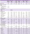
A total of 368 subjects with suspected E-TB were prospectively enrolled, and the T-SPOT.TB test was performed on blood samples. Of these patients, 196 (53.3%) were classified as having TB, including 119 (32.3%) with confirmed, 34 (9.2%) with probable, and 43 (11.7%) with possible TB, and 172 (46.7%) were classified as non-TB. Of the 368 patients, 223 (60.6%) had positive results on the T-SPOT.TB assay, and 113 (30.7%) had negative results. Finally, 32 (8.7%, 95% CI, 6.0% to 11.7%) patients had ITRs; of these, 13 (40.6%) had negative responses in the positive control wells, and 19 (59.4%) had high numbers of background spots in the nil-control wells. Of these 32 patients, 26 (81.2%) underwent tuberculin skin tests (TSTs), with 6 (23.1%) having positive results. There were no HIV-infected individuals in the ITR group. The baseline clinical characteristics of the patients with ITRs are presented in Table 1.
2. Risk factors for indeterminate test results in T-SPOT.TB
We evaluated the risk factors associated with ITRs. ITRs were not significantly associated with old age (≥70 years). In addition, underlying diseases, immunosuppressive states, and immunosuppressive treatment were not significantly associated with ITRs. Lymphocyte counts were not significantly different between the ITR group and non-ITR group (median 1,401 cells/mm3 [interquantile range (IQR), 1,034-2,013 cells/mm3] vs. median 1,279 cells/mm3 [IQR, 880-1,802 cells/mm3], P=0.16). Furthermore, we detected no significant differences between the two groups in the clinical manifestations of E-TB-namely, miliary TB, disseminated TB, and lymph node TB-or in the severity of E-TB. In the multivariate analysis, old age, underlying diseases, immunosuppressive states, and lymphopenia were not significantly associated with ITRs (Table 2). Subgroup analyses, including ITRs due to a low mitogen response and ITRs due to a high nil response, did not reveal any significant associations with old age, underlying diseases, immunosuppressive states, severity of E-TB, or clinical manifestations of E-TB (Table 1).
Discussion
This large prospective study revealed that ITRs in the T-SPOT.TB test are relatively frequent in routine clinical practice (8.7%, 95% CI, 6.0 to 11.7). This frequency is consistent with the frequencies reported in previous studies on the T-SPOT.TB [18-20] and QFT-TB [21-26] tests. However, we found that the traditional risk factors for ITRs identified in the QFT-TB test, such as old age, underlying diseases, immunosuppressive treatment, and lymphopenia, were not associated with ITRs in the T-SPOT.TB assay.
There have been several previous studies on risk factors for ITRs in the QFT-TB test. Kobashi et al. reported that ITRs were significantly affected by very old age, immunosuppressive therapy, lymphopenia, and hypoalbuminemia [24]. Other researchers reported that neutropenia and underlying diseases influencing cellular immunity, such as chronic renal disease, chronic lung disease, and autoimmune disease, were associated with ITRs in the QFT-TB test [21, 22, 25, 26]. However, the ELISPOT assay, on which the T-SPOT.TB test is based, is methodologically different from the ELISA used in the QFT-TB test [22]. For example, while PBMCs (250,000 cells per well) are used in the T-SPOT.TB assay, whole blood is used in the QFT-TB test, regardless of the leukocyte number. Thus, theoretically, it is possible that the T-SPOT.TB test is less affected than the QFT-TB test by lymphopenia. However, to the best of our knowledge, only one previous study has systematically evaluated the risk factors for ITRs in the T-SPOT.TB assay [20]. In that study, Beffa et al. found that the frequency of ITRs in the T-SPOT.TB test was 2.0%, and they reported that old age (> 75 years) and the season in which the samples were collected were related to ITRs [20]. However, they found no association between immunosuppression or lymphopenia and ITRs, in agreement with our findings [20]. It seems that lymphopenia may have little impact on the T-SPOT.TB test because, unlike the QFT-TB test, technically skilled personnel directly count the PBMCs in this test [18, 19]. The depression of cellular immunity in advanced age, which is a well-known risk factor for ITRs in the QFT-TB test, could be implicated in the higher frequency of ITRs in the T-SPOT.TB test [28]. However, Ferrara et al. noted that the T-SPOT.TB test tended to be less influenced by immunosuppressive treatment than the QFT-TB test, although they did not evaluate the association between ITRs in IGRAs and immunosuppression [21]. Therefore, this may explain our finding that extreme old age is not associated with ITRs in the T-SPOT.TB test.
It is counterintuitive that, in our study, the frequency of ITRs in the T-SPOT.TB test was similar to that of ITRs in the QFT-TB test in previous studies [21, 22, 24] despite the fact that the traditional risk factors for ITRs in the QFT-TB test are not risk factors for ITRs in the T-SPOT.TB test. Because our hospital is a tertiary referral hospital, however, we assume that more immunosuppressed patients were included in our study than in previous studies. Hence, direct comparison between previous studies and ours is difficult. Indeed, some factors, such as lymphopenia or immunosuppression, may actually be associated with ITRs in the T-SPOT.TB test. Alternatively, other factors-such as technical problems, including isolation of lymphocytes.may be associated with ITRs in this test. In addition, our study may not have sufficient power to detect these associations. However, because our study involved a relatively large number of patients (n = 368), it seems likely that traditional risk factors such as old age, lymphopenia, and immunosuppression do not significantly affect ITRs in the T-SPOT.TB test, or their effects on ITRs are minimal in routine clinical practice. Further studies on the risk factors for ITRs in the T-SPOT.TB test are needed to confirm our findings.
The current literature supports the possibility that the time required to transport samples to the laboratory and process them could influence the incidence of ITRs [20]. In addition, the freeze-thaw process in the T-SPOT.TB assay, in general, makes it difficult to recover enough cells for the test [29, 30]. However, as most of our samples were examined in a laboratory near the hospital within 4 to 6 hours and without freeze-thawing, we were not able to evaluate the processing time and freeze-thaw process as potential risk factors for ITR.
Some researchers have suggested that ITRs could be resolved by re-testing the affected patients, as most ITRs likely are caused by technical errors during transport or processing of the samples [26, 31]. It has been reported that valid results were obtained in 17% to 77% of patients with ITRs in IGRAs by re-testing them within a few months [20, 24]. Therefore, a possible limitation of the present study is that we did not evaluate the clinical utility of re-testing. However, it is likely that our study involved few technical and measurement errors, as the processing and transport times were relatively short and the T-SPOT.TB test results were interpreted by experts.
In conclusion, ITRs in the T-SPOT.TB test appear not to be affected by age, underlying disease, immunosuppressive conditions, lymphopenia, or the clinical manifestation of E-TB, unlike ITRs in QFT-TB. Further studies are needed to evaluate the predictive factors of ITRs in the T-SPOT.TB test.

 XML Download