

 PDF
PDF ePub
ePub Citation
 Print
Print


Abstract
Kikuchi's disease is a benign disease characterized mainly by fever and cervical lymphadenitis. We report a case of Kikuchi's disease that manifested as intra-abdominal lymphadenitis. A 39 year old woman presented with fever that had persisted for one week. Her history and physical examination were unremarkable. The laboratory findings revealed mild leukopenia and increased C-reactive protein. Abdominal CT revealed multiple lymph node enlargements in the mesenteric root and around the ileocecal valve. Positron emission tomography-computed tomography (PET-CT) revealed increased 18-fluoro-deoxyglucose(FDG) uptake in the lymph nodes observed by abdominal CT. A laparoscopic excisional biopsy of the lymph node was performed for a confirmatory diagnosis and the pathology findings were compatible with Kikuchi's disease. Although intra-abdominal Kikuchi's disease is a rare disease, it should be considered in a differential diagnosis of intra-abdominal lymphadenopathy with increased FDG uptake on PET-CT.
Figures and Tables
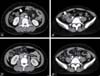 | Figure 1Abdominal CT scan shows enlarged (A) mesenteric root lymph nodes and (B) ileocolic lymph nodes. After one month, (C) enlarged mesenteric lymph nodes and (D) ileocolic lymph nodes had disappeared.
|
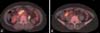 | Figure 2PET-CT scan shows multiple lymph nodes in the mesenteric root (A) and ileocolic area (B) with 18-fluorodeoxyglucose (FDG) uptake (standardized uptake value, SUV 11.3 and 6.8).
|

References
1. Kikuchi M. Lymphadenitis showing focal reticulum cells hyperplasia with nuclear debris and phagocytosis : A clinicopathological study. Acta Hematol Jpn. 1972. 35:379–380.
2. Dorfman RF, Berry GJ. Kikuchi's histiocytic necrotizing lymphadenitis: an analysis of 108 cases with emphasis on differential diagnosis. Semin Diagn Pathol. 1988. 5:329–345.
3. Rudin C, Wernli R, Rutishauser M, Ohnacker H. Mesenterial histiocytic necrotizing lymphadenitis. Case report. Helv Paediatr Acta. 1987. 42:35–40.
4. Yabe H, Sinzato I, Hashimoto K. Necrotizing lymphadenitis presenting as mesenteric lymphadenopathy. Rinsho Ketsueki. 1999. 40:658–662.
5. Kita Y, Kikuchi M, Nakae T, Nakai Y, Okamura A, Ohshima T, Hara T. A case of Kikuchi's disease with abdominal manifestations. Surgery. 1997. 122:962–963.

6. Rudniki C, Kessler E, Zarfati M, Turani H, Bar-Ziv Y, Zahavi I. Kikuchi's necrotizing lymphadenitis: a cause of fever of unknown origin and splenomegaly. Acta Haematol. 1988. 79:99–102.
7. Yoon YR, Lim JY, Park CH, Choi MB, Woo HO, Youn HS. Necrotizing lymphadenitis mimicking acute appendicitis affecting mesenteric lymph node. Korean J Pediatr Gastroenterol Nutr. 2003. 6:68–72.
8. Oh BJ, Choi WJ, Lim KS, Kim W. Kikuchi-Fujimoto disease: three cases presenting as acute abdomen. J Korean Soc Emerg Med. 2005. 16:194–199.
9. Ito K, Morooka M, Kubota K. Kikuchi disease: 18F-FDG positron emission tomography/computed tomography of lymph node uptake. Jpn J Radiol. 2010. 28:15–19.
10. Tsujikawa T, Tsuchida T, Imamura Y, Kobayashi M, Asahi S, Shimizu K, Tsuji K, Okazawa H, Kimura H. Kikuchi-Fujimoto disease: PET/CT assessment of a rare cause of cervical lymphadenopathy. Clin Nucl Med. 2011. 36:661–664.
11. Turner RR, Martin J, Dorfman RF. Necrotizing lymphadenitis. A study of 30 cases. Am J Surg Pathol. 1983. 7:115–123.
12. Huh J, Chi HS, Kim SS, Gong G. A study of the viral etiology of histiocytic necrotizing lymphadenitis (Kikuchi-Fujimoto disease). J Korean Med Sci. 1998. 13:27–30.
13. Ohshima K, Shimazaki K, Kume T, Suzumiya J, Kanda M, Kikuchi M. Perforin and Fas pathways of cytotoxic T-cells in histiocytic necrotizing lymphadenitis. Histopathology. 1998. 33:471–478.
14. Pileri SA, Facchetti F, Ascani S, Sabattini E, Poggi S, Piccioli M, Rondelli D, Vergoni F, Zinzani PL, Piccaluga PP, Falini B, Isaacson PG. Myeloperoxidase expression by histiocytes in Kikuchi's and Kikuchi-like lymphadenopathy. Am J Pathol. 2001. 159:915–924.
15. Famularo G, Giustiniani MC, Marasco A, Minisola G, Nicotra GC, De Simone C. Kikuchi Fujimoto lymphadenitis: case report and literature review. Am J Hematol. 2003. 74:60–63.
16. Han HS, Kim GH, Cho YS, Joo HJ, Lee OJ, Ryu DH, Lee KH, Kim ST. Intra-abdominal Kikuchi's disease mimicking malignant lymphoma on FDG PET-CT. Nucl Med Mol Imaging. 2009. 43:363–365.
17. Liao AC, Chen YK. Cervical lymphadenopathy caused by Kikuchi disease: positron emission tomographic appearance. Clin Nucl Med. 2003. 28:320–321.
18. Kaicker S, Gerard PS, Kalburgi S, Geller MD, Hailoo D. PET-CT scan in a patient with Kikuchi disease. Pediatr Radiol. 2008. 38:596–597.
19. Zhang MJ, Xiao L, Zhu YH, Jiang JJ, Jiang MS, He W. Lymph node uptake of 18F-fluorodeoxyglucose detected with positron emission tomography/computed tomography mimicking malignant lymphoma in a patient with Kikuchi disease. Clin Lymphoma Myeloma Leuk. 2010. 10:477–479.
20. Kim CH, Hyun OJ, Yoo IeR, Kim SH, Sohn HS, Chung SK. Kikuchi Disease Mimicking Malignant Lymphoma on FDG PET/CT. Clin Nucl Med. 2007. 32:711–712.
 XML Download