

 PDF
PDF ePub
ePub Citation
 Print
Print


Abstract
Background
Pelvic inflammatory disease (PID) is a common genital tract infection in reproductive women. This study aimed to determine the frequency of Neisseria gonorrheae, Chlamydia trachomatis, Ureaplasma urealyticum, and Mycoplasma hominis in Pelvic inflammatory disease (PID), and to further sub-analyze the clinical characteristics in patients diagnosed with Fitz-Hugh-Curtis syndrome (FHCS).
Material and Methods
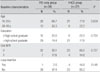
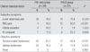
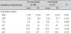
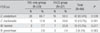
Sixty-six patients diagnosed clinically as PID were recruited from April, 2007 to February, 2011. Retrospective chart review was performed for investigating the characteristics of the clinical manifestation, laboratory findings, and image findings. And then all subjects were classified into two groups, the PID-only group and the FHCS group, depending on whether or not computed tomography showed increased perihepatic enhancement. Samples obtained in endocervical swabs were tested using Roche COBAS Amplicor Polymerase-chain reaction (PCR) for N. gonorrheae, C. trachomatis, U. urealyticum, and M. hominis.
Results
The 66 PID patients ranged in age from 19 to 49 years. Thirty nine patients were diagnosed as having an inflammation localized only in the lower abdomen (PID only), and 27 patients were diagnosed as FHCS. According to results of PCR, U. urealyticum was found most commonly in both the PID-only group and the FHCS group (66.7% and 59.3%, respectively).
Figures and Tables
References
1. Holmes KK, Handsfield HH. Braunwald E, Fauci AS, Kasper DL, Hauser SL, Longo DL, Jameson JL, editors. Sexually transmitted disease: overview and clinical approach. Harrison's principles of internal medicine. 2004. 16th ed. New York: McGraw-Hill;770.
2. Workowski KA, Berman SM. Center for Disease Control and prevention. Sexually transmitted disease treatment guidelines, 2006. MMWR Recomm Rep. 2006. 55:1–94.
3. Holmes KK, Sparling PF, Mardh PA, Lemon SM, Stamm WE, Pilot P, Wasserheit JN. Sexually transmitted disease. 1998. 3rd ed. New York: McGraw-Hill;738–809.
4. Hager WD. Follow-up of patients with tubo-ovarian abscess (es) in association with salpingitis. Obstet Gynecol. 1983. 61:680–684.
5. Sørbye IK, Jerve F, Staff AC. Reduction in hospitalized women with pelvic inflammatory disease in Oslo over the past decade. Acta Obstet Gynecol Scand. 2005. 84:290–296.

6. Simms I, Eastick K, Mallinson H, Thomas K, Gokhale R, Hay P, Herring A, Rogers PA. Association between Mycoplasma genitalium, Chlamydial trachomatis and pelvic inflammatory disease. J Clin Pathol. 2003. 56:616–618.
7. Cho MK. Update on the management of pelvic inflammatory disease. Korean J Obstet Gynecol. 2010. 53:961–966.
8. Patten RM, Vincent LM, Wolner-Hanssen P, Thorpe E Jr. Pelvic inflammatory disease. Endovaginal sonography with laparoscopic correlation. J Ultrasound Med. 1990. 9:681–689.
9. Sam JW, Jacobs JE, Birnbaum BA. Spectrum of CT findings in acute pyogenic pelvic inflammatory disease. Radiographics. 2002. 22:1327–1334.
10. Jung SI, Kim YJ, Park HS, Heon HJ, Jeong KA. Acute pelvic inflammatory disease: diagnostic performance of CT. J Obstet Gynaecol Res. 2011. 37:228–235.
11. Joo SH, Kim MJ, Lim JS, Kim JH, Kim KW. CT diagnosis of Fitz-Hugh and Curtis syndrome: value of arterial scan. Korean J Radiol. 2007. 8:40–47.
12. Ness RB, Soper DE, Holley RL, Peipert J, Randall H, Sweet RL, Sondheimer SJ, Hendrix SL, Amortegui A, Trucco G, Songer T, Lave JR, Hillier SL, Bass Dc, Kelsey SF. Effectiveness of inpatient and outpatient treatment strategies for women with pelvic inflammatory disease: results from the pelvic inflammatory disease evaluation and clinical health (PEACH) Randomized trial. Am J Obstet Gynecol. 2002. 186:929–937.
13. Boom R, Sol CJ, Salimans MM, Jnsen CL, Wertheim van Diller PM, van der Noordaa J. Rapid and simple method for purification of nucleic acids. J Clin Microbiol. 1990. 28:495–503.
14. Blanchard A, Henschel J, Duffy L, Baldus K, Cassel GH. Detection of Ureaplasma urealyticum by polymerase chain reaction in the urogenital tract of adult, in amniotic fluid, and in the respiratory tract of newborn. Clin Infect Dis. 1993. 17:Suppl 1. S148–S153.
15. Lee HH, Ju KS, Lee KH, Won NH. Detection of chlamydia trachomatis, Mycoplasma hominis and Ureaplasma urealyticum in the cervical swab and paraffin tissue with female genital tract infection. Korean J Obstet Gynecol. 1999. 42:549–555.
16. Zhou B, Cong L, Sha Y. Pathogens of transmitted disease in the pathogenesis of acute pelvic inflammatory disease. Zhonghua Fu Chan Ke Za Zhi. 2001. 36:539–541.
17. Rodrigues MM, Fernandes PÁ, Haddad JP, Paiva MC, Souza Mdo C, Andrade TC, Fernandes AP. Frequency of Chlamydia trachomatis, Neisseria gonorrhoeae, Mycoplasma genitalium, Mycoplasma hominis and Ureaplasma species in cervical samples. J Obstet Gynaecol. 2011. 31:237–241.
18. Núñez-Troconis JT. Mycoplasma hominis and ureaplasma urealyticum in different gynecologic disease. Invest Clin. 1999. 40:9–24.
19. Grześko J, Elias M, Maczyńska B, Kasprzykowska U, Tłaczała M, Goluda M. Frequency of detection of ureaplasma urealyticum and Mycoplasma hominis in cervical canal and Douglas pouch of infertile and fertile women. Med Dosw Mikrobiol. 2007. 59:169–175.
20. Mårdh PA, Weström L. T-mycoplasma in genito-urinary tract of the female. Acta Pathol Microbiol Scand B Microbiol Immunol. 1970. 78:367–374.
21. Møller BR, Sparre Jørgensen A, From E, Stenderup A. Chlamydia, mycoplasma, ureaplasmas, and yeasts in the lower genital tract of female. Comparison between a group attending a venereal disease clinic and a control group. Acta Obstet Gynecol Scand. 1985. 64:145–149.
22. Tully JG, Taylor-Robinson D, Cole RM, Rose DL. A newly discovered mycoplasma in the human urogenital tract. Lancet. 1981. 1:1288–1291.
23. Idriss WM, Patton WC, Taymor ML. On the etiologic role of ureaplasma urealyticum (T-mycoplasma) infection in infertility. Fertil Steril. 1978. 30:293–296.
24. Foulon W, Naessen A, Cammu H, Gossens A, Lauwers S. Epidemiology and pathogenesis of ureaplasma urealyticum in spontaneous abortion and early premature labor. Acta Obstet Gynecol Scand. 1987. 66:513–516.
25. Embree JE, Krause VW, Embril JA, MacDonald S. Placental infection with Mycoplasma hominis and Ureaplasma urealyticum: clinical correlation. Obstet Gynecol. 1980. 56:475–481.
26. Kim SY, Lee YJ, Huh M, Lee SH, Park IY, Ahn HY, Moon HB, Shin JC, Kim SP. Analysis of Mycoplasma hominis and Ureaplasma urealyticum infection in preterm labor and PROM patients. Korean J Obstet Gynecol. 2004. 47:1469–1473.
27. Division of Health Prevention and Disease Prevention. Preventing low birthweight. 1985. Washington DC: National Academy Press;4–14.
 XML Download