Abstract
Purpose
The purpose of this study is to evaluate the accuracy of surgical stent according to the supported type.
Materials and methods
5 sets of dental models which have tooth supported edentulous area and tooth-tissue supported edentulous area were made. Dental model were scanned with model scanner, and CBCT was taken. CT data and model scan data were overlapped using In2Guide software, implant were virtually planned in the software. Surgical stents are fabricated by 3D printing. The implant fixture were installed using the surgical stent, CBCT were retaken. CBCT before surgery and after surgery were overlapped, and the differences (angle difference, coronal difference, apical difference) were evaluated using statistical analysis.
Results
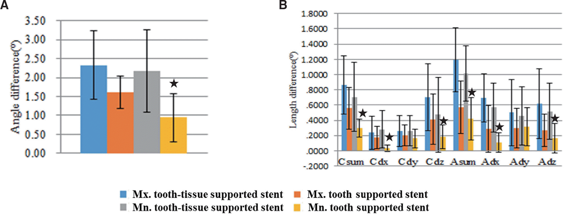
In the assessment of the accuracy of surgical guides according to arch type, there are no statistically significant differences between maxilla and mandible. In the case of support type, tooth supported stents showed lower angle difference and length difference than tooth-tissue supported stents, which are statistically significant.
Go to : 
REFERENCES
1.Van Steenberghe D., Malevez C., Van Cleynenbreugel J., Bou Serhal C., Dhoore E., Schutyser F., Suetens P., Jacobs R. Accuracy of drilling guides for transfer from three-dimensional CT-based planning to placement of zygoma implants in human cadavers. Clin Oral Implants Res. 2003. 14:131–6.

2.Ramasamy M., Giri ., Raja R., Subramonian ., Karthik ., Naren-drakumar R. Implant surgical guides: From the past to the present. J Pharm Bioallied Sci. 2013. 5:S98–S102.

3.Malo P., de Araujo Nobre M., Lopes A. The use of computer-guided flapless implant surgery and four implants placed in immediate function to support a fixed denture: preliminary results after a mean follow-up period of thirteen months. J Prosthet Dent. 2007. 97:S26–34.

4.Somogyi-Ganss E., Holmes HI., Jokstad A. Accuracy of a novel prototype dynamic computer-assisted surgery system. Clin Oral Implants Res. 2015. 26:882–90.

5.Stumpel LJ. Deformation of stereolithographically produced surgical guides: an observational case series report. Clin Implant Dent Relat Res. 2012. 14:442–53.

6.Karami D., Alborzinia HR., Amid R., Kadkhodazadeh M., Yousefi N., Badakhshan S. In-Office Guided Implant Placement for Prosthetically Driven Implant Surgery. Craniomaxillofac Trauma Reconstr. 2017. 10:246–54.

7.Nickenig HJ., Eitner S. Reliability of implant placement after virtual planning of implant positions using cone beam CT data and surgical (guide) templates. J Craniomaxillofac Surg. 2007. 35:207–11.

8.Widmann G., Widmann R., Widmann E., Jaschke W., Bale R. Use of a surgical navigation system for CT-guided template production. Int J Oral Maxillofac Implants. 2007. 22:72–8.
9.Holst S., Blatz MB., Eitner S. Precision for computer-guided implant placement: using 3D planning software and fixed intraoral reference points. J Oral Maxillofac Surg. 2007. 65:393–9.

10.Di Giacomo GA., Cury PR., de Araujo NS., Sendyk WR., Sendyk CL. Clinical application of stereolithographic surgical guides for implant placement: preliminary results. J Periodontol. 2005. 76:503–7.
11.Van Assche N., van Steenberghe D., Guerrero ME., Hirsch E., Schutyser F., Quirynen M., Jacobs R. Accuracy of implant placement based on pre-surgical planning of three-dimensional cone-beam images: a pilot study. J Clin Periodontol. 2007. 34:816–21.

12.Tardieu PB., Vrielinck L., Escolano E., Henne M., Tardieu AL. Computer-assisted implant placement: scan template, sim-plant, surgiguide, and SAFE system. Int J Periodontics Restorative Dent. 2007. 27:141–9.
13.Park C., Raigrodski AJ., Rosen J., Spiekerman C., London RM. Accuracy of implant placement using precision surgical guides with varying occlusogingival heights: an in vitro study. J Prosthet Dent. 2009. 101:372–81.

Go to : 
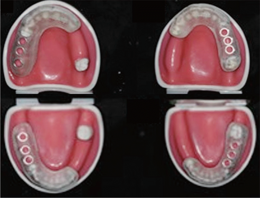
 | Fig. 1.The dental model was shown. (A) Frontal view, (B) Lateral view, (C) Occlusal view of maxilla, (D) Occlusal view of mandible. |
 | Fig. 2.Model scanning data. (A) Occlusal view of maxilla, (B) Lateral view of maxilla, (C) Occlusal view of mandible, (D) Lateral view of mandible. |
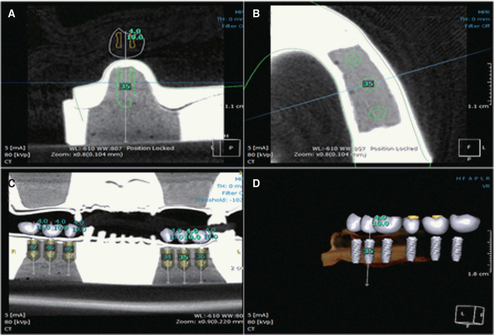
 | Fig. 3.Planning of implant installation at dental CT software. (A) CT view of proper position of implants, (B) Proper location of implants, (C, D) The proper path and position of implants were confirmed by using CT. |
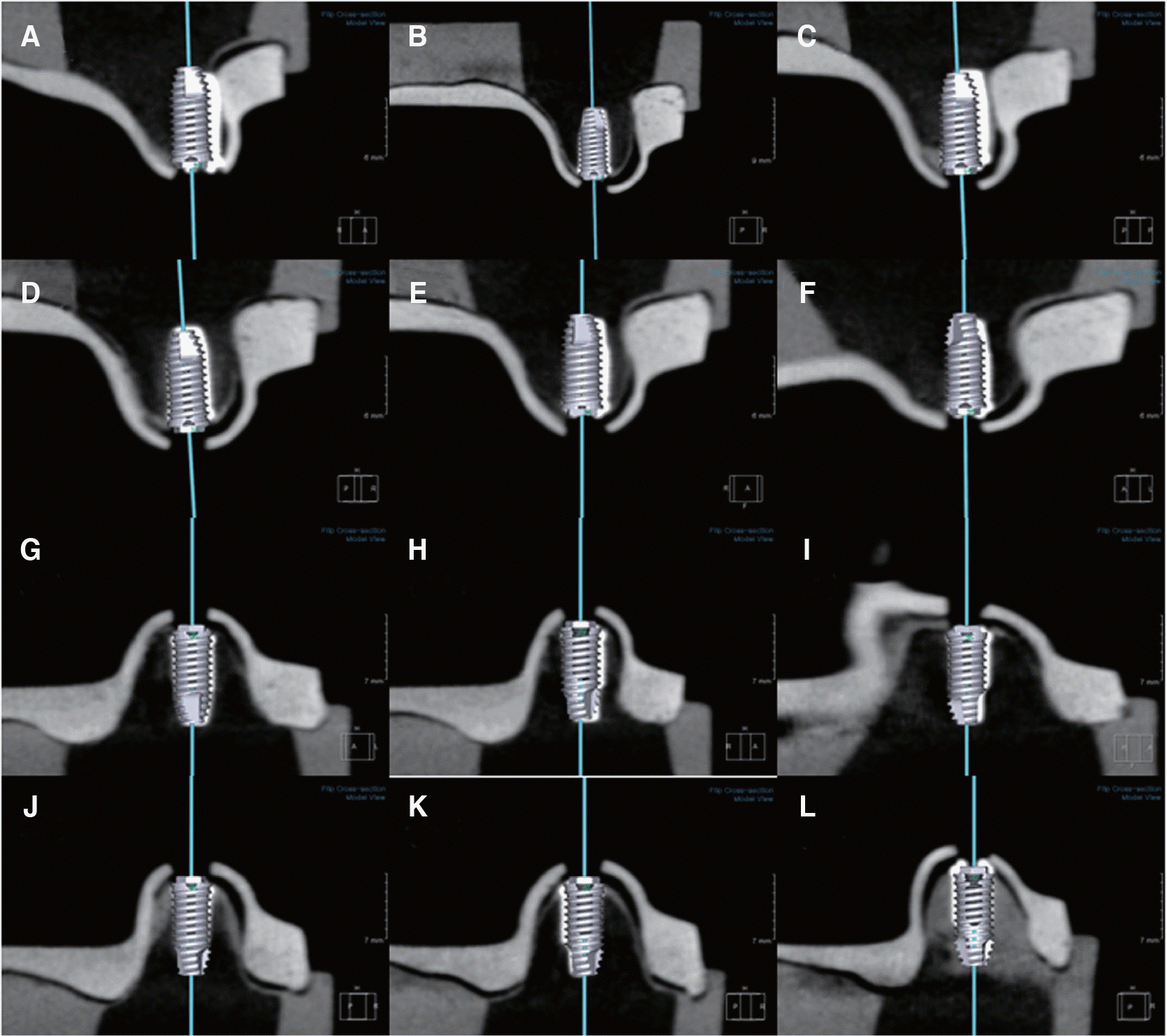
 | Fig. 5.Overlapping observation between the implant planning data and implant installation data. (A) #15i, (B) 16i, (C) 17i, (D) 24i, (E) 25i, (F) 26i, (G) 34i, (H) 35i, (I) 36i, (J) 45i, (K) 46i, (L) 47i. |
 | Fig. 6.Accuracy result of guided surgery according arch type. (A) Angle difference results, (B) Length difference results. |
 | Fig. 7.Accuracy result of guided surgery according supported type (★ significant at P < .05). (A) Angle difference results, (B) Length difference results. |
 | Fig. 8.Accuracy result of guided surgery accroding to arch type and support type (★ significant at P < .05). (A) Angle difference result, (B) Length difference. |
Table 1.
The accuracy result between maxilla and mandible
Table 2.
Accuracy result of guided surgery according to support type
Table 3.
Accuracy result of guided surgery according to arch type and support type


 PDF
PDF ePub
ePub Citation
 Print
Print


 XML Download