

 PDF
PDF ePub
ePub Citation
 Print
Print


Ju-Nam An1 , Jung-Jin Lee1, Jae-Min Seo1, Kyoung-A Kim2
, Jung-Jin Lee1, Jae-Min Seo1, Kyoung-A Kim2
Abstract
Prosthetic treatment using implants in fully edentulous patients includes implant-supported fixed prosthesis, implant hybrid prosthesis, implant retained- or supported-overdenture and implant supported fixed prosthesis has advantages such as psychological stability, pronunciation. If an implant supported fixed prosthesis is planned, the implants should be placed in consideration of pronunciation, esthetics, and oral hygiene. For this, clinical and radiological diagnosis is indispensable. When placing the prosthetic driven implant at the site determined from the diagnosis, a sufficient amount of alveolar bone and soft tissue support are required. If these requirements found to be insufficient, a wide range of bone grafting should be performed in advance. In this case, a fully edentulous patient with severe alveolar bone resorption due to periodontal disease was treated with a full mouth rehabilitation using implant-supported fixed prosthesis preceding maxillary sinus graft and alveolar bone augmentation. We report this patient were satisfied with esthetic and function.
Figures and Tables
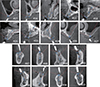
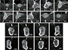
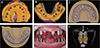
 | Fig. 4Occlusal view of clinical images at bone graft. (A) Pre-operative view of maxillary left area, (B) After sinus graft and alveolar bone augmentation of maxillary left area, (C) Flap closure of maxillary left area, (D) Pre-operative view of maxillary right area, (E) After alveolar bone augmentation of maxillary right area, (F) Flap closure of maxillary right area, (G) Pre-operative view of mandible, (H, I) After alveolar bone augmentation of mandibular posterior region, (J) Flap closure of mandible.
|
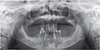
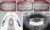
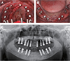 | Fig. 6(A) Implant surgery on maxilla, (B) Implant surgery on mandible, (C) Panorama view was taken after implant placement in maxilla and mandible.
|
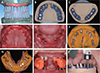 | Fig. 7(A) Computer aided design of individualized abutment, (B) Individualized zirconia and titanium abutments was connected to working model of maxilla, (C) Individualized titanium abutments was connected to working model of mandible, (D) Bonnet crown are connected to implant abutment of maxilla, (E) Bonnet crown are connected to implant abutment of mandible, (F) Pick-up impression of maxillary implant bonnet crown, (G) Pick-up impression of mandibular implant bonnet crown, (H) Registration of inter-occlusal relationship with auto-polymerized acrylic resin bite jig & silicone bite registration material, (I) Additional milling of individualized implant abutment on newly created working model.
|
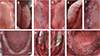
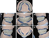 | Fig. 8Full contour wax-up for provisional restorations. (A) Maxillary occlusal view, (B) Right lateral view, (C) Frontal view (D) Left lateral view, (E) Lateral movement - right side, (F) Mandibular occlusal view, (G) Lateral movement - left side.
|
 | Fig. 10(A) Final impression taking of maxilla, (B) Final impression taking of mandible, (C) Final working model of maxilla, (D) Final working model of mandible, (E) Registration of inter-occlusal relationship with auto-polymerized acrylic resin bite jig, (F) Fabrication of customized anterior incisal guide table.
|
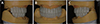
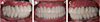
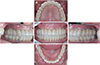 | Fig. 11Definitive prosthesis. (A) Maxillary occlusal view, (B) Right lateral view, (C) Frontal view, (D) Left lateral view, (E) Mandibular occlusal view.
|
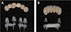 | Fig. 12Fabricating abutment replica with hot melt adhesive material. (A) Abutment replica of maxillary anterior prosthesis, (B) Abutment replica of mandibular anterior prosthesis.
|
 | Fig. 13Eccentric occlusion with definitive prosthesis. (A) Anterior movement: posterior teeth of both side were disoccluded, (B) Lateral movement - right side: group function, (C) Lateral movement - left side: group function, (D) Occlusal analysis using T-scan III: Equal distribution of occlusal force for whole dentition, (E) Post-treatment panoramic radiograph.
|

References
1. Misch CE. Dental implant prosthetics. 1st ed. Mosby;2004. p. 43–52.
2. Pozidi G, Lamprinoudi A, Papavasileiou G, Kamposiora P, Pallis D. Implant rehabilitation of the edentulous maxilla. A case series of different treatment options. Clin Oral Impl Res. 2016; 27:389.
3. Gallucci GO, Bernard JP, Belser UC. Treatment of completely edentulous patients with fixed implant-supported restorations: three consecutive cases of simultaneous immediate loading in both maxilla and mandible. Int J Periodontics Restorative Dent. 2005; 25:27–37.
4. Beretta M, Cicci M, Poli PP, Rancitelli D, Bassi G, Grossi GB, Maiorana C. A retrospective evaluation of 192 implants placed in augmented bone: Long-term follow-up study. J Oral Implantol. 2015; 41:669–674.

5. Lekovic V, Kenney EB, Weinlaender M, Han T, Klokkevold P, Nedic M, Orsini M. A bone regenerative approach to alveolar ridge maintenance following tooth extraction. Report of 10 cases. J Periodontol. 1997; 68:563–570.
6. Albrektsson T, Zarb G, Worthington P, Eriksson AR. The long-term efficacy of currently used dental implants: a review and proposed criteria of success. Int J Oral Maxillofac Implants. 1986; 1:11–25.
7. Hürzeler MB, Kirsch A, Ackermann KL, Quiñones CR. Reconstruction of the severely resorbed maxilla with dental implants in the augmented maxillary sinus: a 5-year clinical investigation. Int J Oral Maxillofac Implants. 1996; 11:466–475.
8. Misch CE. Contemporary Implant Dentistry. 3rd ed. St. Louis: CV Mosby;2009. p. 367–388.
9. Seo CW, Han AR, Seo JM, Lee JJ. A technique for fabricating abutment replica with hot melt adhesive material to minimize residual cement in implant restoration: a case report. J Dent Rehabil Appl Sci. 2016; 32:240–245.
10. Sadowsky SJ. The implant-supported prosthesis for the edentulous arch: design considerations. J Prosthet Dent. 1997; 78:28–33.
11. Henry PJ. An alternative method for the production of accurate casts and occlusal records in osseointegrated implant rehabilitation. J Prosthet Dent. 1987; 58:694–697.
12. Assif D, Marshak B, Schmidt A. Accuracy of implant impression techniques. Int J Oral Maxillofac Implants. 1996; 11:216–222.
13. Vigolo P, Fonzi F, Majzoub Z, Cordioli G. An evaluation of impression techniques for multiple internal connection implant prostheses. J Prosthet Dent. 2004; 92:470–476.
14. Cheng CW, Chien CH, Chen CJ, Papaspyridakos P. Complete-mouth implant rehabilitation with modified monolithic zirconia implant-supported fixed dental prostheses and an immediate-loading protocol: a clinical report. J Prosthet Dent. 2013; 109:347–352.
15. Baldissara P, Llukacej A, Ciocca L, Valandro FL, Scotti R. Translucency of zirconia copings made with different CAD/CAM systems. J Prosthet Dent. 2010; 104:6–12.
 XML Download