

 PDF
PDF ePub
ePub Citation
 Print
Print


Introduction
Tooth wear refers to loss of tooth surface by causes other than dental caries, dental trauma or developmental disability.1 Tooth wear can be categorized into physiological wear and pathological wear. Physiological wear occurs gradually through a lifetime and it is accepted as a natural phenomenon. However pathological wear occurs at a certain time with an excessive amount.23
An important consideration in restoring worn dentition is to determine the amount of occlusal vertical dimension (OVD) that has been lost. In the case of physiological wear, it is known that OVD can be maintained due to the eruption of the teeth and the compensatory growth of the alveolar bone.45 On the other hand, pathological tooth wear is faster than the compensatory mechanism, which accompanies loss of OVD. As a result, side effects occur such as esthetic problems, decreased masticatory function, and joint disease due to loss of anterior and lateral guidance.6
Turner and Missirlian recommend to set up a treatment plan considering the amount of decreased OVD and interocclusal space required for prosthetic rehabilitaion.7 It is often necessary to restore decreased OVD in order to secure interocclusal space for prosthetic restoration and improve occlusion and esthetics.78 Increment of OVD should be carefully determined through overall analysis of the patient. When increasing the OVD, its effects to the temporomandibular joints, neuromuscular activity, and pronunciation must be monitored carefully.9
The patient in this case had excessive wear of the mandibular anterior teeth due to loss of mandibular posterior teeth for a long time, moreover the opposing maxillary dentition were porcelain prosthesis. This clinical report describes that a satisfactory clinical result was achieved with an improvement in functions and esthetics by full mouth rehabilitation restoring the OVD using removable partial dentures and few implants.
Case report
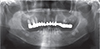
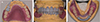
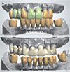
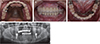
The patient was a 75-year-old female who was referred from the Department of Periodontics for prosthetic consultation. Her chief complaint was that it was inconvenient to eat because mandibular posterior teeth have been lost for a long time and mandibular anterior teeth were worn out. The patient was taking hypertension drugs and had history of bisphosphonate injections due to osteoporosis. A 14-unit one-piece fixed partial denture (FPD) on maxillary teeth was made 10 years ago and the mandibular molars were extracted about 5 years ago. Porcelain fracture on the left premolar area of maxillary porcelain fused metal (PFM) prosthesis and decementation with secondary caries on posterior molars were observed (Fig. 1). Missing teeth on mandible were right first and second molars, left second premolar, first and second molars. Severe occlusal wear was observed in all remaining mandibular teeth. Caries and pulp exposure were accompanied on several mandibular teeth. Radiographic examination revealed wears on occlusal surface of the mandibular teeth and severe secondary caries with peri-apical lesions on the maxillary posterior teeth (Fig. 2). There were no suspicious radiologic findings on temporomandibular joint, and mandibular opening movement was also in a normal range. There was no history of para-functional habit or TMJ disease. The patient had a brachycephalic face with slightly developed masseter muscles.
Preliminary impression was obtained to make diagnostic casts. The diagnostic casts were mounted on a semi-adjustable articulator (Hanau Modular Articulator, Whip Mix Corp., Louisville, KY, USA) with a face-bow record and a centric relation registration record. There was not enough interocclusal space to restore the missing mandibular molars. As the result, it was decided that full mouth rehabilitation with OVD increment for functional and esthetic restoration. The patient's freeway space was 5 mm, being greater than the normal value, 2 – 4 mm. Based on the analysis of patient's facial appearance, freeway space and interocclusal space for restoration, increment of OVD was 2 mm for functional and esthetical prosthesis. According to Turner and Missirlian's classification of tooth wear, this patient was in category 1 which indicates excessive tooth wear with loss of OVD.7 In this category, reduced OVD should be restored with trial restorations to evaluate patient's adaptation and function at the increased OVD, and then definitive restoration should be processed.
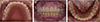
On the semi-adjustable articulator, OVD was elevated at 2 mm with incisal pin, and diagnostic wax up was made (Fig. 3). Temporary shells were prefabricated using autopolymerizing acrylic resin (Alike, GC America Inc., Chicago, IL, USA) based on the diagnostic wax-up. The maxillary one-piece FPD was removed, and maxillary first and second molars on both sides which were hopeless teeth were extracted. Then an 8-unit provisional FPD with maxillary right second premolar, right lateral incisor, left lateral incisor and canine abutment was delivered. Mandibular right first and second premolar and left first premolar were extracted and provisional restorations were delivered to the remained teeth after preparation. Provisional removable partial denture was fabricated for posterior edentulous area of maxilla and mandible (Fig. 4). After provisional restoration placement, reduced freeway space to 2 mm was verified which is in a normal range.
The patient used provisional restorations for 6 weeks. The patient was well adapted to the provisional prosthesis with raised OVD and did not show temporomandibular joint discomfort. Thus, definitive restorations were designed to reproduce OVD in provisional restorations. The patient wanted removable partial dentures and implants which are covered by national health insurance for definitive restorations, and did not want complicated surgery such as bone grafting. Therefore, definitive treatment plan was to make removable partial dentures (RPD) with surveyed crowns. In addition, placement of two implants in the mandibular posterior region was planned to obtain additional retention and support for the mandibular RPD.
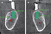
Cone-beam computed tomography was taken with diagnostic stent prepared by duplicating the provisional removable partial denture for implant placement (Fig. 5). In selecting positions for implant placement, it was considered that selecting mandibular premolar site to make Kennedy class I RPD, but alveolar bone was so narrow that guided bone regeneration was required for implant placement at planned position. On the other hand, the second molar site had sufficient bone width for an implant with a diameter of 5 mm. Considering the distance to the inferior alveolar nerve, internal connection implants (TSIII SA; Osstem implant Co., Busan, Korea) were placed on mandible, diameter of 5.0 and a height of 8.5 mm on right second molar, diameter of 5.0 and a height of 10 mm on left second molar. Root canal treatment and post and core build up was performed on mandibular right lateral incisor, canine, left lateral incisor, canine, first premolar and maxillary right second premolar due to hypersensitivity.
After osseointegration of implants, the definitive impression was made. The autopolymerizing acrylic resin (SR Ivoren, Ivoclar Vivadent, Schaan, Liechtenstein) was used to make individual trays. The pick-up impression coping was fastened to the mandibular implant and definitive preparation of teeth was done. Definitive impression was made with polyvinyl siloxane impression material (Imprint II Garant, 3M ESPE, St. Paul, MN, USA) for surveyed crowns. The master casts were fabricated with a Type IV dental stone (GC Fujirock EP, GC Europe N. V., Leuven, Belgium). Occlusal rims were fabricated with visible light-curing acrylic resin (Triad TruTray VLC, Dentsply Inc., York, PA, USA) and baseplate wax (Truwax Baseplate Wax, Dentsply Inc., York, PA, USA). Interocclusal registration was made with occlusal rim and polyvinylsiloxane occlusal registration material (O-bite, DMG, Hamburg, Germany). The casts were mounted on a semi-adjustable articulator (Hanau Modular Articulator, Whip Mix Corp., Louisville, KY, USA) with a face-bow record. On the mounted working casts, appropriate size of stock abutments was selected and milled.
The definitive fixed restorations were PFM surveyed crowns that were designed and fabricated in a digital method. 3D laser scanner (3Shape D800, 3Shape A/S, Copenhagen, Denmark) was used to make digital impressions of mater casts and interocclusal relation. Computer-aided design program (3Shape Dental Manager, 3Shape A/S, Copenhagen, Denmark) was used to make digital wax-ups for crowns (Fig. 6A). Then the digital wax-ups were cut back to allow for porcelain application (Fig. 6B). PFM frameworks were made of cobalt-chrome alloy through laser-sintering technique using 3D printer (EOSINT M270, Electro Optical Systems GmbH, Munich, Germany). An 8-unit FPD in maxilla and single crowns in mandible were fabricated. The metal frameworks were tried in and confirmed good fit and marginal adaptation. The metal frameworks were veneered with porcelain. The definitive fixed prostheses were cemented with resin modified glass ionomer cement (FujiCEM II, GC Corp., Tokyo, Japan).
Polyvinyl siloxane impression material (Exadenture, GC Corp., Tokyo, Japan) with individual tray was used for the definitive impression of RPD. The maxillary RPD was Kennedy Class I which uses palatal plate as major connector, and the mandibular RPD was Kennedy Class III which uses lingual bar as major connector. RPD frameworks were fabricated with the conventional casting technique. Fit of RPD frameworks was examined intraorally with silicone material (Fit-checker, GC, Tokyo, Japan), which showed good adaptation and satisfactory retention. On top of the frameworks, baseplate wax (Truwax Baseplate Wax, Dentsply Inc., York, PA, USA) was build up to make occlusal wax rim and inter-occlusal relation was recorded with bite record material (O-bite, DMG, Hamburg, Germany). Master casts were mounted on articulator and denture teeth were aligned by mutually protected occlusion. After the trial of wax dentures, curing with pink denture resin (Rapid simplified, Vertex, Zeist, Netherlands) were carried on. Maxillary and mandibular removable partial dentures were delivered to the patient (Fig. 7). Oral hygiene instruction and regular follow up check were administered. After follow-up checks at one week, one month, six months, and 1 year interval, the result was functionally and esthetically satisfying.
Discussion
Loss of posterior support has been identified as the cause for severe anterior attrition and decreased OVD.10 In this case, it is considered that the pathological teeth wear has progressed as the mandibular natural anterior teeth are occluded with the maxillary PFM restorations. The reduced OVD was restored and the posterior support was established through full mouth rehabilitation using surveyed crowns, removable partial dentures, and implants. Freeway space, interocclusal space for restoration and facial appearance were carefully evaluated to determine the amount of OVD increment, and OVD was increased by 2 mm through prosthetic rehabilitation. In general, increasing OVD up to 5 mm is considered to be a feasible alteration and whether an alteration of OVD exceeds the capacity of the neuromuscular system should be evaluated through provisional restorations or removable appliances.11 The patient used the provisional restorations for about 4 months and there were no discomforts, temporomandibular joint disorders, and wears or fractures of the provisional restoration. In the definitive restorations, the anterior guidance was established by fixed restorations on the anterior teeth and uniform occlusal contact of the posterior teeth was achieved in the centric occlusion, preventing recurrence of wears and blocking lateral forces to the posterior implants. No abnormal findings were found during the follow-up period of 1 year. It is necessary to confirm the maintenance of occlusion through periodic observation, and occlusal adjustment or denture relining may be required.
In this case, the laser-sintering technique used in the fabrication of metal frameworks of PFM restorations is a new computer aided design/computer aided manufacturing (CAD/CAM) method. Based on the three-dimensional CAD data, high intensity laser can rapidly fuse base metal alloy powder. This technique can save manufacturing time and costs compared to the lost wax casting technique and good results have been reported in respect of internal fit, bond strength with porcelain, and strength.1213 Because of the risk of casting distortion for maxillary long-span PFM framework, laser-sintering technique was used in this case and good marginal fit of the frameworks was confirmed.
The mandibular Kennedy Class I RPD requires periodic denture relining to establish posterior support, because the alveolar ridge under the distal extension resorbs. Therefore, a cost-effective method to solve this problem has been reported by several authors, which is to establish the posterior support by placing implants in the posterior region.1415 This treatment which provides additional support and retention to removable partial denture using a small number of implants is referred to as implant-assisted removable partial denture (IARPD).16
In this case, the patient wanted an economical treatment and rejected complicated surgery, so IARPD was fabricated and delivered by placing implants at the mandibular second molar sites. Since 2016, in Korea, dentures and implants for elderly patients over 65 years old have been covered by national health insurance. Because up to two implants including upper prosthesis are covered by national health insurance, two implants were placed in the mandibular left and right second molar sites, and implant surveyed crowns were fabricated. Therefore, the effect of converting the mandibular Kennedy Class I RPD to the Kennedy Class III RPD was obtained.
The technique using surveyed crowns on implants in IARPD has been reported only in case reports and all cases were about distal extension RPD.17181920 On the other hand, in this case, two single implants were placed on the mandibular posterior molars, and the occlusal rests and the retention arms were designed on the implant surveyed crowns, so it was suspected that functional force will be exerted on the implant during insertion, removal and function. However, the bone quality of implants insertion site was good, and the measured implant stability quotient (ISQ) values were 88 (mandibular left implant) and 86 (mandibular right implant). It is also expected that the implants will have a good prognosis because the occlusal force on the implants will not be large due to the opposing maxillary distal extension RPD and because the implant is protected during lateral & protrusive excursion by canine guidance.
Conclusion
In this case, the OVD of the patient was evaluated by examining freeway space, interocclusal space for restoration and analyzing facial appearance. Based on this, diagnostic wax-up was made to determine occlusal restoration, and a temporary prosthesis was fabricated to evaluate the patient's adaptation to the increased OVD. After confirming patient's comfort, full mouth rehabilitation with combination of removable partial denture and few implants was completed. This resulted in economical, functional, and esthetically pleasing outcome.

 XML Download