

 PDF
PDF ePub
ePub Citation
 Print
Print


Cheol-Keun Kang , Seong-Joo Heo, Seong-Kyun Kim, Jai-Young Koak
, Seong-Joo Heo, Seong-Kyun Kim, Jai-Young Koak
Abstract
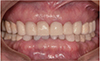
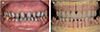
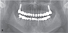
Severe dental attrition causes pathological changes of the tooth, collapsed occlusion, and functional and aesthetic complications and can also result in a decrease in occlusal vertical dimension. Before increasing the vertical dimension with full-mouth rehabilitation, it is important to determine the amount of vertical dimension through accurate diagnosis. In this case, a 77 year old elderly male patient on anticoagulant medication with generalized attrition and fracture of teeth was treated with full-mouth rehabilitation in order to recover vertical dimension and aesthetics. Accurate clinical and radiographic examination, diagnostic, wax-up, and occlusal vertical dimension evaluation were step by step performed considering pre-medical history and old age. Patient adaptability was evaluated using an occlusal splint and interim restoration. After 3 months of stabilization with interim restoration, definitive prostheses were fabricated. Satisfactory functional and esthetic outcomes are observed after 6 months of follow up.
Figures and Tables
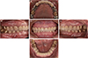
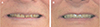
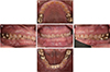 | Fig. 1Intraoral photograph before treatment. (A) Maxillary, (B) Right, (C) Frontal, (D) Left, (E) Mandibular.
|
 | Fig. 6Cross articulation. (A) Mounting of provisional restoration, (B) Customized guide table, (C) Mounting of working cast.
|


References
1. Dawson PE. Functional Occlusion: from TMJ to smile design. St. Louis; MO: Mosby;2007. p. 430–452.
2. Lerner J. A systematic approach to full-mouth reconstruction of the severely worn dentition. Pract Proced Aesthet Dent. 2008; 20:81–87.
3. Dombrady L. Investigation into the transient instability of the rest position. J Prosthet Dent. 1966; 16:479–490.

4. Tallgren A, Lang BR, Walker GF, Ash MM Jr. Changes in jaw relations, hyoid position, and head posture in complete denture wearers. J Prosthet Dent. 1983; 50:148–156.
5. Murphy T. Compensatory mechanisms in facial height adjustment to functional tooth attrition. Australian Dent J. 1959; 4:312–323.
6. Berry DC, Poole DF. Attrition: possible mechanisms of compensation. J Oral Rehabil. 1976; 3:201–206.
7. Rivera-Morales WC, Mohl ND. Restoration of the vertical dimension of occlusion in the severely worn dentition. Dent Clin North Am. 1992; 36:651–664.
8. Turner KA, Missirlian DM. Restoration of the extremely worn dentition. J Prosthet Dent. 1984; 52:467–474.
9. McGEE GF. Use of facial measurements in determining vertical dimension. J Am Dent Assoc. 1947; 35:342–350.
10. Willis FM. Features of the face involved in full denture prosthesis. Dent Cosmos. 1935; 77:851–854.
11. Oh SC, Jung JH. Morphology and size of clinical crown of permanent upper anterior teeth in Korean adult. J Korean Acad Stomatognathic Funct Occlusion. 2001; 17:37–42.
12. Kwon KR. The prosthetic approach for collapsed vertical dimensions of occlusion. J Dent Rehabil Appl Sci. 2004; 20:169–181.
13. Nelson SJ. Wheeler's dental anatomy, physiology and occlusion. 9th ed. St. Louis: Sanunders Elsevier Health Sciences;2009. p. 99–139.
14. Grippo JO, Simring M, Schreiner S. Attrition, abrasion, corrosion and abfraction revisited: a new perspective on tooth surface lesions. J Am Dent Assoc. 2004; 135:1109–1118.
15. Ahmad I. Geometric considerations in anterior dental aesthetics: restorative principles. Pract Periodontics Aesthet Dent. 1998; 10:813–822.
16. Wood GN. Centric relation and the treatment position in rehabilitating occlusions: a physiologic approach. Part II: The treatment position. J Prosthet Dent. 1988; 60:15–18.
 XML Download