

 PDF
PDF ePub
ePub Citation
 Print
Print


Hae-Yong Jeong , Yu-Sung Choi
, Yu-Sung Choi
Abstract
It is reported that the causes of unaesthetic proportion of anterior teeth vary widely. Especially, when the unaesthetic tooth proportion of the mandibular incisors arises due to the wear of the anterior teeth accompanied by the compensation of the alveolar bone, it may cause serious functional and aesthetic problems. In such case, it should be considered that the evaluation of vertical dimension and tooth proportion as well as smile line, soft tissue and hard tissue morphology. And, increase of vertical dimension or clinical crown lengthening followed by prosthodontic restorations is needed to improve the interdental mesial/distal, width/length ratio considering the anterior guidance. This case report demonstrates functional and aesthetic improvements through systematic diagnosis and treatment procedures in a 48-year-old male patient with unaesthetic anterior teeth proportion because of tooth wear accompanied by the compensation of alveolar bone and defect of several central incisors due to chronic periodontitis.
Figures and Tables
 | Fig. 1Initial extra-oral photographs in frontal and lateral view. (A) Facial midline with the midline of central incisors, (B) Rickett's E-plane, (C) Naso-labial angle.
|
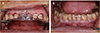
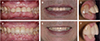
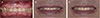 | Fig. 2Frontal view of initial photograph. (A) Intra-oral photograph, (B) Slight smile view, (C) Maximum smile view.
|
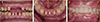
 | Fig. 3Analysis of teeth width and length. (A) Maxillary anterior teeth, (B) Mandibular anterior teeth.
|
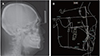 | Fig. 4Analysis of cephalometric projection. (A) Lateral cephalometric radiograph, (B) Tracing of lateral cephalometric radiograph.
|
 | Fig. 5Analysis of Inter-occlusal distance. (A) Rest vertical dimension, (B) Occlusal vertical dimension.
|
 | Fig. 6Analysis of Periodontal tissue and crown/root ratio for clinical crown lengthening. (A) Bone sounding, (B) Analysis of anatomic crown/root ratio.
|
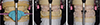
 | Fig. 7Semi-articulator mounting procedures. (A) Face-bow transfer, (B) Mounting the diagnostic model to an articulator in lateral view, (C) Mounting the diagnostic model to an articulator in frontal view.
|
 | Fig. 8Fabrication of the custom anterior guide table. (A) Right excursion in frontal view, (B) Left excursion in frontal view, (C) Protrusive excursion in lateral view.
|
 | Fig. 9Diagnostic wax-up in diagnostic model. (A) Wax-up on the mandibular anterior teeth, (B) Duplication of mandibular wax-up model, (C) Wax-up on the palatal surface of the maxillary anterior teeth, (D) Facial bite recording, (E) Re-mounting according to facial bite, (F) Wax-up on the labial surface of the maxillary anterior teeth.
|
 | Fig. 10Fabrication of the surgical template and the provisional restoration. (A) Full contour wax-up, (B) Surgical template, (C) Provisional restoration.
|
 | Fig. 11Surgical template application and clinical crown lengthening procedure. (A) Application of surgical template on the maxillary anterior teeth, (B) Application of surgical template on the mandibular anterior teeth.
|
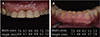
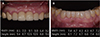
 | Fig. 12Teeth preparation and provisional restoration application. (A) Frontal view after teeth preparation, (B) Occlusal view after teeth preparation, (C) Frontal view after provisional restoration delivery.
|
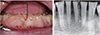
 | Fig. 13Individual tray fabrication and final impression taking. (A) Cord packing of maxillary anterior teeth, (B) Cord packing of mandibular anterior teeth, (C) Final impression of maxillary teeth, (D) Final impression of mandibular teeth.
|
 | Fig. 14Cross mounting of the master model with diagnostic model. (A) Mounting of the diagnostic models, (B) Mounting of the diagnostic model and master model, (C) Mounting of the master models.
|
References
1. Johansson A, Johansson AK, Omar R, Carlsson GE. Rehabilitation of the worn dentition. J Oral Rehabil. 2008; 35:548–566.

2. Berry DC, Poole DF. Attrition: possible mechanisms of compensation. J Oral Rehabil. 1976; 3:201–206.
3. Briggs P, Bishop K. Fixed prostheses in the treatment of tooth wear. Eur J Prosthodont Restor Dent. 1997; 5:175–180.
4. Hemmings KW, Darbar UR, Vaughan S. Tooth wear treated with direct composite restorations at an increased vertical dimension: results at 30 months. J Prosthet Dent. 2000; 83:287–293.
5. Dawson PE. Functional occlusion from TMJ to smile design. 1st ed. Amsterdam: Elsevier;2008. p. 114–122.
6. Turner KA, Missirlian DM. Restoration of the extremely worn dentition. J Prosthet Dent. 1984; 52:467–474.
7. Ochsenbein C, Ross S. A reevaluation of osseous surgery. Dent Clin North Am. 1969; 13:87–102.
8. Schuyler CH. The function and importance of incisal guidance in oral rehabilitation. 1963. J Prosthet Dent. 2001; 86:219–232.
9. Araujo NS, Moda MD, Silva EA, Zavanelli AC, Mazaro JV, Pellizzer EP. Survival of all-ceramic restorations after a minimum follow-up of five years: A systematic review. Quintessence Int. 2016; 47:395–405.
10. Ahmad I. Anterior dental aesthetics: facial perspective. Br Dent J. 2005; 199:15–21.
11. Dong JK, Jin TH, Cho HW, Oh SC. The esthetics of the smile: a review of some recent studies. Int J Prosthodont. 1999; 12:9–19.
12. Monteith B. The role of the free-way space in the generation of muscle pain among denture-wearers. J Oral Rehabil. 1984; 11:483–498.
13. Orthlieb JD, Laurent M, Laplanche O. Cephalometric estimation of vertical dimension of occlusion. J Oral Rehabil. 2000; 27:802–807.
14. Park JH, Jeong CM, Jeon YC, Lim JS. A study on the occlusal plane and the vertical dimension in Korean adults with natural dentition. J Korean Acad Prosthodont. 2005; 43:41–51.
15. Willis FM. Features of the face involved in full denture prosthesis. Dent Cosmos. 1935; 77:851–854.
16. Mohamed SE, Christensen LV. Mandibular reference positions. J Oral Rehabil. 1985; 12:355–367.
17. Silverman MM. The speaking method in measuring vertical dimension. 1952. J Prosthet Dent. 2001; 85:427–431.
18. Coslet JG, Vanarsdall R, Weisgold A. Diagnosis and classification of delayed passive eruption of the dentogingival junction in the adult. Alpha Omegan. 1977; 70:24–28.
 XML Download