

 PDF
PDF ePub
ePub Citation
 Print
Print


Abstract
The most important factor in the treatment of fully edentulous patients using implants is the shape of the definitive prosthesis. After the shape of the definitive prosthesis is determined, residual bone analysis and selection of the implant type, number and position should be followed. In this case, for restoration of an edentulous patient fully implanted (except the maxillary right lateral incisor) without considering definitive prosthesis, facial esthetics and possibility of fixed type prosthesis were evaluated using complete denture. It was determined that the fixed type prosthesis was possible. Implants that could not be used for the definitive prosthesis were excluded from the treatment plan and fixed type provisional restorations were fabricated. After four months of provisional restorations, the patient showed stable occlusion and esthetic satisfaction. Definitive prosthesis was made of zirconia using CAD/CAM (computer aided design and computer aided manufacturing). The results were satisfactory during the 3 months of follow-up period after termination of treatment. (J Korean Acad Prosthodont 2017;55:427-35)
Go to : 
REFERENCES
1.Almog DM., Sanchez R. Correlation between planned prosthetic and residual bone trajectories in dental implants. J Prosthet Dent. 1999. 81:562–7.

2.Misch CE. Dental implant prosthetics. 2nd ed.St. Louis: Mosby; Elsevier;2015. p. 187.
3.Turner KA., Missirlian DM. Restoration of the extremely worn dentition. J Prosthet Dent. 1984. 52:467–74.
4.Mericske-Stern RD., Taylor TD., Belser U. Management of the edentulous patient. Clin Oral Implants Res. 2000. 11:108–25.
5.Sadowsky SJ. The role of complete denture principles in implant prosthodontics. J Calif Dent Assoc. 2003. 31:905–9.
6.Neves FD., Mendonça G., Fernandes Neto AJ. Analysis of influence of lip line and lip support in esthetics and selection of maxillary implant-supported prosthesis design. J Prosthet Dent. 2004. 91:286–8.
7.Savabi O., Nejatidanesh F. Interocclusal record for fixed implant-supported prosthesis. J Prosthet Dent. 2004. 92:602–3.
8.Tarnow DP., Cho SC., Wallace SS. The effect of inter-implant distance on the height of inter-implant bone crest. J Periodontol. 2000. 71:546–9.
9.Carlsson GE. Dental occlusion: modern concepts and their application in implant prosthodontics. Odontology. 2009. 97:8–17.
10.Wittneben JG., Millen C., Brägger U. Clinical performance of screw-versus cement-retained fixed implant-supported reconstructions-a systematic review. Int J Oral Maxillofac Implants. 2014. 29:84–98.
11.Rajan M., Gunaseelan R. Fabrication of a cement- and screw-retained implant prosthesis. J Prosthet Dent. 2004. 92:578–80.
12.Abdulmajeed AA., Lim KG., Närhi TO., Cooper LF. Complete-arch implant-supported monolithic zirconia fixed dental prostheses: A systematic review. J Prosthet Dent. 2016. 115:672–7.
13.Misch CE. Dental Implant Prosthetics. 2nd ed.St. Louis: Mosby; Elsevier;2015. p. 277.
14.Linkevicius T., Puisys A., Vindasiute E., Linkeviciene L., Apse P. Does residual cement around implant-supported restorations cause peri-implant disease? A retrospective case analysis. Clin Oral Implants Res. 2013. 24:1179–84.
15.Chee WW., Duncan J., Afshar M., Moshaverinia A. Evaluation of the amount of excess cement around the margins of cement-retained dental implant restorations: the effect of the cement application method. J Prosthet Dent. 2013. 109:216–21.
Go to :
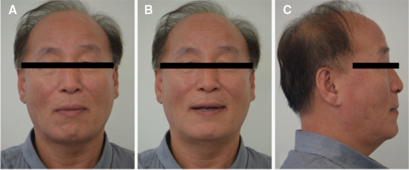 | Fig. 1.Initial extra-oral view (old denture wearing state); long face and lack of maxillary lip support. (A) Frontal view in centric occlusion, (B) Frontal view at rest position, (C) Lateral view. |
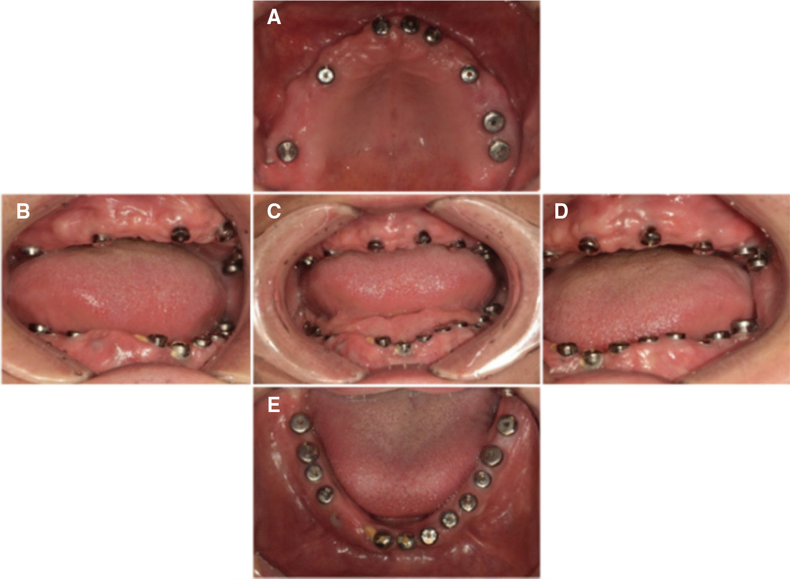 | Fig. 2.Initial intra-oral view. (A) Occlusal view of maxilla, (B) Lateral view (right side), (C) Frontal view, (D) Lateral view (left side), (E) Occlusal view of mandible. |
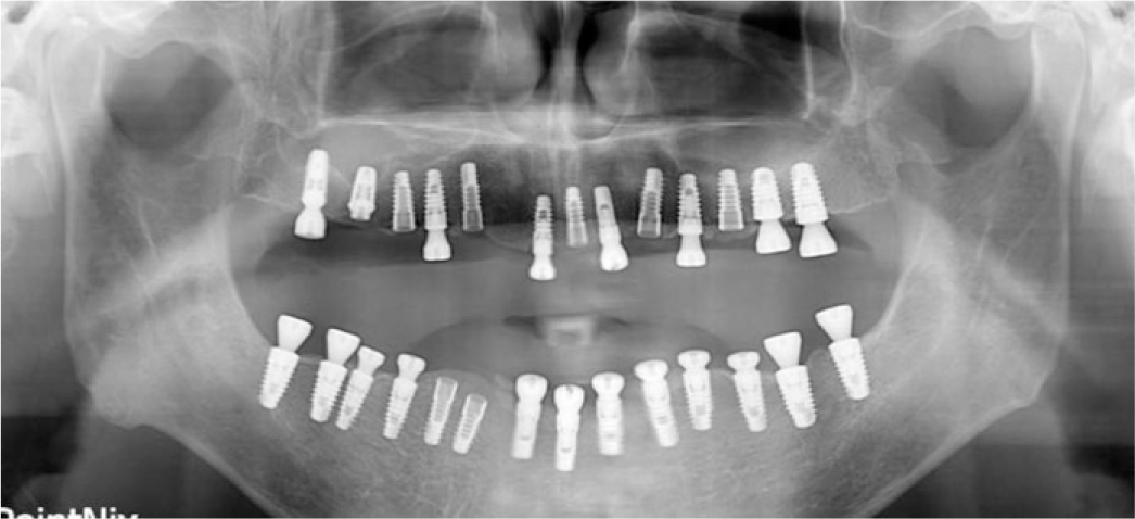 | Fig. 3.Initial panoramic view; various types of implant fixture fully implanted except right maxillary lateral incisor. |
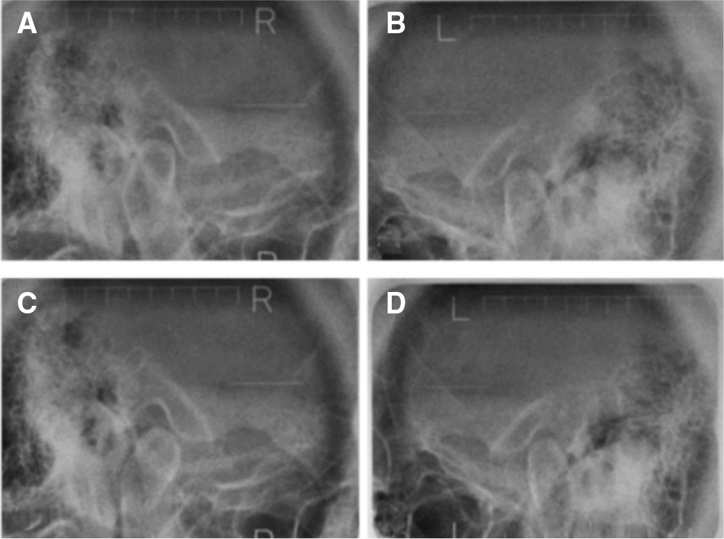 | Fig. 4.Initial trans-cranial view. (A) Right TMJ at rest position, (B) Left TMJ at rest position, (C) Right TMJ in MIP (maximal intercuspal position); condylar anterior displacement, (D) Left TMJ in MIP. |
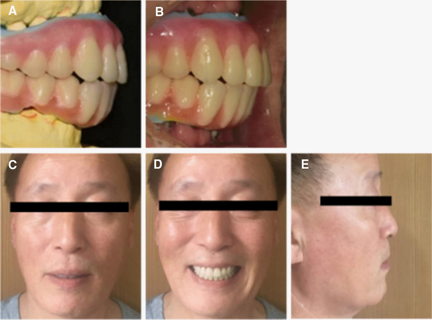 | Fig. 5.Evaluation of fixed type prostheses availability. (A) Duplication of complete denture with labial flange removed, (B) Try-in of duplicated denture with labial flange removed, (C) Evaluation of incisor exposure, (D) Evaluation of smile line, (E) Evaluation of facial support. |
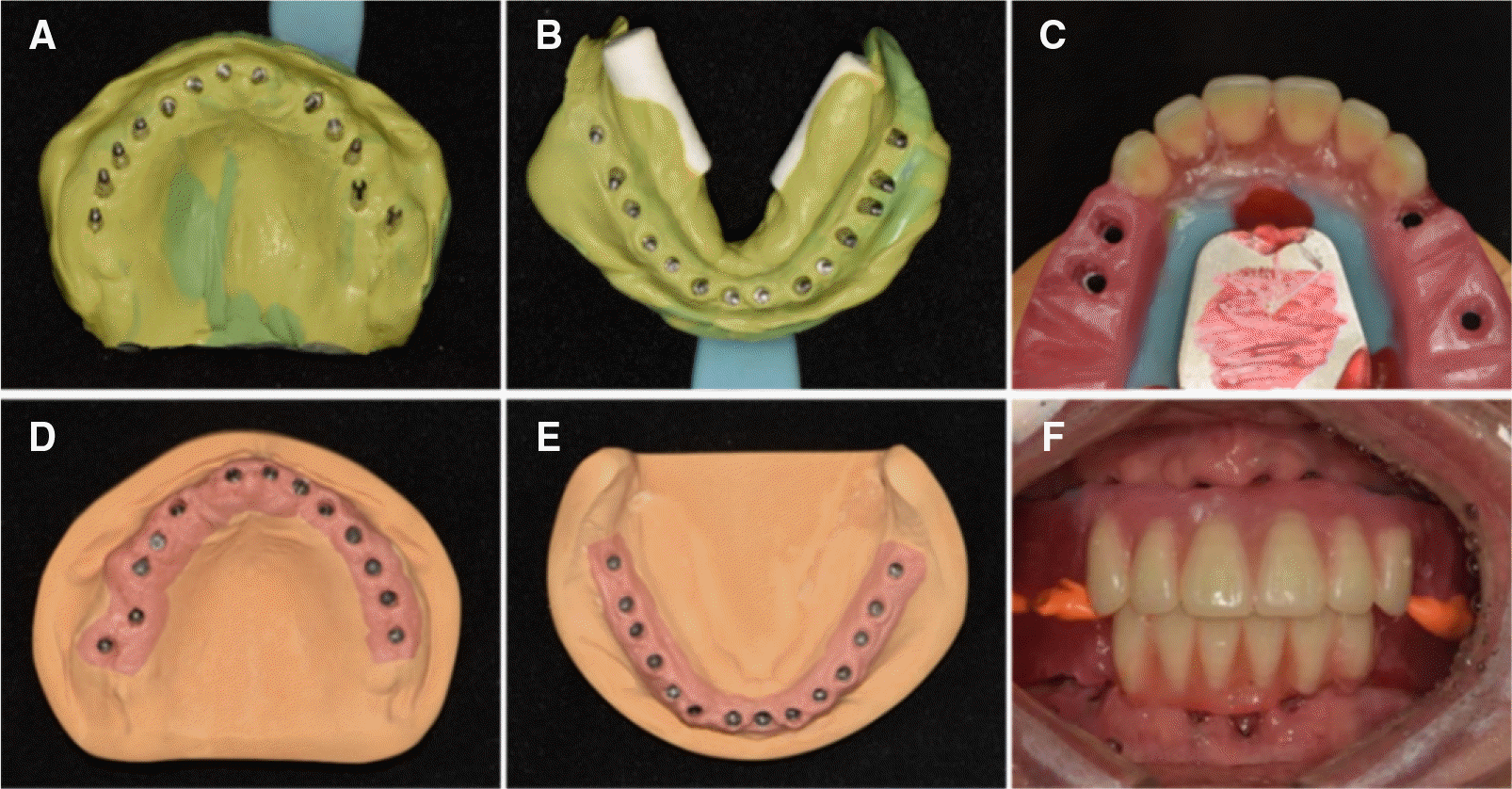 | Fig. 6.(A) Impression of maxillary implants, (B) Impression of mandibular implants, (C) Gothic arch tracing and implant supported record base, (D) Maxillary working model, (E) Mandibular working model, (F) Interocclusal record taking. |
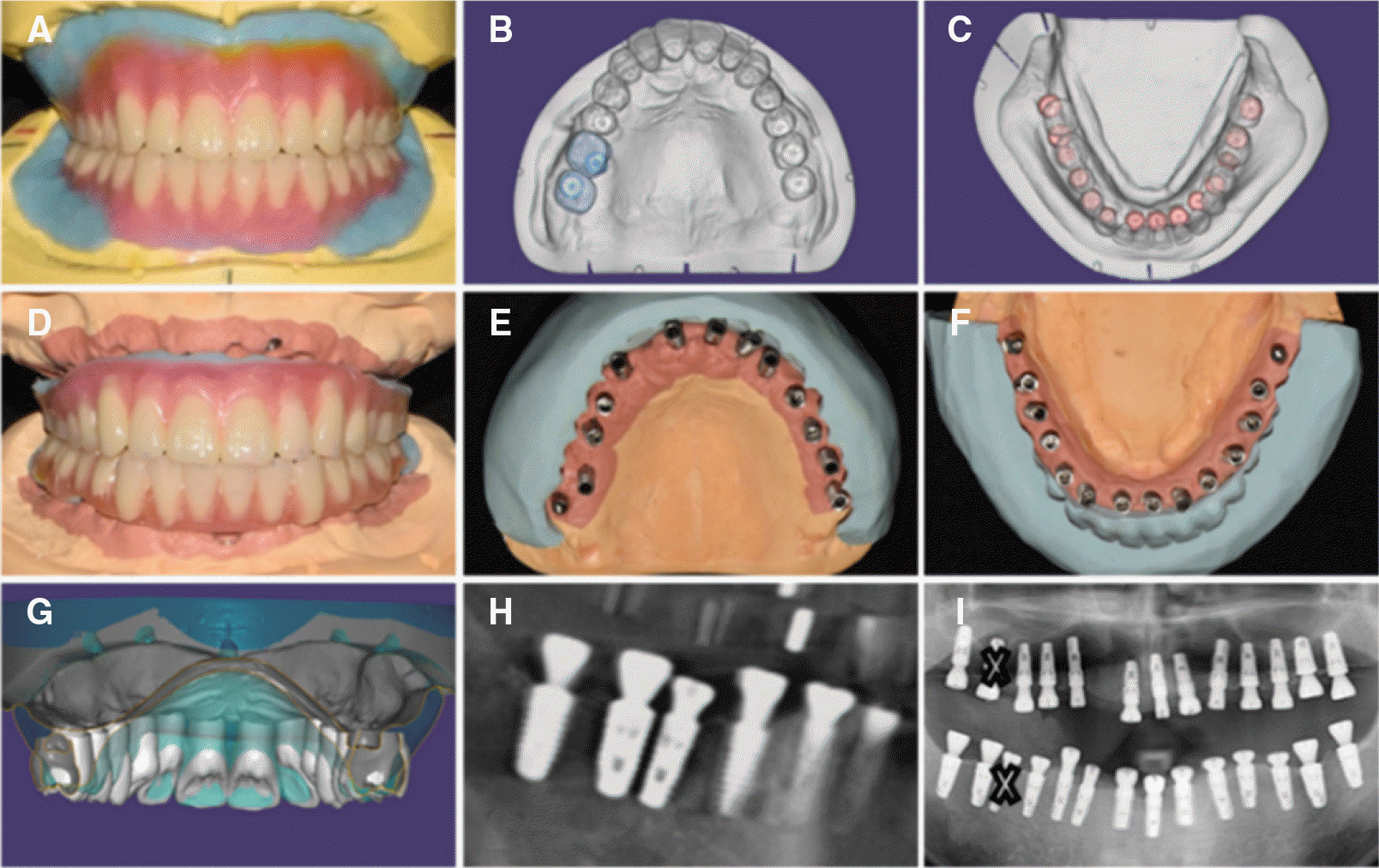 | Fig. 7.(A) Diagnostic model and wax denture, (B) Maxillary implants location analysis using CAD, (C) Mandibular implants location analysis using CAD, (D) Working model and wax denture, (E) Maxillary implants path analysis using silicone index, (F) Mandibular implants path analysis using silicone index, (G) Implant of the right maxillary first molar invasive of lingual space, (H) Implants of the right mandibular second premolar and first molar were too closely located mesio-distally, (I) Implants of the right maxillary first molar and right mandibular second premolar are excluded from treatment plan. |
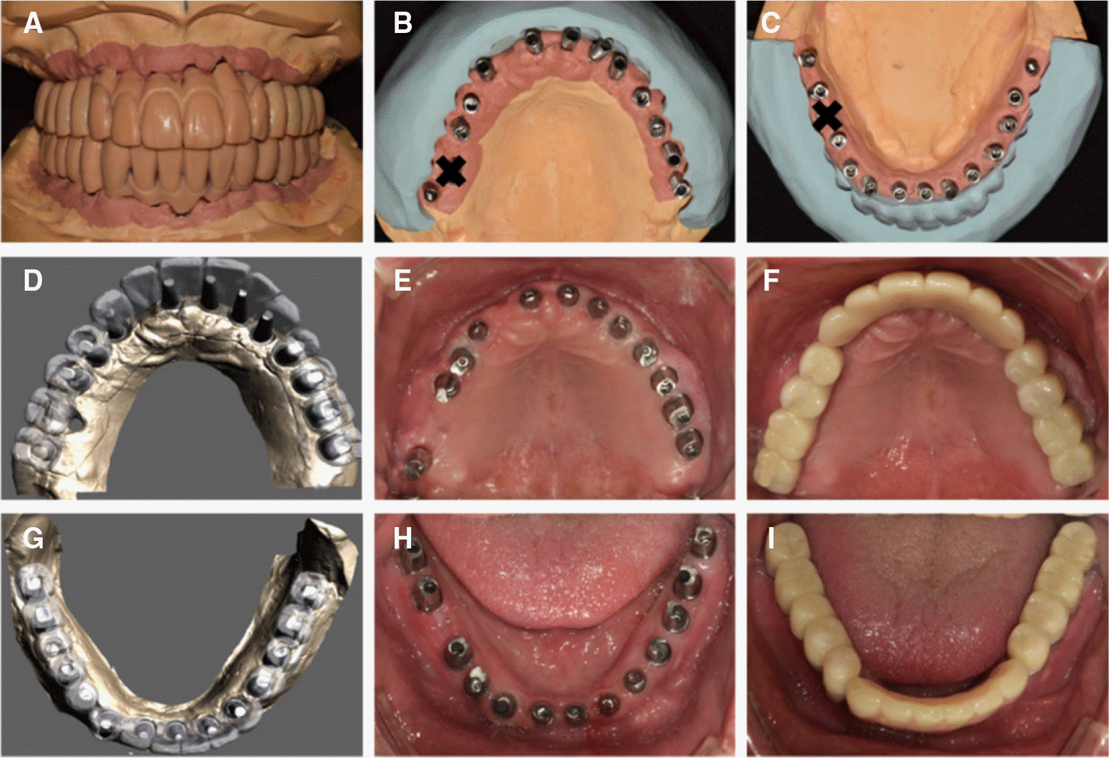 | Fig. 8.1st fixed type provisional prostheses. (A) Wax pattern for provisional prostheses, (B) Implants path analysis using silicon index (maxilla), (C) Implants path analysis using silicon index (mandible), (D) Computer aided design of customized abutment (maxilla), (E) Customized abutment setting (maxilla), (F) Fixed type provisional prostheses (maxilla), (G) Computer aided design of customized abutment (mandible), (H) Customized abutment setting (mandible), (I) Fixed type provisional prostheses (mandible). |

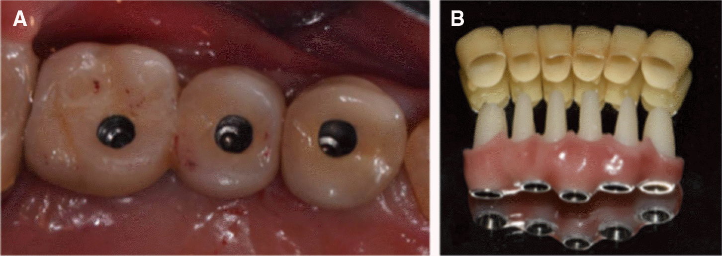 | Fig. 10.(A) SCRP type monolithic zirconia crown, (B) Screw type zirconia substructure and cement type monolithic zirconia crown. |
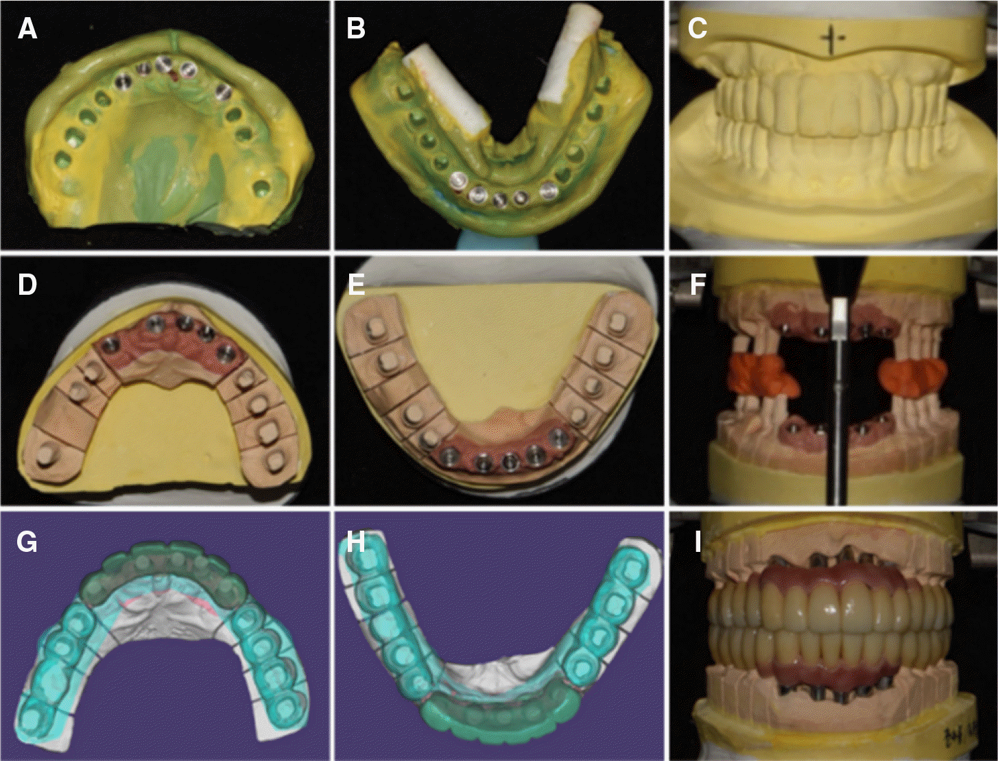 | Fig. 11.(A) Final impression of maxilla, (B) Final impression of mandible, (C) Model of provisional prostheses, (D) Working model of maxilla, (E) Working model of mandible, (F) Cross mounting, (G) Computer aided design (CAD) of Definitive prostheses (maxilla), (H) CAD of definitive prostheses (mandible), (I) Definitive prostheses fabrication using computer aided manufacturing (CAM). |
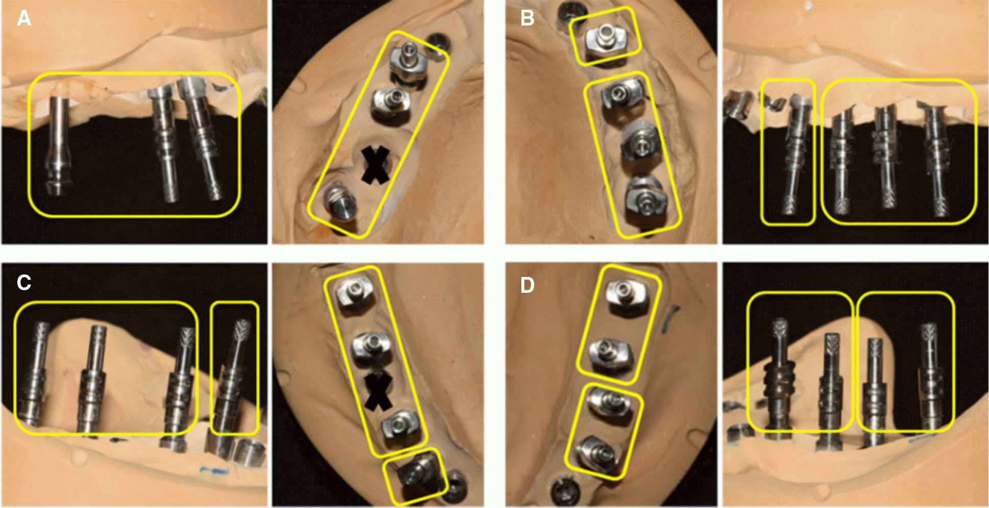 | Fig. 12.Segmentaion for retrievability of posterior prostheses. (A) Right maxillary posterior implants, (B) Left maxillary posterior implants, (C) Right mandibular posterior implants, (D) Left mandibular posterior implants. |
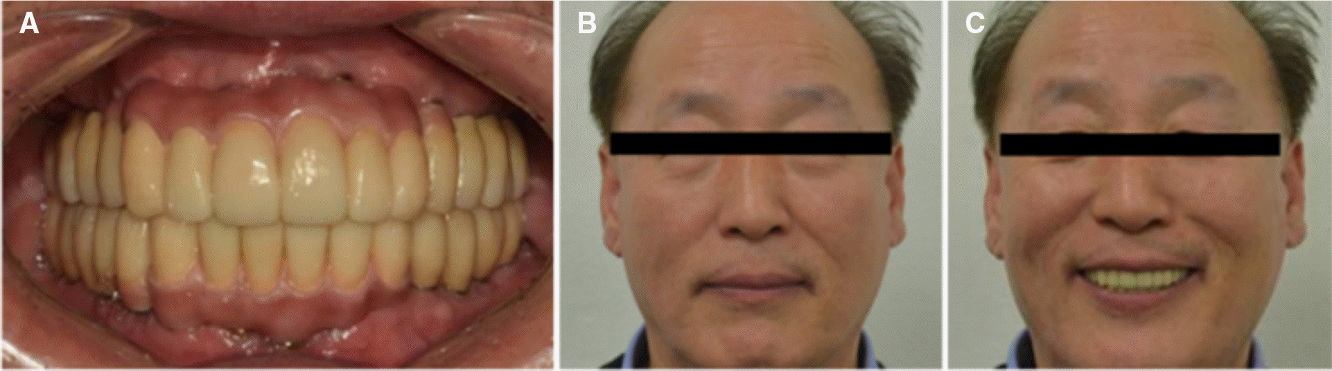 | Fig. 13.Definitive prostheses. (A) Intraoral view, (B) Extraoral view in centric occlusion, (C) Extraoral view on smile. |
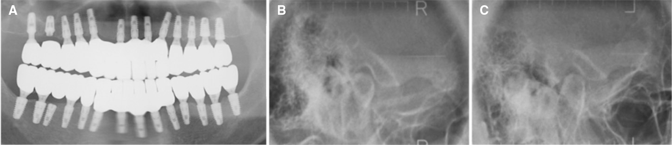 | Fig. 14.Evaluation of definitive prostheses. (A) Panoramic view, (B) Right trans-cranial view, (C) Left trans-cranial view. |
Table 1.
Distribution of implant type and abutment connection type
 XML Download