

 PDF
PDF ePub
ePub Citation
 Print
Print


Mi-Sun Shin1 , Jang-Hoon Lee2
, Jang-Hoon Lee2
Abstract
Purpose
This study was conducted to compare the marginal and internal fit of all ceramic crown using the replica technique and the triple-scan protocol.
Materials and methods
Twenty zirconia ceramic crowns were fabricated using titanium abutment model. All crowns were divided into two groups of 10 each, depending on the replica technique and the triple-scan protocol. The internal and marginal fit of 10 zirconia ceramic crowns were measured at 17 points for each specimen using the replica technique. The other 10 ceramic crowns were measured using the triple-scan protocol. Statistical analysis was performed by t-test (α= .05).
Results
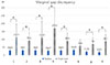
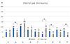
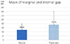
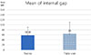
The mean and standard deviation of marginal and internal fit were significantly different between the replica technique (49.86 ± 29.69 µm) and triple-scan protocol (75.35 ± 64.73 µm, P<.001). The mean and standard deviation of internal fit except marginal fit were 58.38 ± 31.77 µm and 64.00 ± 46.43 µm, respectively (P>.343).
Figures and Tables
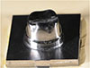 | Fig. 1Master model with titanium block made by using CAD/CAM system (Juyoung Dental Lab, Seoul, Korea).
|
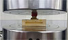 | Fig. 3The constant seating force (40 N) was maintained using a universal testing machine for 5 minutes.
|
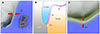
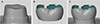 | Fig. 4Scanned three dimensional surface data. (A) Titanium master die, (B) zirconia crown, (C) superimposed crown on the master die.
|
 | Fig. 6Cross-section view of replica specimens. (A) Buccolingual section, ×50. AI: Axial internal gap. (B) AMD: Absolute marginal discrepancy, MG: Marginal gap. (C) Buccolingual section, ×50. OI: Occlusal internal gap.
|
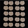
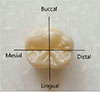
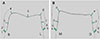 | Fig. 7Measurement points for marginal gap (1, 2, 8, 9, a, b, g, h) and internal gap (3, 4, 5, 6, 7, c, d, e, f) (A) Buccolingual section, (B) Mesiodistal section.
|


References
1. Jacobs MS, Windeler AS. An investigation of dental luting cement solubility as a function of the marginal gap. J Prosthet Dent. 1991; 65:436–442.

2. Knoernschild KL, Campbell SD. Periodontal tissue responses after insertion of artificial crowns and fixed partial dentures. J Prosthet Dent. 2000; 84:492–498.
3. Sailer I, Fehér A, Filser F, Gauckler LJ, Lüthy H, Hämmerle CH. Five-year clinical results of zirconia frameworks for posterior fixed partial dentures. Int J Prosthodont. 2007; 20:383–388.
4. Ko IS, Kim JM, Cho HW. Comparison of micro CT and crosssection technique for evaluation of marginal and internal fit of lithium disilicate crowns. J Korean Acad Prosthodont. 2016; 54:226–233.
5. Contrepois M, Soenen A, Bartala M, Laviole O. Marginal adaptation of ceramic crowns: a systematic review. J Prosthet Dent. 2013; 110:447–454.e10.
6. Rahme HY, Tehini GE, Adib SM, Ardo AS, Rifai KT. In vitro evaluation of the “replica technique” in the measurement of the fit of Procera crowns. J Contemp Dent Pract. 2008; 9:25–32.
7. Holst S, Karl M, Wichmann M, Matta RE. A new triple-scan protocol for 3D fit assessment of dental restorations. Quintessence Int. 2011; 42:651–657.
8. Planitz BM, Maeder AJ, Williams JA. The correspondence framework for 3D surface matching algorithms. Comput Vis Image Underst. 2005; 97:347–383.
9. Besl PJ, McKay ND. A method for registration of 3-D shapes. IEEE Trans Pattern Anal Mach Intell. 1992; 14:239–256.
10. Akca D. A new algorithm for 3D surface matching. In : 20th ISPRS Congress; July 12-23, 2004; Istanbul, Turkey. IAPRS& SIS;2004. 35(B7):p. 960–965.
11. Holmes JR, Bayne SC, Holland GA, Sulik WD. Considerations in measurement of marginal fit. J Prosthet Dent. 1989; 62:405–408.
12. Moon BH, Yang JH, Lee SH, Chung HY. A study on the marginal fit of all-ceramic crown using CCD camera. J Korean Acad Prosthodont. 1998; 36:273–292.
13. McLean JW, von Fraunhofer JA. The estimation of cement film thickness by an in vivo technique. Br Dent J. 1971; 131:107–111.
14. Gulker I. Margins. N Y State Dent J. 1985; 51:213–215.
15. Moldovan O, Rudolph H, Quaas S, Bornemann G, Luthardt RG. Internal and external fit of CAM-made zirconia bridge frameworks-a pilot study. Dtsch Zahnärztl Z. 2006; 61:38–42.
16. Molin MK, Karlsson SL, Kristiansen MS. Influence of film thickness on joint bend strength of a ceramic/resin composite joint. Dent Mater. 1996; 12:245–249.
17. Mörmann WH, Bindl A, Lüthy H, Rathke A. Effects of preparation and luting system on all-ceramic computer-generated crowns. Int J Prosthodont. 1998; 11:333–339.
18. Sorensen JA, Munksgaard EC. Interfacial gaps of resin cemented ceramic inlays. Eur J Oral Sci. 1995; 103:116–120.
19. Huh JB, Kim U, Kim HY, Kim JE, Lee JY, Kim YS, Jeon YC, Shin SW. Marginal and internal fitness of three-unit zirconia cores fabricated using several CAD/CAM systems. J Korean Acad Prosthodont. 2011; 49:236–244.
20. Groten M, Girthofer S, Pröbster L. Marginal fit consistency of copy-milled all-ceramic crowns during fabrication by light and scanning electron microscopic analysis in vitro. J Oral Rehabil. 1997; 24:871–881.
21. Kim H, Pae A. Marginal and internal fit of lithium disilicate crowns assessed by the triple-scan protocol. Kyunghee University;2017. MSD thesis.
22. Pimenta MA, Frasca LC, Lopes R, Rivaldo E. Evaluation of marginal and internal fit of ceramic and metallic crown copings using x-ray microtomography (micro-CT) technology. J Prosthet Dent. 2015; 114:223–228.
23. Matta RE, Schmitt J, Wichmann M, Holst S. Circumferential fit assessment of CAD/CAM single crowns-a pilot investigation on a new virtual analytical protocol. Quintessence Int. 2012; 43:801–809.
24. Lee DH. Digital approach to assessing the 3-dimensional misfit of fixed dental prostheses. J Prosthet Dent. 2016; 116:836–839.
25. Park JM, Hämmerle CH, Benic GI. Digital technique for in vivo assessment of internal and marginal fit of fixed dental prostheses. J Prosthet Dent. 2017; 03. 23. pii: S0022-3913(17)30008-2.
26. Pelekanos S, Koumanou M, Koutayas SO, Zinelis S, Eliades G. Micro-CT evaluation of the marginal fit of different In-Ceram alumina copings. Eur J Esthet Dent. 2009; 4:278–292.
27. Neves FD, Prado CJ, Prudente MS, Carneiro TA, Zancopé K, Davi LR, Mendonça G, Cooper LF, Soares CJ. Micro-computed tomography evaluation of marginal fit of lithium disilicate crowns fabricated by using chairside CAD/CAM systems or the heat-pressing technique. J Prosthet Dent. 2014; 112:1134–1140.
 XML Download