

 PDF
PDF ePub
ePub Citation
 Print
Print


Abstract
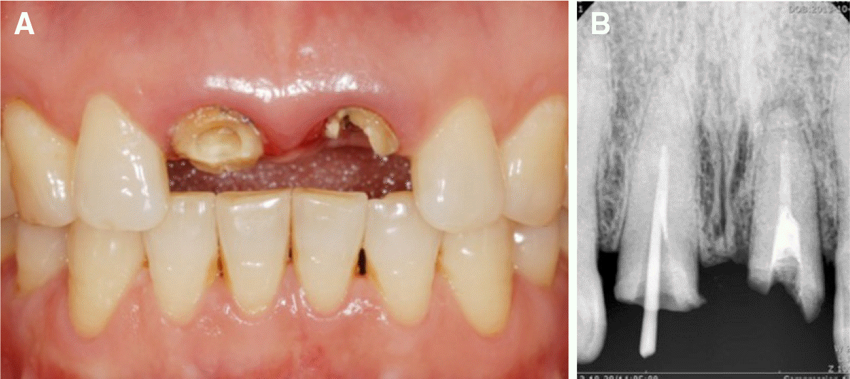
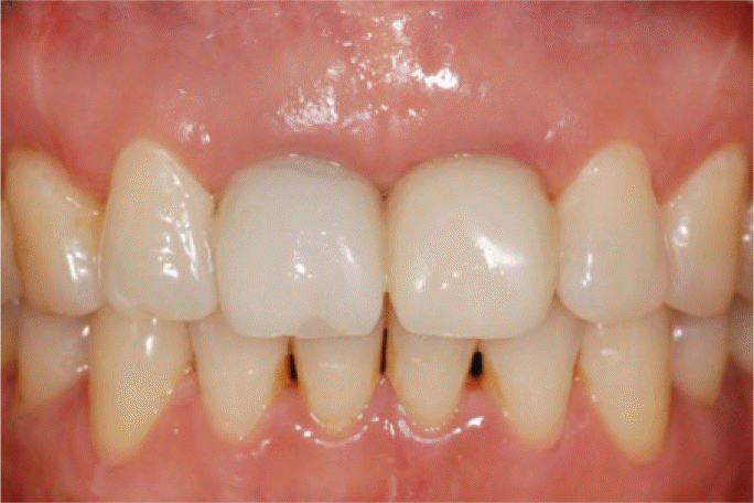
Maintaining the blood supply of the interdental alveolar bone is crucial for preserving the interdental papilla. Rebuilding the interimplant papilla between adjacent implants is more difficult than rebuilding the interdental papilla between the natural tooth and implant. Therefore, preserving the interimplant tissue is necessary when adjacent implants are closely placed. In this case report, three effective methods for maintaining the surrounding tissue, namely strategic serial extraction, immediate implantation, and provisionalization of adjacent maxillary central incisors, were performed. The marginal gingiva and interimplant papilla were well maintained for 24 months. (J Korean Acad Prosthodont 2017;55:286-91)
Go to : 
REFERENCES
1.Belser UC., Schmid B., Higginbottom F., Buser D. Outcome analysis of implant restorations located in the anterior maxilla: a review of the recent literature. Int J Oral Maxillofac Implants. 2004. 19:30–42.
2.Phillips K., Kois JC. Aesthetic peri-implant site development. The restorative connection. Dent Clin North Am. 1998. 42:57–70.
3.Tarnow DP., Magner AW., Fletcher P. The effect of the distance from the contact point to the crest of bone on the presence or absence of the interproximal dental papilla. J Periodontol. 1992. 63:995–6.

4.Chen MC., Liao YF., Chan CP., Ku YC., Pan WL., Tu YK. Factors influencing the presence of interproximal dental papillae between maxillary anterior teeth. J Periodontol. 2010. 81:318–24.
5.Salama H., Salama M., Kelly J. The orthodontic-periodontal connection in implant site development. Pract Periodontic Aesthet Dent. 1996. 8:923–32.
6.Tarnow D., Elian N., Fletcher P., Froum S., Magner A., Cho SC., Salama M., Salama H., Garber DA. Vertical distance from the crest of bone to the height of the interproximal papilla between adjacent implants. J Periodontol. 2003. 74:1785–8.
7.Greenstein G., Cavallaro J Jr. Serial extraction protocol: transitioning a hopeless dentition to a full-arch reconstruction. Compend Contin Educ Dent. 2008. 29:526–34.
8.Attard NJ., Zarb GA. Immediate and early implant loading protocols: a literature review of clinical studies. J Prosthet Dent. 2005. 94:242–58.
9.Arau′jo MG., Linder E., Lindhe J. Bio-Oss collagen in the buccal gap at immediate implants: a 6-month study in the dog. Clin Oral Implants Res. 2011. 22:1–8.
10.Tarnow DP., Cho SC., Wallace SS. The effect of interimplant distance on the height of interimplant bone crest. J Periodontol. 2000. 71:546–9.
11.Kan JY., Rungcharassaeng K. Interimplant papilla preservation in the esthetic zone: a report of six consecutive cases. Int J Periodontic Restor Dent. 2003. 23:249–59.
12.Salama H., Salama MA., Garber D., Adar P. The interproximal height of bone: a guidepost to predictable aesthetic strategies and soft tissue contours in anterior tooth replacement. Pract Periodontic Aesthet Dent. 1998. 10:1131–41.
13.Cooper LF., Raes F., Reside GJ., Garriga JS., Tarrida LG., Wiltfang J., Kern M., de Bruyn H. Comparison of radiographic and clinical outcomes following immediate provisionalization of single-tooth dental implants placed in healed alveolar ridges and extraction sockets. Int J Oral Maxillofac Implants. 2010. 25:1222–32.
14.Beagle JR. Surgical reconstruction of the interdental papilla: case report. Int J Periodontic Restor Dent. 1992. 12:145–51.
15.Salama H., Salama M. The role of orthodontic extrusive remodeling in the enhancement of soft and hard tissue profiles prior to implant placement: a systematic approach to the management of extraction site defects. Int J Periodontic Restor Dent. 1993. 13:312–33.
16.Gargiulo AW., Wentz FM., Orban B. Dimensions and relations of the dentogingival junction in humans. J Periodoltol. 1961. 32:261–7.
17.Kois JC. Altering gingival levels: the restorative connection part I: biologic variables. J Esthet Restor Dent. 1994. 6:3–7.
Go to :
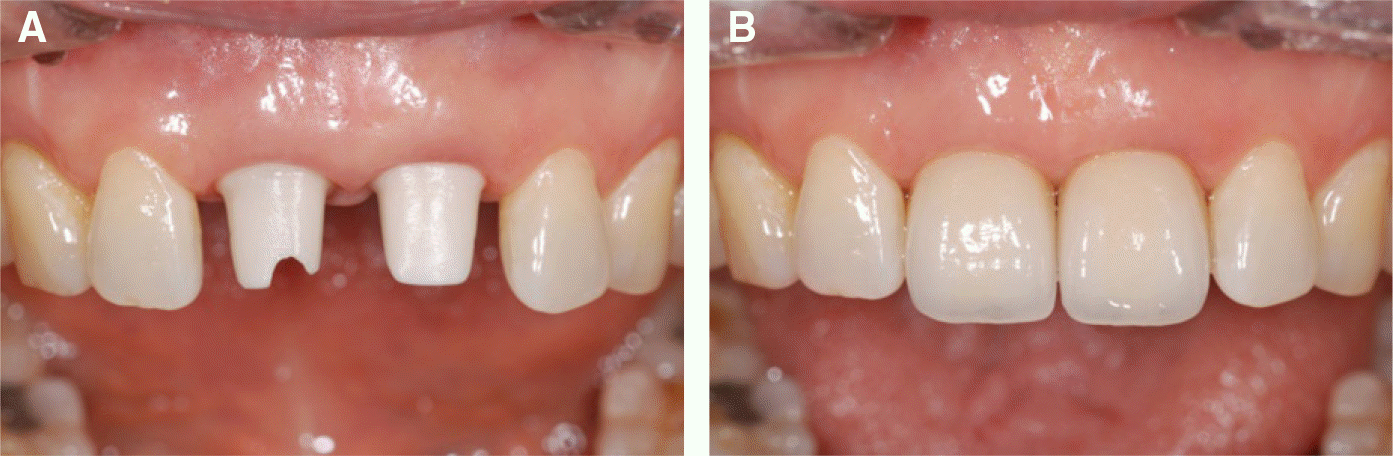
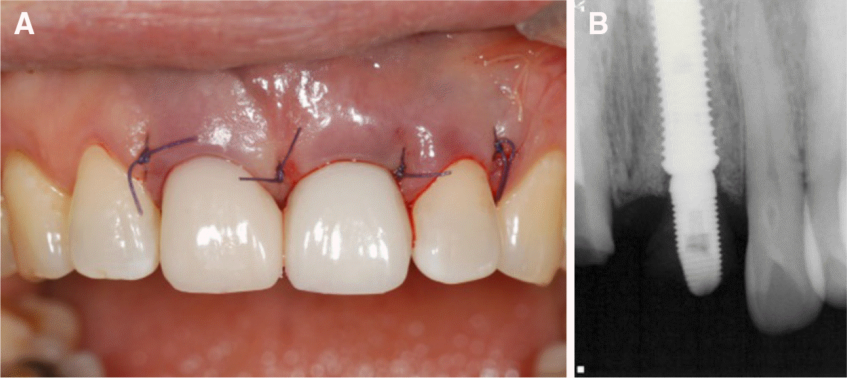 | Fig. 2.(A) Immediate implant was placed and provisional restoration was connected on the left maxillary central incisor, (B) Periapical radiograph after implantation on the left maxillary central incisor. |
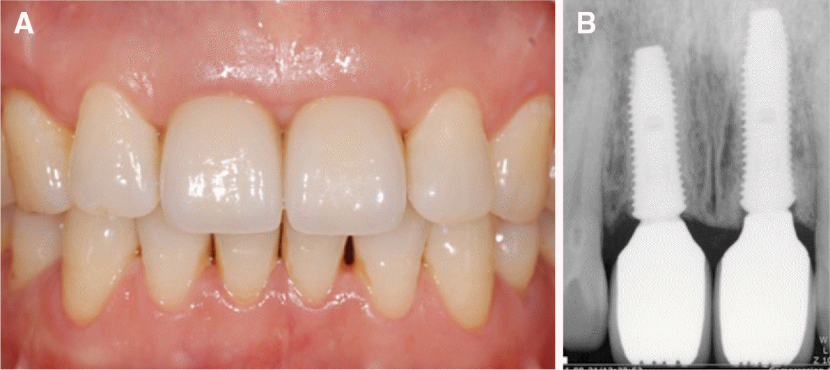
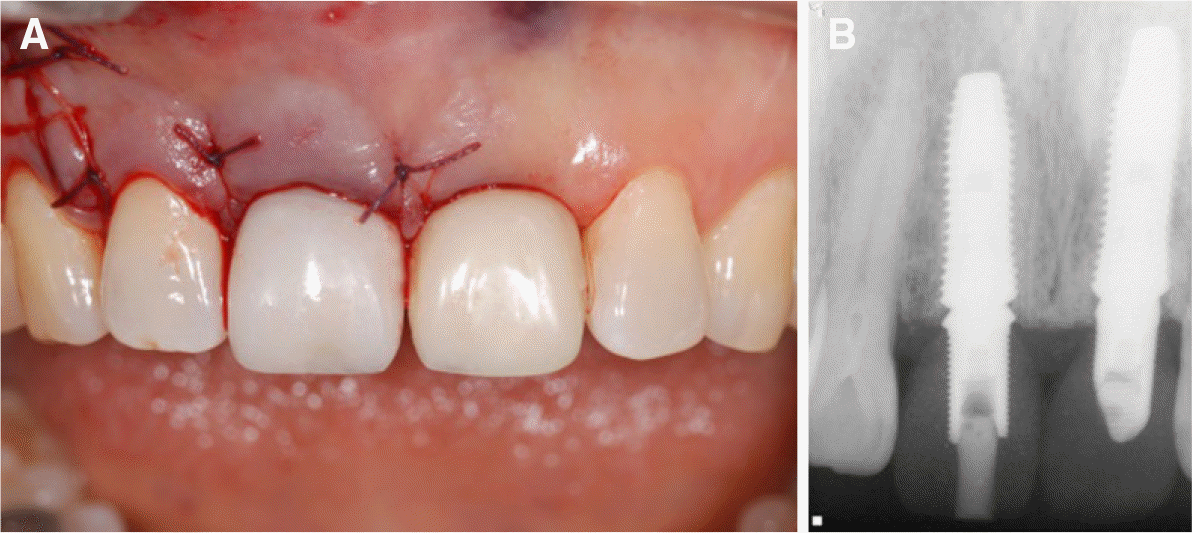 | Fig. 3.(A) Immediate implant was placed and provisional restoration was connected on the right maxillary central incisor, (B) Periapical radiograph after provisional restorations were connected. |
 XML Download