Abstract
Purpose
In this study, the retrospective radiographic study is executed to evaluate amount of bone loss of various conditions in patients using customized abutment for 4 years of follow-up.
Materials and methods
The subjects of this study were implant fixed dental prosthesis using CAD/CAM customized abutments. CAD/CAM customized abutment and fixed dental prosthesis were manufactured by the Prosthodontics Department of Chosun University Dental Hospital from August 1, 2011 to July 31, 2012. Radiological assessments were performed on the patients who were treated by the fixed prosthodontics. After each treatment, a retrospective study was performed for a total of 4 years at 3 months, 6 months, 1 year, 2 years, 3 years, and 4 years.
Results
As a result of the study, the customized abutment using CAD/CAM showed less bone loss than the results of existing research. There was no statistically significant differences at alveolar bone loss between splinting group and non-splinting group (respectively 0.27 mm, 0.5 mm). Also, there were statistically significant differences at alveolar bone loss in mx. anterior, mx. posterior, mn. anterior and mn. posterior part (respectively 1.37 mm, 0.39 mm, 0.00 mm, 0.30 mm).
Go to : 
REFERENCES
1.Goto T., Nishinaka H., Kashiwabara T., Nagao K., Ichikawa T. Main occluding area in partially edentulous patients: changes before and after implant treatment. J Oral Rehabil. 2012. 39:677–83.

2.Mericske-Stern RD., Taylor TD., Belser U. Management of the edentulous patient. Clin Oral Implants Res. 2000. 11:108–25.
3.Brånemark PI., Adell R., Breine U., Hansson BO., Lindström J., Ohlsson A. Intra-osseous anchorage of dental prostheses. I. Experimental studies. Scand J Plast Reconstr Surg. 1969. 3:81–100.
4.Albrektsson T., Brånemark PI., Hansson HA., Lindström J. Osseointegrated titanium implants. Requirements for ensuring a long-lasting, direct bone-to-implant anchorage in man. Acta Orthop Scand. 1981. 52:155–70.
5.Le Guéhennec L., Soueidan A., Layrolle P., Amouriq Y. Surface treatments of titanium dental implants for rapid osseointegration. Dent Mater. 2007. 23:844–54.

6.Hebel KS., Gajjar RC. Cement-retained versus screw-retained implant restorations: achieving optimal occlusion and esthetics in implant dentistry. J Prosthet Dent. 1997. 77:28–35.

7.Döring K., Eisenmann E., Stiller M. Functional and esthetic considerations for single-tooth Ankylos implant-crowns: 8 years of clinical performance. J Oral Implantol. 2004. 30:198–209.

8.Brunski JB. Biomaterials and biomechanics in dental implant design. Int J Oral Maxillofac Implants. 1988. 3:85–97.
9.Gross M., Abramovich I., Weiss EI. Microleakage at the abutment-implant interface of osseointegrated implants: a comparative study. Int J Oral Maxillofac Implants. 1999. 14:94–100.
10.Duke ES. The status of CAD/CAM in restorative dentistry. Compend Contin Educ Dent. 2001. 22:968–72.
11.Lewis S., Beumer J 3rd., Hornburg W., Moy P. The "UCLA" abutment. Int J Oral Maxillofac Implants. 1988. 3:183–9.
12.Fuster-Torres MA., Albalat-Estela S., Alcañiz-Raya M., Peñarrocha-Diago M. CAD/CAM dental systems in implant dentistry: update. Med Oral Patol Oral Cir Bucal. 2009. 14:E141–5.
13.Yoo HS., Kang SN., Jeong CM., Yun MJ., Huh JB., Jeon YC. Effects of implant collar design on marginal bone and soft tissue. J Korean Acad Prosthodont. 2012. 50:21–8.

14.Albrektsson T., Zarb G., Worthington P., Eriksson AR. The longterm efficacy of currently used dental implants: a review and proposed criteria of success. Int J Oral Maxillofac Implants. 1986. 1:11–25.
15.Porter JA., von Fraunhofer JA. Success or failure of dental implants? A literature review with treatment considerations. Gen Dent. 2005. 53:423–32.
16.Priest G. Virtual-designed and computer-milled implant abutments. J Oral Maxillofac Surg. 2005. 63:22–32.

17.Miyazaki T., Hotta Y., Kunii J., Kuriyama S., Tamaki Y. A review of dental CAD/CAM: current status and future perspectives from 20 years of experience. Dent Mater J. 2009. 28:44–56.

18.Galindo-Moreno P., Leo′n-Cano A., Monje A., Ortega-Oller I., O'Valle F., Catena A. Abutment height influences the effect of platform switching on peri-implant marginal bone loss. Clin Oral Implants Res. 2016. 27:167–73.

19.Cho YB., Moon SJ., Chung CH., Kim HJ. Resorption of labial bone in maxillary anterior implant. J Adv Prosthodont. 2011. 3:85–9.

20.Vigolo P., Zaccaria M. Clinical evaluation of marginal bone level change of multiple adjacent implants restored with splinted and nonsplinted restorations: a 5-year prospective study. Int J Oral Maxillofac Implants. 2010. 25:1189–94.
Go to : 
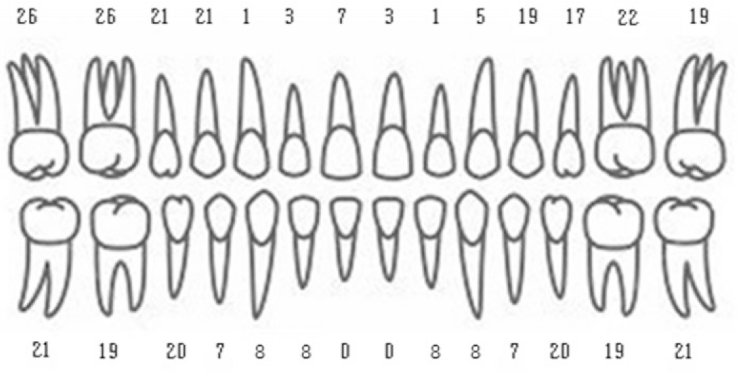
 | Fig. 1.Number of implants placed in each location (eg. 26 maxillary right 1st molar were replaced with implant supported single tooth prostheses). |
Table 1.
Causes of missing tooth
| Group | Periodontal disease | Caries | Trauma | Congenital missing | Periodontal disease & Caries | Total |
|---|---|---|---|---|---|---|
| No. of patient | 89 | 32 | 18 | 7 | 24 | 170 |
| No. of tooth | 207 | 62 | 31 | 10 | 47 | 357 |
Table 2.
Number of implant classified by implant company
| Classification of implant fixture | ||||||
|---|---|---|---|---|---|---|
| Astra | Biomet 3i | Zimmer | Dentis | Osstem | Total | |
| No. of fixture | 80 | 83 | 34 | 53 | 107 | 357 |
Table 3.
Total number of dental archs, number of fixtures and splinted fixtures
| No. of dental arch | No. of fixture | No. of splinted fixtures | Proportion of splinted fixtures | |
|---|---|---|---|---|
| Mx. | 85 | 199 | 161 | 81.0% |
| Mn. | 79 | 158 | 119 | 75.3% |
Table 4.
Vertical bone loss of around the implants classified by splinting
Table 5.
Vertical bone loss of around the implants classified by location of implant


 PDF
PDF ePub
ePub Citation
 Print
Print


 XML Download